Pressure and time dependence of the
cardiopulmonary reflex response in
patients with hypertensive cardiomyopathy
Unidade de Hipertensão, Instituto do Coração, Faculdade de Medicina, Universidade de São Paulo, São Paulo, SP, Brasil
M.E.B. Otto, F.M. Consolim-Colombo, C.R.M. Rodrigues Sobrinho and E.M. Krieger
Abstract
The first minutes of the time course of cardiopulmonary reflex control evoked by lower body negative pressure (LBNP) in patients with hypertensive cardiomyopathy have not been investigated in detail. We studied 15 hypertensive patients with left ventricular dysfunction (LVD) and 15 matched normal controls to observe the time course response of the forearm vascular resistance (FVR) during 3 min of LBNP at -10, -15, and -40 mmHg in unloading the cardiopulmonary receptors. Analysis of the average of 3-min intervals of FVR showed a blunted response of the LVD patients at -10 mmHg (P = 0.03), but a similar response in both groups at -15 and -40 mmHg. However, using a minute-to-minute analysis of the FVR at -15 and -40 mmHg, we observed a similar response in both groups at the 1st min, but a marked decrease of FVR in the LVD group at the 3rd min of LBNP at -15 mmHg (P = 0.017), and -40 mmHg (P = 0.004). Plasma norepineph-rine levels were analyzed as another neurohumoral measurement of cardiopulmonary receptor response to LBNP, and showed a blunted response in the LVD group at -10 (P = 0.013), -15 (P = 0.032) and -40 mmHg (P = 0.004). We concluded that the cardiopulmonary reflex response in patients with hypertensive cardiomyopathy is blunted at lower levels of LBNP. However, at higher levels, the cardiopulmonary reflex has a normal initial response that decreases progressively with time. As a consequence of the time-dependent response, the cardio-pulmonary reflex response should be measured over small intervals of time in clinical studies.
Correspondence
M.E.B. Otto
Unidade de Hipertensão, InCor Faculdade de Medicina, USP Av. Dr. Eneas C. Aguiar, 44 05403-000 São Paulo, SP Brasil
Fax: +55-11-3069-5048 E-mail: maria.otto@zerbini.org.br
Research supported by the E. Zerbini Foundation and FAPESP (No. 01/00009-0).
Received May 11, 2004 Accepted September 3, 2004
Key words
•Baroreflex
•Essential hypertension •Heart failure
•Cardiopulmonary reflex •Baroreceptors
Introduction
Hypertension is considered to be a major health problem, with clear evidence that it is one of the most important causes of heart failure (1). Indeed, heart failure (2-7) and hypertension (8-13) are associated with ab-normalities in cardiovascular reflexes and neurohumoral regulation, each of which are
2,5-7) and hypertension (9,10,25,26). How-ever, most studies of cardiopulmonary re-flex control in humans have used data aver-aged over several minutes, without includ-ing the 1st min of progressive levels of lower body negative pressure (LBNP) to analyze the nature of this impairment in patients with hypertension (9,10,25) and LVD (5,7). We are not aware of studies that analyzed the cardiopulmonary reflex minute-to-minute, including the 1st min of stimulation, which could be an important parameter to under-stand the mechanism of reflex impairment because the neural reflex response is very rapid at its onset. Therefore, the analysis of the average of several minutes might mask transient changes.
More recently, Hisdal et al. (27) observed transient changes in cardiovascular variables after the rapid onset and release of low levels of LBNP in normal volunteers, which clearly demonstrates that the intensity and time of stimulus application are important to the activation of the different receptors. Further-more, the reflex response is very rapid at its onset, and when the overall response is ana-lyzed as an average over several minutes, transient changes in the reflex response can-not be detected.
Our objective was to determine in pa-tients with hypertensive cardiomyopathy whether minute-to-minute analysis of the forearm vascular resistance (FVR) in com-parison with averaging over several minutes, as reported by many studies, is able to detect differences in the time course of the cardio-pulmonary reflex and to provide information about the mechanism of the reflex.
Subjects and Methods
Subjects
The study included 15 patients (8 men and 7 women), 35 to 59 years of age, mean body mass index 27.3 ± 2.5 kg/m2, with
mild-to-moderate LVD determined by
cal-culation of the ejection fraction by echocar-diography using Simpson’s method (group with LVD). The ejection fractions ranged from 36 to 50%, and the mean left ventricle diameter was 63 ± 2.3 mm. All patients were asymptomatic at rest and during exercise and none showed evidence of lung conges-tion on the day of the study. The LVD group was compared to 15 healthy controls (7 men and 8 women), mean body mass index 26.5 ± 2.0 kg/m2, 31 to 58 years of age (normal
control group). All subjects had normal labo-ratory tests that included a complete blood count as well as blood glucose, blood urea nitrogen, and serum creatinine levels. Total cholesterol levels (normal = 203 ± 7 vs LVD = 197 ± 11, P = NS) and triglyceride levels (normal = 106 ± 8 vs LVD = 107 ± 8, P = NS) were also similar in both groups. None of the patients were diabetic, had any other associ-ated disease or used any medications, in-cluding hormonal therapy for the women in both groups. Written consent was obtained from all subjects before the study, and the Medical Ethics Committee of the University of São Paulo Medical School, São Paulo, Brazil, approved the study protocol.
Hemodynamic measurements
The following hemodynamic measure-ments were performed: arterial blood pres-sure, heart rate, central venous pressure (CVP), forearm blood flow (FBF), and FVR. All measurements were recorded simulta-neously on a Gould strip-chart recorder (RS 3800; Gould Inc., Recording System Divi-sion, Valley View, OH, USA) and on a com-puter (Gateway 2000 4DX2-66V) with a CODAS system for data analysis (Computer Operated Data Acquisition Software: AT-CODAS; DATAQ Instruments, Inc., Akron, OH, USA).
2300, Monitoring Systems, Englewood, CO, USA). Heart rate was calculated by analysis of the peak systolic interval of the arterial pressure curves obtained with the digital photoplethysmograph. CVP (mmHg) was obtained with a polyethylene catheter (Intra-cath, 16 gauge/24"; Jonhson & Jonhson, Waterloo, Belgium) inserted percutaneously through an antecubital vein into the left arm and advanced to the right atrium under fluo-roscopy in the Hemodynamic Laboratory. This device was connected to a Gould P23 transducer in the Hypertension Laboratory, and the CVP values were determined; the zero reference was estimated at the subjects’ midaxillary level in the fourth intercostal space. FBF (ml/100 ml forearm volume per minute) was measured in the right arm by venous occlusion plethysmography using a double-stranded mercury-in-silastic strain gauge designed by Whitney (28). The venous occlusion pressure was 35 to 40 mmHg. The circulation in the hand was blocked by in-flating a wrist cuff above the systolic pres-sure for 1 min before limb blood flow was determined. Venous occlusion was per-formed every 10 s and 3 curves of FBF per min at baseline and increasing levels of LBNP were determined. The average was then calcu-lated as the mean value of data collected dur-ing 1 min of each stimulus. FVR (units) was calculated as mean arterial pressure/FBF.
Humoral measurements
Humoral measurements consisted of de-termining norepinephrine concentration in blood samples withdrawn from the right atrium at the end of the baseline period and at each level of LBNP. Blood was stored in pre-chilled tubes and processed at low tem-peratures by high performance liquid chro-matography.
Protocol
Hypertensive patients had their
medica-tion regimens changed with the progressive withdrawal of beta-blockers, angiotensing convertig enzyme inhibitors and central al-pha-blockers over a 3-week period. During the last week before the experiment, they received only vasodilators with short half-lives (nifedipine or hydralazine) and loop diuretics (furosemide). Administration of these medications was discontinued 24 h before the study. Patients did not consume alcohol or caffeine 24 h before the protocol, and no smokers were included in the study. The experimental protocol was similar to that used in a previous study performed in our laboratory (29). Hemodynamic variables were continuously measured during the 3-min baseline period and during sequential applications of LBNP at -10, -15, and -40 mmHg, with a 5-min rest between stimuli. Each LBNP stimulus was preceded by 1 min of baseline measurements to confirm that these parameters were stable.
Statistical analysis
Data were processed with JMP 5.0 soft-ware (a business unit from the SAS Insti-tute). Baseline values, the average of 3 min of each stimulus, and the norepinephrine response were compared between groups by the unpaired Student t-test. Analysis of vari-ance for repeated measures was used to com-pare the minute-to-minute responses of the FVR during the 3 min of LBNP at -10, -15 and -40 mmHg. P values of <0.05 were considered to be significant. Data are re-ported as means ± SEM.
Results
Baseline hemodynamic data
group, FBF decreased and FVR increased significantly from baseline (Table 3). The decrease in FBF variation and the percent increase in FVR variation were markedly reduced in the LVD group (Table 3).
LBNP at -15 and -40 mmHg. The de-crease in CVP was similar in both groups at -15 and -40 mmHg of LBNP (Table 2). Sys-tolic, diasSys-tolic, and mean blood pressures did not change significantly from baseline values in either group (Table 2). However, heart rate increased significantly from base-line in both groups at -40 mmHg of LBNP (Table 2). The percent variation in FBF de-crease and FVR inde-crease did not differ sig-nificantly from baseline for either group (Table 3).
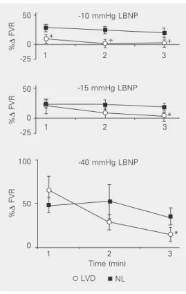
Hemodynamic responses to LBNP: analysis of the 1-min average data
Minute-to-minute FVR analysis showed that the LVD group has a blunted response during all 3 min at -10 mmHg of LBNP without differences between minutes (Fig-ure 1). Conversely, at -15 and -40 mmHg of LBNP, the LVD group had a normal re-sponse in the 1st min, but a marked decrease in FVR up to the 3rd min of each stimulus (Figure 1).
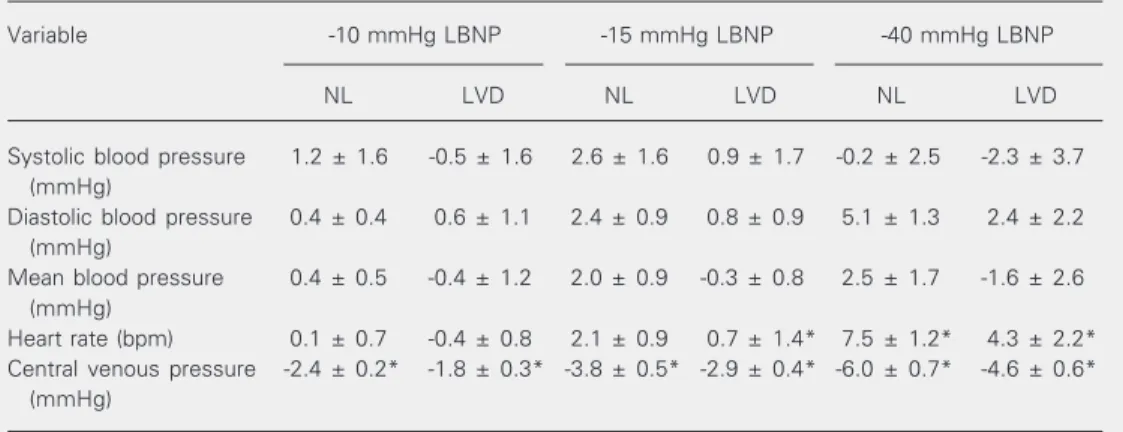
Table 2. Absolute changes in cardiovascular variables during lower body negative pressure.
Variable -10 mmHg LBNP -15 mmHg LBNP -40 mmHg LBNP
NL LVD NL LVD NL LVD
Systolic blood pressure 1.2 ± 1.6 -0.5 ± 1.6 2.6 ± 1.6 0.9 ± 1.7 -0.2 ± 2.5 -2.3 ± 3.7
(mmHg)
Diastolic blood pressure 0.4 ± 0.4 0.6 ± 1.1 2.4 ± 0.9 0.8 ± 0.9 5.1 ± 1.3 2.4 ± 2.2
(mmHg)
Mean blood pressure 0.4 ± 0.5 -0.4 ± 1.2 2.0 ± 0.9 -0.3 ± 0.8 2.5 ± 1.7 -1.6 ± 2.6
(mmHg)
Heart rate (bpm) 0.1 ± 0.7 -0.4 ± 0.8 2.1 ± 0.9 0.7 ± 1.4* 7.5 ± 1.2* 4.3 ± 2.2*
Central venous pressure -2.4 ± 0.2* -1.8 ± 0.3* -3.8 ± 0.5* -2.9 ± 0.4* -6.0 ± 0.7* -4.6 ± 0.6*
(mmHg)
Data have been compared to those in Table 1. Data are reported as means ± SEM for 15 subjects in each group. LBNP = lower body negative pressure; NL = normotensive control subjects; LVD = left ventricular dysfunction patients.
*P < 0.05 compared to baseline values (unpaired Student t-test).
Table 1. Baseline hemodynamic data of normo-tensive controls and hypernormo-tensive patients with left ventricular dysfunction.
Variable NL LVD
Systolic blood pressure 134 ± 3 181 ± 10*
(mmHg)
Diastolic blood pressure 73 ± 2 105 ± 7*
(mmHg)
Mean blood pressure 93 ± 2 129 ± 8*
(mmHg)
Heart rate (bpm) 65 ± 3 74 ± 3*
CVP (mmHg) 6 ± 1 8 ± 1
FBF (ml/100 ml tissue 3 ± 0.3 5 ± 1
per minute)
FVR (U) 37 ± 5 33 ± 4
Data are reported as mean ± SEM for 15 subjects in each group. NL = normotensive control sub-jects; LVD = left ventricular dysfunction patients; CVP = central venous pressure; FBF = forearm blood flow; FVR = forearm vascular resistance.
*P < 0.05 compared to NL (unpaired Student
t-test).
Hemodynamic responses to LBNP: analysis of the 3-min average data
Hormonal responses to LBNP
Plasma norepinephrine responses to LBNP at -10, -15, and -40 mmHg were meas-ured in 13 patients of the LVD group and 9 patients of the normal group. There was a trend to higher baseline plasma levels of norepinephrine (in pg/ml) in the LVD group (LVD: 342.6 ± 43.5 vs normal: 245.0 ± 23.2; P = 0.06). Percent variations in plasma nor-epinephrine during LBNP were attenuated in the LVD group at -10 mmHg (LVD: 7 ± 6
vs normal: 38 ± 9%, P = 0.013), -15 mmHg (LVD: 35 ± 6 vs normal: 54 ± 11%, P = 0.032), and -40 mmHg (LVD: 40 ± 7 vs
normal: 104 ± 13%, P = 0.004).
Discussion
The most important finding of this study was the accurate minute-by-minute charac-terization of the time course of the cardio-pulmonary reflex control during 3 min of LBNP in hypertensive cardiomyopathy. Im-portant information was lost when the anal-ysis averaged 3-min data. In contrast to pre-vious studies, by using a minute-to-minute analysis of FVR we observed a sustained blunted response in the LVD group during 3 min of LBNP at -10 mmHg, but a normal initial response at the 1st min of -15 and -40 mmHg of LBNP, with a marked decrease up to the 3rd min for each stimulus.
The present study is the first to character-ize the temporal behavior of the cardiopul-monary reflex response in patients with hy-pertensive cardiomyopathy using a minute-to-minute analysis. We included data for the 1st min of the stimulus, in contrast to the studies which used the average of several minutes and which did not include the 1st min of LBNP (8-10).
Normal volunteers submitted to rapid low levels of LBNP showed a decrease in mean arterial blood pressure during the first sec-onds of receptor unloading (27). These pre-mature hemodynamic alterations caused by
Table 3. Three-minute interval average measurements of variations in forearm vascu-lar resistance and forearm blood flow during lower body negative pressure.
Variable -10 mmHg LBNP (%) -15 mmHg LBNP (%) -40 mmHg LBNP (%)
NL LVD NL LVD NL LVD
FBF -15.1±5.2 0.8±5.0* -10.7±5.5 -0.1±5.3 -22.6±4.4 -18.4±6.1
FVR 23.8±6.2 4.2±6.0 20.8±6.5 10.4±9.5 45.2±10.7 36.0±14.1
Data have been compared to those in Table 1. Data are reported as means ± SEM for 15 subjects in each group. FBF = forearm blood flow; FVR = forearm vascular resistance; LVD = left ventricle dysfunction patients; NL = normal control subjects.
*P < 0.05 between LVD and NL (unpaired Student t-test).
LBNP may cause an early deactivation of the cardiopulmonary reflex. The present data revealed the importance of minute-to-minute analysis of the response from the 1st min for a better characterization of the physiology and pathophysiology of the cardiopulmo-nary reflex regulation. In the present data, unlike the minute-to-minute analysis of FVR, the analysis of the average of 3 min failed to show the impairment of the reflex response indicated by a blunted norepinephrine
re-%
∆
FVR
50
0
-25
%
∆
FVR
50
0
-25
%
∆
FVR
100
0 50
1 2 3
1 2 3
1 2 3
-15 mmHg LBNP
-40 mmHg LBNP -10 mmHg LBNP
LVD
Figure 1. Minute-to-minute changes in forearm vascular re-sistance during the first 3 min at -10, -15 and -40 mmHg of lower body negative pressure. Each minute was compared to data presented in Table 1. FVR = forearm vascular resistance; LBNP = lower body negative pressure; LVD = left ventricular dysfunction group; NL = normal
group. +P < 0.05 compared to
NL; *P < 0.05 compared to pre-vious minutes (analysis of vari-ance for repeated measures).
NL Time (min) +
+ +
*
sponse at -15 and -40 mmHg of LBNP in the LVD group.
The similar decrease in CVP observed during the application of increasing levels of LBNP indicates an equivalent receptor un-loading in both groups. Changes in FVR were observed without significant changes in arterial blood pressure or heart rate during -10 mmHg of LBNP, suggesting that the vascular responses were mainly due to the cardiopulmonary reflex. During -40 mmHg of LBNP, the small increase in heart rate indicated that the arterial baroreceptors, in addition to the cardiopulmonary barorecep-tors, were unloaded, in agreement with pre-vious reports that the application of LBNP higher than -20 mmHg also engages arterial baroreceptors in the final response (23,30). Nevertheless, this small increase does not exclude the participation of the arterial baroreceptors in the hemodynamic responses induced even by low levels of LBNP, such as -15 mmHg (23,30).
The impaired norepinephrine responses to LBNP in the LVD group constitute addi-tional evidence for an altered cardiopulmo-nary reflex response. Similar findings of an impaired norepinephrine response to LBNP were obtained in patients with LVD (5) and hypertension (10,25).
The minute-to-minute analyses of FVR at -10, -15 and -40 mmHg of LBNP were able to demonstrate a different pattern of response in the LVD group. There was a parallel, but blunted response of FVR in the LVD group compared to control at lower levels of LBNP (-10 mmHg), when the re-flex response is mainly a consequence of cardiopulmonary receptor unloading. How-ever, at higher levels of LBNP (-15 and -40 mmHg), when the arterial baroreceptors in addition to the cardiopulmonary receptors are participating in the reflex response, the LVD group had an increase in FVR similar to the control group in the 1st min, but a significant decrease over 3 min. In contrast, the control group showed a small change in
the reflex response along minutes, probably as a consequence of normal interaction of the two reflexes and a normal sympathetic activation response.
The mechanisms associated with impair-ment of the cardiopulmonary reflex and baro-reflex sensitivity (2,31-33) in patients with LVD and hypertension are not clear. To understand the mechanism underlying the present findings, we speculate that at higher levels of LBNP, the unloading of arterial baroreceptors could initially compensate the impairment of the cardiopulmonary reflex resulting in the initial normal response. How-ever, as a consequence of the limitations of the methodology of reflex analysis applied to humans, it is not possible to precisely separate the responses of cardiopulmonary receptors and baroreceptors and the exact time of their initial influence on the final response. In addition, the impaired response observed in hypertensive patients with left ventricle dysfunction might be also the con-sequence of alterations in the neurohumoral regulation of other substances, such as atrial natriuretic peptide and renin, each of which is also regulated by the cardiopulmonary reflex (34,35).
There is clear evidence that LBNP is a good test for orthostatic tolerance (36), and that the cardiopulmonary response is attenu-ated in these patients. Especially consider-ing the characteristics of progressive dete-rioration, the prevalence of orthostatic intol-erance in these patients could be increased. Therefore, frequently used medications to treat hypertension and LVD, such as vasodi-lators and diuretics, should be prescribed with caution to patients whose cardiopulmo-nary reflex is impaired.
periods of LBNP and the normal initial re-sponse at higher levels of LBNP, showing the importance to monitor the time-course of
References
1. Vasan RS & Levy D (1996). The role of hypertension in the
patho-genesis of heart failure. A clinical mechanistic overview. Archives of
Internal Medicine, 156: 1789-1796.
2. Zucker IH, Schultz HD, Li YF, Wang Y, Wang W & Patel KP (2004). The origin of sympathetic outflow in heart failure: the roles of
angiotensin II and nitric oxide. Progress in Biophysics and Molecular
Biology, 84: 217-232.
3. Zucker IH, Wang W, Brandle M, Schultz HD & Patel KP (1995). Neural regulation of sympathetic nerve activity in heart failure. Progress in Cardiovascular Diseases, 37: 397-414.
4. Packer M (1992). The neurohormonal hypothesis: a theory to
ex-plain the mechanism of disease progression in heart failure. Journal
of the American College of Cardiology, 20: 248-254.
5. Mohanty PK, Arrowood JA, Ellenbogen KA & Thames MD (1989). Neurohumoral and hemodynamic effects of lower body negative
pressure in patients with congestive heart failure. American Heart
Journal, 118: 78-85.
6. Modesti PA, Polidori G, Bertolozzi I, Vanni S & Cecioni I (2004). Impairment of cardiopulmonary receptor sensitivity in the early
phase of heart failure. Heart, 90: 30-36.
7. Ferguson DW, Abboud FM & Mark AL (1984). Selective impairment of baroreflex-mediated vasoconstrictor responses in patients with
ventricular dysfunction. Circulation, 69: 451-460.
8. Trimarco B, De Luca N, Ricciardelli B, Cuocolo A, Rosiello G, Lembo G & Volpe M (1986). Effects of lower body negative pressure in
hypertensive patients with left ventricular hypertrophy. Journal of
Hypertension, 4 (Suppl): S306-S309.
9. Trimarco B, De Luca N, Ricciardelli B, Cuocolo A, De Simone A, Volpe M, Mele AF & Condorelli M (1986). Impaired responsiveness of the ventricular sensory receptor in hypertensive patients with left
ventricular hypertrophy. Circulation, 74: 980-990.
10. Grassi G, Giannattasio C, Cleroux J, Cuspidi C, Sampieri L, Bolla GB & Mancia G (1988). Cardiopulmonary reflex before and after
regres-sion of left ventricular hypertrophy in essential hypertenregres-sion.
Hyper-tension, 12: 227-237.
11. Mancia G, Grassi G & Giannattasio C (1988). Cardiopulmonary
re-ceptor reflex in hypertension. American Journal of Hypertension, 1:
249-255.
12. Grassi G, Giannattasio C, Seravalle G, Cattaneo BM, Cleroux J & Mancia G (1990). Cardiogenic reflexes and left ventricular
hypertro-phy. European Heart Journal, 11 (Suppl G): 95-99.
13. Zanchetti A & Mancia G (1991). Cardiovascular reflexes and
hyper-tension. Hypertension, 18: III-13-III-21.
14. Moe GW, Rouleau JL, Charbonneau L, Proulx G, Arnold JM, Hall C, de Champlain J, Barr A, Sirois P & Packer M (2000). Neurohormonal activation in severe heart failure: relations to patient death and the
effect of treatment with flosequinan. American Heart Journal, 139:
587-595.
15. Cohn JN (1995). Plasma norepinephrine and mortality. Clinical
Car-diology, 18: I-9-I-12.
16. Omland T, Aarsland T, Aakvaag A, Lie RT & Dickstein K (1993). Prognostic value of plasma atrial natriuretic factor, norepinephrine
and epinephrine in acute myocardial infarction. American Journal of
Cardiology, 72: 255-259.
17. Packer M, Lee WH, Kessler PD, Gottlieb SS, Bernstein JL & Kukin ML (1987). Role of neurohormonal mechanisms in determining
survival in patients with severe chronic heart failure. Circulation, 75:
IV-80-IV-92.
18. Hirsch AT, Dzau VJ & Creager MA (1987). Baroreceptor function in congestive heart failure: effect on neurohumoral activation and
regional vascular resistance. Circulation, 75: IV-36-IV-48.
19. Chobanian AV, Bakris GL, Black HR et al. (2003). Seventh report of the Joint National Committee on Prevention, Detection, Evaluation,
and Treatment of High Blood Pressure. Hypertension, 42:
1206-1252.
20. Thoren P (1979). Role of cardiac vagal C-fibers in cardiovascular
control. Reviews of Physiology, Biochemistry and Pharmacology,
86: 1-94.
21. Salgado HC & Krieger EM (1981). Baroreceptor resetting during
pressure recovery from hypotension. Hypertension, 3: II-147-II-150.
22. Krieger EM, Salgado HC & Michelini LC (1982). Resetting of the
baroreceptors. International Review of Physiology, 26: 119-146.
23. Johnson JM, Rowell LB, Niederberger M & Eisman MM (1974). Human splanchnic and forearm vasoconstrictor responses to
re-ductions of right atrial and aortic pressures. Circulation Research,
34: 515-524.
24. Grassi G, Giannattasio C, Cuspidi C, Bolla GB, Cleroux J, Ferrazzi P, Fiocchi R & Mancia G (1988). Cardiopulmonary receptor regulation
of renin release. American Journal of Medicine, 84: 97-104.
25. Trimarco B, Lembo G, De Luca N, Volpe M, Ricciardelli B, Condorelli G, Rosiello G & Condorelli M (1989). Blunted sympathetic response to cardiopulmonary receptor unloading in hypertensive patients with left ventricular hypertrophy. A possible compensatory role of atrial
natriuretic factor. Circulation, 80: 883-892.
26. Uggere TA, Abreu GR, Sampaio KN, Cabral AM & Bissoli NS (2000). The cardiopulmonary reflexes of spontaneously hypertensive rats are normalized after regression of left ventricular hypertrophy and
hypertension. Brazilian Journal of Medical and Biological Research,
33: 589-594.
27. Hisdal J, Toska K & Walloe L (2001). Beat-to-beat cardiovascular
responses to rapid, low-level LBNP in humans. American Journal of
Physiology, 281: R213-R221.
28. Whitney RJ (1953). The measurement of volume changes in human
limbs. Journal of Physiology, 121: 1-27.
29. Consolim-Colombo FM, Filho JA, Lopes HF, Sobrinho CR, Otto ME, Riccio GM, Mady C & Krieger EM (2000). Decreased
cardiopulmo-nary baroreflex sensitivity in Chagas’ heart disease. Hypertension,
36: 1035-1039.
30. Pannier B, Slama MA, London GM, Safar ME & Cuche JL (1995). Carotid arterial hemodynamics in response to LBNP in normal
sub-jects: methodological aspects. Journal of Applied Physiology, 79:
1546-1555.
31. Zucker IH, Wang W & Schultz HD (1995). Cardiac receptor activity in heart failure: implications for the control of sympathetic nervous
outflow. Advances in Experimental Medicine and Biology, 381: 109-124.
32. Ferguson DW, Berg WJ, Roach PJ, Oren RM & Mark AL (1992). Effects of heart failure on baroreflex control of sympathetic neural
activity. American Journal of Cardiology, 69: 523-531.
33. Dibner-Dunlap M (1992). Arterial or cardiopulmonary baroreflex
con-trol of sympathetic nerve activity in heart failure. American Journal
of Cardiology, 70: 1640-1642.
34. Thomas CJ & Woods RL (2003). Guanylyl cyclase receptors
medi-ate cardiopulmonary vagal reflex actions of ANP. Hypertension, 41:
279-285.
35. Egan B, Fitzpatrick MA & Julius S (1987). The heart and the regula-tion of renin. Circularegula-tion, 75: I-130-I-133.
36. Hyatt KH, Jacobson LB & Schneider VS (1975). Comparison of 70 degrees tilt, LBNP, and passive standing as measures of orthostatic
tolerance. Aviation Space and Environmental Medicine, 46: