Characterization of nerve-induced
relaxation of gastrointestinal
sphincteric smooth muscle in
a South American opossum
(
Didelphis albiventris
)
Departamentos de 1Farmacologia e 2Clínica Médica, Faculdade de Medicina de Ribeirão Preto,
Universidade de São Paulo, 14049-900 Ribeirão Preto, SP, Brasil N.M. Matsuda1,
R.B. Oliveira2 and G. Ballejo1
Abstract
The presence of inhibitory nonadrenergic noncholinergic (NANC) intrinsic innervation of the circular muscle of the gastrointestinal sphincters of the South American (SA) opossum was investigated in vitro. Isolated circular muscle strips from the esophagogastric and ileocolonic junctions but not from the gastroduodenal (pylorus) region developed spontaneous tension. Tetrodotoxin (TTX, 1 µM) aug-mented the spontaneous tension only in the ileocolonic junction strips. Electrical field stimulation of esophagogastric and ileocolonic junc-tion strips caused frequency-dependent responses consisting of a relaxation at lower frequencies (<1 Hz) and a biphasic response or contraction at higher frequencies. In the strips from the pyloric region electrical field stimulation abolished the spontaneous activity at lower frequencies and induced contractions at higher frequencies. The re-sponses elicited by electrical field stimulation in the three sphincters were abolished by TTX (1 µM). Electrical field-induced contractions were reduced while relaxations were enhanced by atropine (1 µM). In the presence of atropine (1 µM) and guanethidine (3 µM), electrical field stimulation, nicotine and ATP induced frequency- or concentra-tion-dependent relaxations of the three sphincters that were abolished by TTX (1 µM). Isoproterenol and sodium nitroprusside caused concentration-dependent relaxations which were TTX-resistant. These findings indicate that the sphincteric circular muscle of the SA opos-sum gastrointestinal tract is relaxed by the activation of intrinsic NANC nerves and therefore can be used as a model for the study of the mechanisms involved in these responses.
Correspondence
N.M. Matsuda
Departamento de Farmacologia FMRP, USP
Av. Bandeirantes, 3900 14049-900 Ribeirão Preto, SP Brasil
E-mail: [email protected]
Research supported by FAPESP (No. 95/2544-8) and CNPq. N.M. Matsuda is the recipient of a Postdoctoral fellowship from FAPESP (No. 94/2752-7). R.B. Oliveira and G. Ballejo are recipients of CNPq fellowships.
Received March 8, 1996 Accepted April 7, 1997
Key words
•Nonadrenergic noncholinergic nerves
•Gastrointestinal sphincter relaxation
Introduction
Nerve-induced relaxation of gastrointes-tinal smooth muscle is involved in important physiological processes of the digestive tract such as lower esophageal sphincter relax-ation following swallowing, accommodrelax-ation to distention of the proximal stomach and descending inhibition (1). Evidence accu-mulated during the last three decades indi-cates that nerves mediating such responses employ neither noradrenaline nor acetylcho-line as a neurotransmitter and consequently are referred to as nonadrenergic noncholin-ergic (NANC) nerves (2). Experiments de-signed to elucidate the mediator and mecha-nisms involved in NANC nerve-induced re-laxations of sphincteric muscle in the gas-trointestinal tract have been performed mainly in North American (NA) opossums (Didelphis virginiana) (3-8) or in dogs (9-12). The properties of opossum gastrointes-tinal sphincters are closer to the human ones as compared to dogs and cats (13) but NA opossums are not readily available in Brazil. Consequently, it became of interest to
char-acterize in vitro the neurally induced
re-sponses of gastrointestinal sphincters from
Didelphis albiventris, an opossum species readily available in South America (SA), in order to determine whether it could be an alternative model to investigate the inhibito-ry innervation of sphincteric muscle from the gastrointestinal tract.
Material and Methods
South American opossums (Didelphis
albiventris) of both sexes, weighing 500-1500 g, were anesthetized with
pentobar-bital (50 mg/kg, ip). Laparotomy was
per-formed and the distal esophagus, stomach and duodenum as well as the distal ileum and
proximal colon were resected en bloc and
placed in Krebs solution containing 118.5
mM NaCl, 4.7 mM KCl, 2.5 mM CaCl2, 1.2
mM MgCl2, 1.2 mM KH2PO4, 23.8 mM
NaHCO3 and 11.1 mM glucose at 37oC and
bubbled with 95% O2/5% CO2. After
re-moval of the mesentery and fat tissue, the esophagogastric, gastroduodenal and ileo-colonic junctions were opened longitudinally and cleaned and the mucosa was removed. Mucosa-free strips (10 x 3 x 3 mm) of the circular muscle were cut and suspended in an isolated organ bath filled with 10 ml of
modified Krebs solution at 37oC bubbled
continuously with 95% O2/5% CO2. One end
of the strip was anchored inside the organ chamber and the other was connected to a force transducer for the measurement of iso-metric force on a four-channel Hewlett-Packard polygraph (model 7754A). Length-passive tension relationship experiments were performed by stretching the strips to different lengths (monitored by the basal tension) and recording the tension after an equilibration period of 30-45 min to estab-lish the optimal length. Once the optimal length was determined, in all the experi-ments in which the responses to electrical field stimulation (EFS) and drugs were char-acterized the strips were submitted to an initial tension of 1.5 g and an equilibration period of 45 min. Responses to EFS were elicited by applying 0.5-ms square wave pulses, supramaximal voltage (40 V) and varying frequencies (0.1-8 Hz) for 10 s de-livered with a Grass S4C stimulator through two platinum ring electrodes placed 20 mm apart above and below the muscle strip. The interval between successive stimuli was at least 2 min.
Results
Circular muscle strips from the lower esophageal sphincter and ileocolonic junc-tion always developed spontaneous tension, i.e., tension rose above the passively applied tension, the magnitude of which was related to the initial length of the strip. Maximum tension was developed when the initial length was set at 1.5 g (about 200% of the strip length). In contrast, though the pyloric strips did not develop spontaneous tension they also did not exhibit any relaxation after ap-plication of the initial tension. In addition, pyloric strips always showed spontaneous phasic activity while ileocolonic junction strips exhibited spontaneous activity that dis-appeared during the equilibration period. Lower esophageal sphincter strips did not show any spontaneous phasic activity. For all the experiments in which the responses to electrical field stimulation and drugs were characterized the strips were submitted to an initial tension of 1.5 g.
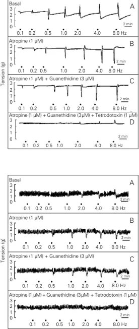
EFS of lower esophageal sphincter and ileocolonic junction strips caused a fre-quency-dependent response consisting of a relaxation at the lower frequencies (<1 Hz) and a biphasic response or contraction at the higher frequencies. In the strips from the pyloric region EFS abolished the spontane-ous activity at the lower frequencies of stim-ulation and induced a contraction at higher frequencies (Figures 1A, 2A and 3A). Addi-tion of atropine (1 µM) did not affect the tension of the strips in any of the sphincters (2.3 g vs 2.4 for lower esophageal sphincter, 1.5 g vs 1.6 g for pylorus and 2.1 g vs 2.0 g for ileocolonic junction) but was associated with a striking reduction in the magnitude of the contractile response and a corresponding enhancement or unmasking of relaxations in the three sphincters (Figures 1B, 2B and 3B). Further addition of guanethidine (3 µM) did not affect the basal tension or the re-sponses to EFS (Figures 1C, 2C and 3C). Similarly, addition of propranolol (10 µM)
Figure 1 - Representative trac-ings showing the response of isolated lower esophageal sphincter strips to different EFS frequencies (0.5 ms, 40 V for 10 s) in the absence (A) or in the presence of atropine (B), pine + guanethidine (C) or atro-pine + guanethidine + tetrodo-toxin (D). The strips were incu-bated with the drugs for at least 20 min.
Figure 2 - Representative trac-ings showing the response of isolated pyloric strips to differ-ent EFS frequencies (0.5 ms, 40 V for 10 s) in the absence (A) or in the presence of atropine (B), atropine + guanethidine (C) or atropine + guanethidine + tetro-dotoxin (D). The strips were in-cubated with the drugs for at least 20 min.
Basal
Tension (g)
3 2 1 0
0.1 0.2 0.5 1.0 2.0 4.0 8.0 Hz 2 min
A
B
C
D Atropine (1 µM) + Guanethidine (3 µM) + Tetrodotoxin (1 µM) Atropine (1 µM)
3 2 1 0
0.1 0.2 0.5 1.0 2.0 4.0 8.0 Hz 2 min
0.1 0.2 0.5 1.0 2.0 4.0 8.0 Hz 2 min Atropine (1 µM) + Guanethidine (3 µM)
0.1 0.2 0.5 1.0 2.0 4.0 8.0 Hz 2 min 3
2 1 0
3 2 1 0
Basal 3 2 1 0
0.1 0.2 0.5 1.0 2.0 4.0 8.0 Hz 2 min
A
B
C
D Atropine (1 µM) + Guanethidine (3 µM) + Tetrodotoxin (1 µM) Atropine (1 µM)
3 2 1 0
0.1 0.2 0.5 1.0 2.0 4.0 8.0 Hz 2 min
0.1 0.2 0.5 1.0 2.0 4.0 8.0 Hz 2 min Atropine (1 µM) + Guanethidine (3 µM)
0.1 0.2 0.5 1.0 2.0 4.0 8.0 Hz 2 min 3
2 1 0
3 2 1 0
Tension (g)
lower esophageal sphincter or the pyloric strips.
In the presence of atropine and guanethi-dine, nicotine induced concentration-depend-ent relaxations which were abolished by TTX (Figure 5) and hexamethonium (10 µM) (data not shown). ATP also caused concentration-dependent relaxations of the lower esophageal sphincter and ileocolonic junction strips in the presence of atropine and guanethidine which were abolished by TTX in the ileocolonic junction but not in lower esophageal sphincter strips (Figure 6). GABA (1-300 µM) did not cause any response in the three sphincters (data not shown).
Isoproterenol caused
concentration-de-pendent relaxations of the lower esophageal sphincter and ileocolonic junction (Figure 7) which were unaffected by TTX (data not shown). Similarly, VIP and SNP caused con-centration-dependent relaxations (data not shown).
Discussion
It is well established that gastrointestinal smooth muscle contractility is influenced by NANC inhibitory nerves (2). Following the description that the smooth muscle composi-tion of the NA opossum lower esophageal sphincter, in contrast to that of more com-mon laboratory animals (rats, cats and dogs), resembles more closely the human lower esophageal sphincter (13) most of the cur-rent concepts regarding the neural control of gastrointestinal sphincters were derived from
in vivo and in vitro studies performed on this animal model (3-8,14,15). Since this opos-sum species is not readily available to us, in
the present study we characterized in vitro
the nerve-induced responses of the circular smooth muscle from gastrointestinal sphinc-ters of the closely related opossum species
Didelphis albiventris, readily available in SA. Our results show that, as described for isolated strips of circular smooth muscle from the esophagogastric and ileocecal junc-tions of other species including humans (14-21), the strips from these regions obtained from the SA opossum developed spontane-ous tension which was not influenced by atropine or adrenergic blockers. The fact that TTX raised the tension in the ileoco-lonic junction but not in the lower esoph-ageal sphincter or pyloric strips indicates the presence of an intrinsic tonic inhibitory in-nervation in this region. Similar responses were reported in the lower esophageal sphinc-ter of the NA opossum (14) as well as in the ileocolonic junction of the cat and NA opos-sum (15,16). Since atropine and guanethi-dine did not affect the basal tension of the ileocolonic junction, such tonic innervation
Figure 3 - Representative trac-ings showing the response of isolated ileocolonic junction strips to different EFS frequen-cies (0.5 ms, 40 V for 10 s) in the absence (A) or in the presence of atropine (B), atropine + nethidine (C) or atropine + gua-nethidine + tetrodotoxin (D). The strips were incubated with the drugs for at least 20 min.
Basal 3 2 1 0
0.1 0.2 0.5 1.0 2.0 4.0 8.0 Hz 2 min
A
B
C
D Atropine (1 µM) + Guanethidine (3 µM) + Tetrodotoxin (1 µM) Atropine (1 µM)
3 2 1 0
0.1 0.2 0.5 1.0 2.0 4.0 8.0 Hz 2 min
0.1 0.2 0.5 1.0 2.0 4.0 8.0 Hz 2 min Atropine (1 µM) + Guanethidine (3 µM)
0.1 0.2 0.5 1.0 2.0 4.0 8.0 Hz 2 min 3
2 1 0
3 2 1 0
Tension (g)
Figure 4 - Relationship between the magnitude of EFS-induced NANC relaxations of isolated lower esophageal sphincter (filled circles) and ileocolonic junction (open circles) strips. Ten-second trains of pulses (0.5 ms and 40 V) were applied at 2-min intervals at different fre-quencies. Data are reported as the mean ± SEM (N = 8). All experiments were performed in the presence of 1 µM atropine and 3 µM guanethidine which were added 20 min before stim-ulation. The relaxation magni-tude is reported as % of that obtained with sodium nitroprus-side (SNP).
Relaxation (% of 10 µM SNP)
100
80
60
20 40
0
0.2 0.5 1.0 2.0 4.0 8.0
Figure 5 - Top, Representative tracings showing the response elicited by 100 µM nicotine on isolated lower esophageal sphincter (left) and ileocolonic junction (right) strips in the ab-sence or in the preab-sence of 1 µM tetrodotoxin (TTX). Bottom, Relationship between the mag-nitude of relaxations induced by different concentrations (M) of nicotine in isolated lower esoph-ageal sphincter (filled circles) and ileocolonic junction (open circles) strips. Data are reported as the mean ± SEM (N = 6). The relaxation magnitude is reported as % of that obtained with so-dium nitroprusside (SNP). All ex-periments were performed in the presence of 1 µM atropine and 3 µM guanethidine.
Relaxation (% of 10 µM SNP)
100
80
60
40
20
0
-6 -5 -4 -3 -2
Nicotine (log M)
Control TTX Control TTX
Nicotine Nicotine Nicotine Nicotine 2 min
1 g
Control TTX Control TTX
ATP ATP ATP ATP
2 min 1 g
Relaxation (% of 10 µM SNP)
60
40
20
0
-5 -4 -3 -2
ATP (log M)
Figure 6 - Top, Representative tracings showing the response elicited by ATP (300 µM) on the isolated lower esophageal sphincter (left) and ileocolonic junction (right) strips in the ab-sence or in the preab-sence of 1 µM tetrodotoxin (TTX). Bottom, Relationship between the mag-nitude of relaxations induced by different concentrations (M) of ATP in isolated lower esoph-ageal sphincter (filled circles) and ileocolonic junction (open circles) strips. Data are reported as the mean ± SEM (N = 6). The relaxation magnitude is reported as % of that obtained with so-dium nitroprusside (SNP). All experiments were performed in the presence of 1 µM atropine and 3 µM guanethidine.
seems to be due to intrinsic NANC nerves as described in the cat and guinea pig (16,17). The isolated strips of the circular muscle obtained from the pyloric region of SA opos-sums did not develop spontaneous tension but always exhibited regular spontaneous phasic activity. These findings are similar to those reported for the cat (18) and humans (19). Interestingly, TTX did not affect the basal tension of the pyloric strips, indicating a lack of tonic neural influences.
The present findings also show that elec-trical field stimulation of isolated circular muscle strips from the lower esophageal sphincter and ileocolonic junction of the SA opossum was associated with relaxation fol-lowed by contraction at the higher frequen-cies of stimulation. Both responses were abolished by TTX indicating that they re-sulted from the activation of intrinsic nerves. The fact that atropine reduced the EFS-in-duced contraction and enhanced the magni-tude of the relaxant responses in both strips while guanethidine and adrenergic blockers failed to affect the relaxations indicates that the latter are caused by the activation of NANC nerves. Inhibitory NANC nerves of the circular smooth muscle from the lower esophageal sphincter and ileocolonic junc-tion have been described in several species including NA opossums, cats, dogs and hu-mans (20-23). The fact that nicotine also caused relaxations of the lower esophageal sphincter and ileocolonic junction in a hexa-methonium- and TTX-sensitive manner sug-gests that the intrinsic NANC neurons of these regions from the SA opossum are acti-vated by nicotinic receptor stimulation. Cir-cular smooth muscle relaxations due to nico-tinic activation of intrinsic NANC neurons have been also reported in the guinea pig taenia coli, rat duodenum and dog ileoco-lonic junction (24-26). Therefore, the SA opossum is also comparable regarding this characteristic to the more widely employed models for studying inhibitory NANC inner-vation of gastrointestinal muscle.
TTX-Figure 7 - Relationship between the magnitude of relaxation in-duced by different concentra-tions (M) of isoproterenol in iso-lated lower esophageal sphinc-ter (filled circles) and ileocolonic junction (open circles) strips. Data are reported as the mean ± SEM (N = 6). The relaxation mag-nitude is reported as % of that obtained with sodium nitroprus-side (SNP). All experiments were performed in the presence of 1 µM atropine and 3 µM guanethi-dine.
Relaxation (% of 10 µM SNP)
100
80
60
20 40
0
-7 -6 -5 -4 -3
Isoproterenol (log M)
resistant manner. This observation is con-sistent with the putative role of ATP as an inhibitory NANC neurotransmitter although it is worth remarking that species differences
occur, since ATP has been shown to induce contractions in isolated human lower esoph-ageal sphincter strips (28). Isoproterenol, as well as the putative NANC neurotransmitter VIP, caused concentration-dependent relax-ations of both lower esophageal sphincter and ileocolonic junction strips in a TTX-resistant manner which is consistent with the potential role of the latter as a NANC neu-rotransmitter.
In conclusion, these findings show that isolated smooth muscle strips from the sphincteric regions of the SA opossum ex-hibit conspicuous inex-hibitory NANC nerve-induced responses and could be useful for studies aimed at the identification of the NANC neurotransmitter as well as to eluci-date the mechanisms involved in these neu-rally induced relaxations.
References
1. Burnstock G & Costa M (1973). Inhibitory innervation of the gut. Gastroenterology, 64: 141-144.
2. Burnstock G (1986). The non-adrenergic non-cholinergic nervous system. Archives Internationales de Pharmacodynamie et de Therapie, 280: 1-15.
3. Goyal RK & Rattan S (1978). Neurohu-moral, hormonal and drug receptors for the lower esophageal sphincter. Gastro-enterology, 74: 598-619.
4. Goyal RK, Rattan S & Said SI (1980). VIP as possible neurotransmitter of noncho-linergic nonadrenergic inhibitory neurons. Nature, 288: 378-380.
5. Tottrup A, Svane D & Forman A (1991). Nitric oxide mediating NANC inhibition in opossum lower esophageal sphincter. American Journal of Physiology, 260: G385-G389.
6. Murray J, Du C, Ledlow A, Bates JN & Conklin JL (1991). Nitric oxide: mediator of nonadrenergic, noncholinergic re-sponses of opossum esophageal muscle. American Journal of Physiology, 261: G401-G406.
7. Thorphy TJ, Fine CF, Burman M, Barnette MS & Ormsbee HS (1986). Lower esoph-ageal sphincter relaxation is associated with increased cyclic nucleotide content. American Journal of Physiology, 251: G786-G793.
8. Rattan S & Thatikunta P (1993). Role of nitric oxide in sympathetic neurotransmis-sion in opossum internal anal sphincter. Gastroenterology, 105: 827-836. 9. Boeckxstaens GE, Pelckmans PA,
Ram-part M, Verbeuren TJ, Herman AG & Van Maercke YM (1990). Nonadrenergic non-cholinergic mechanisms in the ileocolonic junction. Archives Internationales de Phar-macodynamie et de Therapie, 303: 270-281.
10. Grous M, Ormsbee III H & Barnette M (1990). Dimethylphenylpiperazinium (DMPP) induced relaxation and elevation of cyclic GMP content in canine lower esophageal sphincter (LES). Biochemical Pharmacology, 40: 1757-1762.
11. Phillips SF, Quigley EM, Kumar D & Kamath PS (1988). Motility of the ileoco-lonic junction. Gut, 29: 390-406. 12. Ward SM, McKeen ES & Sanders KM
(1992). Role of nitric oxide in nonadrener-gic, noncholinergic inhibitory junction po-tentials in canine ileocolonic sphincter. British Journal of Pharmacology, 105: 776-782.
13. Christensen J & Lund GF (1969). Esoph-ageal responses to distension and electri-cal stimulation. Journal of Clinical Investi-gation, 48: 408-419.
14. Goyal RK & Rattan S (1976). Genesis of basal sphincter pressure: Effect of tetro-dotoxin on lower esophageal sphincter pressure in vivo. Gastroenterology, 71: 62-67.
15. Conklin JL & Christensen J (1975). Local specialization at ileocecal junction of the cat and opossum. American Journal of Physiology, 228: 1075-1081.
16. Cardwell BA, Rubin MR, Snape Jr WJ & Cohen S (1981). Properties of the cat ileo-cecal sphincter muscle. American Journal of Physiology, 241: G222-G226. 17. Kubota M (1982). Electrical and
mechani-cal properties and neuro-effector trans-mission in the smooth muscle layer of the guinea-pig ileocecal junction. Pflügers Archiv, European Journal of Pharmacolo-gy, 394: 353-361.
18. Bertiger G, Reynolds JC, Ouyang A & Cohen S (1987). Properties of the feline pyloric sphincter in vitro. Gastroenterol-ogy, 92: 1965-1972.
19. Schulze-Delrieu K & Shirazi SS (1983). Neuromuscular differentiation of the hu-man pylorus. Gastroenterology, 84: 287-292.
21. Christensen S, Conklin J & Freeman B (1973). Physiological specialization at the esophagogastric junction in three species. American Journal of Physiology, 225: 1265-1270.
22. Tottrup A, Forman A, Funch-Jensen P, Raundahl U & Anderson KE (1990). Ef-fects of transmural field stimulation in iso-lated muscle strips from human esopha-gus. American Journal of Physiology, 258: G344-G351.
23. Tottrup A, Forman A, Uldbjerg N, Funch-Jensen P & Anderson KE (1990). Mechan-ical properties of isolated human esoph-ageal smooth muscle. American Journal of Physiology, 258: G338-G343.
24. Burnstock G, Campbell G & Rand MJ (1966). The inhibitory innervation of the taenia of the guinea-pig caecum. Journal of Physiology, 182: 504-526.
25. Guimarães CRC, Rodrigues LA, Vettore O & Antonio A (1988). The relaxing response of the isolated rat duodenum to nicotine. General Pharmacology, 19: 655-659. 26. Pelckmans PA, Boeckxstaens GE, Van
Maercke Y, Herman AG & Verbeuren TJ (1989). Acetylcholine is an indirect inhibi-tory transmitter in the canine ileocolonic junction. European Journal of Pharmacol-ogy, 170: 235-242.
27. Daniel EE, Crankshaw J & Sarna S (1979). Prostaglandins and tetrodotoxin-insensi-tive relaxation of opossum lower esoph-ageal sphincter. American Journal of Physiology, 236: E153-E172.