www.reumatologia.com.br
REVISTA BRASILEIRA DE
REUMATOLOGIA
Original article
Evaluation of respiratory impairment in patients with
systemic lupus erythematosus with the six-minute walk test
Marivone Arruda Leite, Mônica Corso Pereira, Lílian Tereza Lavras Costallat,
Wander de Oliveira Villalba, Marcos Mello Moreira, Ilma Aparecida Paschoal*
Department of Internal Medicine, Universidade Estadual de Campinas (UNICAMP), Campinas, SP, Brazil
a r t i c l e i n f o
Article history:
Received on 24 April 2013 Accepted on 10 February 2014
Keywords:
Systemic lupus erythematosus Six-minute walk test
Oxygen saturation Respiratory function test Questionnaires
a b s t r a c t
Objective: Evaluate SLE stable patients, without overt respiratory compromise, by means of 6MWT.
Casuistic and methods: Forty-ive stable SLE patients were enrolled. The ATS/ERS protocol for 6MWT, was used and two parameters with cut-off points were chosen.
Results: Forty-two patients were women. The mean age was 39 ± 11.4 years; mean duration of disease, 121 ± 93.1 months; mean value of MRC, 2 ± 0; mean FVC, 85.9 ± 34.2%; mean FEV1, 67.5 ± 21.6%; mean MIP, 82 ± 58.4%; mean MEP, 78 ± 37.3%; mean heart rate at rest, 75 ± 12.8 bpm; mean respiratory rate at rest, 19 ± 5.3 bpm; mean 6MWD, 478 ± 82 m; mean SpO2 at rest was 98 ± 0.8%; mean fall in SpO2, 4 ± 6 points. When the study population was divided according to the 400-m walk distance cut-off value, the heart rate immediately before the test was signiicant lower in those participants who walked less than 400 m (p = 0.0043), just like the value of Borg scale (p = 0.0036); according to the presence of saturation ≥ 4, heart rate at the end of the test was signiicantly higher in those participants who were showing desaturation (p = 0.0170); MEP (p = 0.0282) and 6MWD (p = 0.0291) were signii-cantly lower, and MIP showed a tendency towards being smaller (p = 0.0504). FVC < normal inferior limit was signiicantly associated with the group with desaturation (p = 0.0274). Conclusion: Compared to 6MWD, desaturation was better suited to ind the patients with the most compromised indexes in respiratory function tests.
© 2014 Sociedade Brasileira de Reumatologia. Published by Elsevier Editora Ltda. All rights reserved.
* Corresponding author.
E-mail: [email protected] (I.A. Paschoal).
193
R E V B R A S R E U M A T O L . 2 0 1 4 ;5 4 ( 3 ): 1 9 2 – 1 9 9Avaliação do comprometimento respiratório em pacientes com lúpus eritematoso sistêmico com o teste de caminhada de seis minutos
Palavras-chave:
Lúpus eritematoso sistêmico (LES) Teste de caminhada de seis minutos
Saturação de oxigênio Teste de função respiratória Questionários
r e s u m o
Objetivo: Avaliar pacientes com LES estável, sem comprometimento respiratório evidente, por meio do TC6M.
Casuística e métodos: Foram recrutados 45 pacientes com LES estável. Foi utilizado o protoco-lo ATS/ERS para TC6M, tendo sido escolhidos dois parâmetros com pontos de corte. Resultados: Quarenta e dois dos pacientes eram mulheres. A média de idade foi 39 ± 11,4 anos; a duração média da doença, 121 ± 93,1 meses; valor médio de MRC 2 ± 0; CVF média 85,9 ± 34,2%; VEF1 médio 67,5 ± 21,6%; PIM média 82 ± 58,4%; PEM média 78 ± 37,3%; frequência car-díaca média em repouso 75 ± 12,8 bpm; frequência respiratória média em repouso 19 ± 5,3 bpm; Distância média no TC6M 478 ± 82 m; SpO2 média em repouso 98 ± 0,8%; queda média em SpO2 4 ± 6 pontos. Quando a população em estudo foi dividida de acordo com o valor de corte de 400 m de distância caminhada, a frequência cardíaca imediatamente antes do tes-te foi signiicativamentes-te menor naqueles participantes-tes que caminharam menos de 400 m (p = 0,0043), da mesma forma que o valor da escala de Borg (p = 0,0036). De acordo com a pre-sença de saturação ≥ 4, a frequência cardíaca ao inal do teste estava signiicativamente mais elevada naqueles participantes exibindo dessaturação (p = 0,0170); PEM (p = 0,0282) e TC6M (p = 0,0291) estavam signiicativamente menores e PIM revelou uma tendência para dimi-nuir (p = 0,0504). CVF < limite inferior do normal foi achado signiicativamente associado com o grupo com dessaturação (p = 0,0274).
Conclusão: Comparado com TC6M, a dessaturação foi o indicador mais apropriado para lo-calizar os pacientes com os índices mais comprometidos nos testes de função respiratória. © 2014 Sociedade Brasileira de Reumatologia. Publicado por Elsevier Editora Ltda.
Todos os direitos reservados.
Introduction
Systemic lupus erythematosus (SLE) is a progressive autoim-mune disease of unknown etiology and a broad spectrum of clinical manifestations, that has a chronic course with peri-ods of exacerbation and remission. According to American studies, the disease most frequently affects young women (9-10:1), with its prevalence ranging from 14 to 50/100.000 in-habitants.1 A study involving Brazilian population observed a higher incidence in Caucasian patients2 and also in young women.3 In pulmonary manifestations, we have, by frequen-cy, pleural involvement (pleural effusion, pleuritis). However, there may be vascular manifestations (pulmonary hyperten-sion, alveolar hemorrhage), interstitial disease (interstitial pneumonia with possible progression to pulmonary ibrosis), neurological involvement (phrenic neuropathy and diaphrag-matic paralysis), among other problems.4,5
The activation of the endothelial cells and the immuno-logical deregulation, which leads to the production of many different autoantibodies, are the central pathological distur-bances of the disease.5
The endothelial cells produce substances that control vas-cular tone and activate the immune and coagulation systems which, in their turn, have the same endothelial cells as tar-gets of the inlammation generated by the immune processes and the coagulation cascade. Vascular injury is probably the primary site of lesion in lupus pathogenesis.5 The activation and damage of the endothelial cells of the immune system are capable of explaining the involvement of the renal, central
nervous, cardiovascular and respiratory systems in patients with SLE.6
Apart from serositis, no other pulmonary or respiratory in-volvement appears in the list of diagnostic criteria proposed by the American College of Rheumatology (ACR).7 The diagno-sis of SLE is not simple and requires the fulillment of a mini-mum number of criteria from a set developed by the ACR.8
Various respiratory manifestations of SLE can provoke acute respiratory symptoms such as pleural thickening, pleu-ral effusion and alveolar hemorrhage; however, some may be insidious and dificult to diagnose, such as interstitial lung disease or vascular disease, which are often silent for quite a long time.
Methods
This was a cross-sectional study that enrolled stable SLE pa-tients, diagnosed according to the updated and revised Amer-ican College of Rheumatology (ACR) criteria,7 who attended the SLE outpatient clinic of the Teaching Hospital of the Uni-versity of Campinas, between November 2007 and August 2009.
All patients were evaluated in order to check if there was presence of any respiratory symptom. The patients were clini-cally stable – during the prior three months – using one or a combination of the following drugs: hydroxychloroquine, chloroquine, prednisone, azathioprine, and/or mycopheno-late mofetil. None of them had recently changed their ther-apeuthic regimen. Six months before the study, all patients were radiographically evaluated by a radiologist and, if it had been noticed the presence of pleural thickening or pleural abnormalities suggestive of interstitial involvement or in-creased cardiac area in some of these patients, they would not have been included in the study.
Patients would not be considered eligible for the 6MWT if they had a recent chest X-Ray showing any abnormality, hemoglobin concentration below normal values, complaints that could interfere with the walk, if the oxygen saturation (SpO2) levels in rest were under 90% on ambient air, or the pulse signal on a pulse oximeter was inadequate due to Rayn-aud’s phenomenon. Aiming to ensure an accurate assess-ment of SpO2, the respiratory therapist checked if the pulse oximeter showed an acceptable pulse signal and if the oxim-eter light was green and pulsing in synchrony with the heart rate before beginning all tests.
The study was approved by the Research Ethics Committee of the Faculty of Medical Sciences of the University of Campi-nas, and an informed consent was signed by each patient.
The protocol used for the 6MWT was designed to ensure an accurate assessment of the walking distance and the oxygen desaturation, as proposed by the American Thoracic Society.8 All patients were tested under standardized conditions by the same technician (ML). Baseline blood pressure and heart rate were measured and SpO2 was determined with a Nonin® pulse
oximeter (inger probe) (Nonin Medical, Inc; MN, USA). The walking course had 30 m of length. The patients walked on a level surface and were gently encouraged periodically. As-sessment of dyspnea by the Borg index was performed at the beginning and at end of the test. SpO2 was measured at rest and immediately after the end of the 6-minute period, and the patients were carefully observed to avoid dangerously ex-ceeding their exercise limits.
For the purpose of data analysis, desaturation was deined as a decrease in SpO2 of 4 points or more (Δsat = resting satu-ration – satusatu-ration immediately after the 6-minute period), in comparison with the initial values. Maximal distance was deined as the maximal achieved walking distance on room air 6MWT.
Spirometric maneuvers were performed as recommended by Brazilian guidelines,15 and the curves for forced vital capac-ity (FVC) and slow vital capaccapac-ity (VC) were performed using a low spirometer (microQuark model; COSMED Srl, Rome, Ita-ly). The measured values were compared with those predicted
for age, sex and height for each patient, and the inferior limit of the normal value for FVC was used to diagnose the reduc-tion in FVC.16
British MRC (Medical Research Council) questionnaire modiied and translated to Portuguese was used to assess the degree of shortness of breath (0=o shortness of breath, except with strenuous exercise; 1=troubled by shortness of breath when hurrying or walking up a slight hill; 2=walks slower than people of the same age due to shortness of breath; need to stop for catch their breath when walking at their own pace; 3=stops to breath after walking for approximately 100 m or after a few minutes; 4=show themselves with an excessive shortness of breath; breathless when dressing or undress-ing).17
The static maximum inspiratory pressure (MIP) and maxi-mum expiratory pressure (MEP) were determined using a digital manuvacuometer (MVD30-Globalmed). The maneu-vers were performed as recommended in ATS/ERS statement about respiratory muscle testing18,and the normal values were expressed as percentage of expected values predicted for Brazilians.19
The quantitative variables measured in these groups were submitted to the Anderson-Darling test to deine their dis-tribution. Variables with normal distribution were analyzed using the Student t test. Variables identiied as not having a normal distribution were studied with the Wilcoxon test. The categorical data were compared using chi-square test or Fish-er’s exact test when necessary. The statistical software used was SAS, version 8®. Differences were considered signiicant
in the face of a p-value < 0.05.
Results
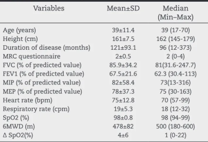
There were forty-ive consecutive patients enrolled who agreed to participate in the study and fulilled the inclusion criteria. There were 42 women with 39 ± 11.4 years, in total. None of the patients were smokers. The duration of the dis-ease was 121 ± 93.1 months in the occasion. The character-istics of the patients and their functional measurements are detailed in Table 1.
The 6MWD was 478 ± 82 m and the SpO2 at rest was 98 ± 0.8%. The fall in SpO2 at the end of the 6MWT was 4 ± 6 points. The spirometric evaluation showed FVC of 85.9 ± 34.2 (% of predicted value) and 21 patients with FVC below the limit of normality. The MIP was 82 ± 58.4 and MEP was 78 ± 37.3 (% of predicted value).
195
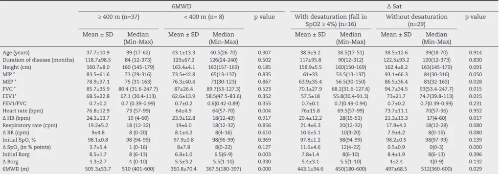
R E V B R A S R E U M A T O L . 2 0 1 4 ;5 4 ( 3 ): 1 9 2 – 1 9 9Borg scale value (p = 0.004). The distance walked by the pa-tients in the two groups was also signiicantly different (p < 0.001): the value in the group ≥ 400 m was 505.3 ± 53.7 m; and in the group < 400 m was 350.8 ± 70.4 m (Table 2).
When the population studied was divided in two groups, according to the presence of desaturation ≥ 4 by the end of the 6MWT,no differences were found between the groups con-cerning age, disease duration, height, FEV1/FVC, initial SpO2, initial heart rate, initial respiratory rate, increase in respira-tory rate, initial value and increase in Borg scale. The heart rate at the end of the test was signiicantly higher in those participants who showed desaturation (p = 0.017). MEP was signiicantly lower in the group with desaturation (p = 0.028) and MIP as well, but it did not reach signiicance (p = 0.050). The distance walked by the patients in the two groups was also signiicantly different (p = 0.029): the value in the group with desaturation was 443.1 ± 94.6 m and in the group with-out desaturation was 497 ± 68.5 m. The ΔSat was also signii-cantly different in the two groups: the value in the group with desaturation was 11.6 ± 4.6 points; and in the one without desaturation, the fall was of 0.5 ± 0.9 points. The inding of a FVC below the limit of the normal expected value was signii-cantly associated with the group with desaturation (p = 0.027) (Table 2).
Discussion
One inding that seems quite relevant in this study is that, within a population of SLE patients without relevant respira-tory symptoms, the 6MWT can give useful information about respiratory compromise, especially if there is a reduction in SpO2 by the end of the test. It was considered a reduction equal to or greater than 4 points as signiicant, based on the indings by Prefaut et al., who validated this cut-off value in a study of exercise-induced hypoxemia during maximal exercise tests in
athletes.20 This 4% fall was deined as accounted for potential inaccuracy of oximetry plus the effects of metabolic acidosis on the hemoglobin saturation curve (a right shift).9
Subjects in this study with ΔSat ≥ 4% showed a signiicant reduction in walking distance (443 m versus 497 m, p = 0.029), although both values were way above the accepted inferior limit for 6MWD. Furthermore, these patients, when compared to those who did not desaturate had a higher heart rate at the end of the 6MWT (p = 0.017), lower MEP (p=0.028), lower MIP (p = 0.050) and a spirometry restrictive defect (FVC below the lower limit of predicted value, p = 0.027, with a normal FEV1/ FVC ratio).
Conversely, those who walked less than 400 m showed no signiicant differences regarding initial saturation or ΔSat ≥ 4%. In addition, there were no signiicant differences between the groups with 6MWD < 400 m and MWD ≥ 400 m in spiro-metric values, heart rate, static pressures or severity of dys-pnea, either.
These indings suggest the hypothesis that desaturation during the 6MWT may be a useful tool to evaluate SLE pa-tients without respiratory symptoms – perhaps more sensi-tive than the 6MWD.
The 6MWT is a standardized submaximal test of exercise capacity that is self-paced, simple, reproducible and inexpen-sive. The measured variables are distance walked in 6 min-utes (6MWD), symptoms and SpO2 at rest and at the end of the test.8 Because of its safety proile, physician attendance is not required, but a health professional, such as physiothera-pist or a nurse, with clinical experience should supervise the patient during the test.
Age, sex, height, weight and ethnicity are important de-terminants of an individual’s 6MWD. In general, men walk further than women; and the distance walked declines with increasing age.21 Equations are available to predict expected normal values of 6MWD, with some variation in the expected distances.21,22 A walking distance of less than 350 m has pre-dictive value of increased mortality in a number of cardio-pulmonary disorders, such as COPD, interstitial lung disease, pulmonary arterial hypertension, cystic ibrosis, congestive heart failure.10,11,14,22-24
Although the 6MWD is a sensitive measurement of walk-ing ability for patients with moderate to severe disease, it is likely that its sensitivity in patients with better preserved exercise tolerance may not be so good. A ceiling effect was reported in patients with pulmonary arterial hypertension whose 6MWD is greater than 450 m, and this observation may be true for patients with other conditions.25
From the studies mentioned above, it can be seen that the cut-off value for the walking distance is not well established; apparently, it is between 350 m and 450 m.
In this study, only 8 patients walked less than 400 m, with median value of 367.5 m and mean value of 350.8 ± 70 m.
For the groups separated by the walking distance, the only statistically signiicant differences were initial heart rate (slower for those who walked less) and degree of dyspnea in Borg scale (smaller for those who walked less). There were no signiicant differences for these variables at the end of the 6MWT. It is hard to have an explanation for these indings, perhaps because of the small number of patients in one of the groups.
Table 1 – Study population characteristics and functional measurements (n = 45)
Variables Mean±SD Median (Min–Max)
Age (years) 39±11.4 39 (17-70)
Height (cm) 161±7.5 162 (145-179) Duration of disease (months) 121±93.1 96 (12-373) MRC questionnaire 2±0.5 2 (0-4) FVC (% of predicted value) 85.9±34.2 81(31.6-247.7) FEV1 (% of predicted value) 67.5±21.6 62.3 (30.4-113) MIP (% of predicted value) 82±58.4 73(13-316) MEP (% of predicted value) 78±37.3 75 (30-163) Heart rate (bpm) 75±12.8 70 (57-99) Respiratory rate (cpm) 19±5.3 18 (12-32)
SpO2 (%) 98±0.8 98 (94-99)
6MWD (m) 478±82 500 (180-600)
Δ SpO2(%) 4±6 1 (0-22)
REV BRAS REUMA
T
OL.
2014;
54(3)
:192–199
Table 2 – Comparison of functional variables and 6MWT parameters between the groups separated by distance and desaturation (n = 45)
6MWD Δ Sat
≥ 400 m (n=37) < 400 m (n= 8) p value With desaturation (fall in SpO2 ≥ 4%) (n=16)
Without desaturation (n=29)
p value
Mean ± SD Median (Min-Max)
Mean ± SD Median (Min-Max)
Mean ± SD Median (Min-Max)
Mean ± SD Median (Min-Max)
Age (years) 37.7±10.9 39 (17-62) 43.1±13.3 40.5(26-70) 0.307 38.9±9.2 38.5(17-51) 38.5±12.6 39(18-70) 0.914
Duration of disease (months) 118.7±98.5 84 (12-373) 129±67.2 126(24-240) 0.502 117±95.8 90(12-312) 122.5±93.2 120(12-373) 0.830
Height (cm) 160.7±8.0 160 (145-179) 163.4±4.1 163(157-169) 0.185 158.9±5.5 160(150-169) 162.4±8.2 163(145-179) 0.091
MIP * 83.5±61.6 73 (29-316) 73.5±42.8 65(13-137) 0.835 61±33 53.5(13-137) 93.1±66.3 84(30-316) 0.050
MEP * 78.9±37.1 75 (31-163) 76.3±40.4 71(30-123) 0.867 63.9±35.4 56.5(30-150) 86.5±36.4 81(32-163) 0.028
FVC * 85.7±35.9 80.4 (31.6-247.7) 87±26.4 89.7(53-127.3) 0.523 70.1±27.9 68.2(31.6-127.6) 94.7±34.5 93(53.4-247.7) 0.015
FEV1* 68.5±22.8 67.1 (30.4-113) 62.6±13.9 58.5(47.5-83.6) 0.352 57.5±18 55.8(30.4-91.3) 73±21.7 74.7(39.8-113) 0.015
FEV1/FVC 0.7±0.2 0.7 (0.39-0.99) 0.7±0.2 0.6(0.42-0.89) 0.355 0.7±0.1 0.7(0.49-0.94) 0.7±0.2 0.7(0.39-0.99) 0.231
Heart rate (bpm) 76.8±12.9 73 (57-99) 64±4.9 64(57-70) 0.004 76±15.8 69.5(57-99) 73.7±11.1 70(57-96) 0.952
Δ HR (bpm) 24.3±13.7 19 (4-60) 23.9±12.8 18(12-49) 0.917 29.4±12.2 28(15-51) 21.3±13.3 17(4-60) 0.017
Respiratory rate (cpm) 19.2±5.2 18 (12-32) 19±6.0 18(12-32) 0.856 21.4±6.3 20(12-32) 17.9±4.2 18(12-28) 0.080
Δ RR (cpm) 9±4.8 8 (0-20) 8.1±4.2 8(4-16) 0.610 10.6±5.1 10(3-20) 7.9±4.2 8(0-16) 0.080
Initial SpO2 % 98.1±0.8 98 (94-99) 97.9±0.8 98(96-99) 0.369 97.8±1.2 98(94-99) 98.2±0.5 98(97-99) 0.139
Δ SpO2 (in % points) 3.7±5.4 1 (0-16) 8±7.8 8(0-22) 0.127 11.6±4.6 12(4-22) 0.5±0.9 0(0-3) 0.000
Initial Borg 8.5±1.7 8 (6-13) 6.8±1.0 6.5(6-9) 0.003 7.8±1.4 8(6-10) 8.4±1.9 8(6-13) 0.396
Δ Borg 4.3±2.7 4 (0-10) 5.5±3.2 5.5(1-10) 0.330 5.4±3.1 5.5(1-10) 4±2.4 4(0-9) 0.132
6MWD (m) 505.3±53.7 510 (401-600) 350.8±70.4 367.5(180-397) 0.000 443.1±94.6 450(180-600) 497±68.5 512(360-600) 0.029
Mean ± SD, mean ± standard deviation; Median (Min-Max), Median (minimum and maximum); 6MWD, 6 minutes walked distance; Δ Sat, Final SpO2 – Initial SpO2; MIP, Maximal inspiratory pressure;
MEP, Maximal expiratory pressure; FVC, Forced Capacity Value; FEV1, Forced Expired Volume in one second; HR, Heart rate; Δ HR, Final HR – Initial HR; cpm, cycles per minute; Δ RR, Final RR – Initial RR; Δ Borg, Final Borg – Initial Borg.
Differences were considered signiicant with a p < 0.05.
197
R E V B R A S R E U M A T O L . 2 0 1 4 ;5 4 ( 3 ): 1 9 2 – 1 9 9The relation between walking distance and mortality is not seen in patients suffering from untreated pulmonary ar-terial hypertension, in whom desaturation during the 6MWT was a better predictor of mortality than walking distance: for every percent decrease in SpO2 there was a 26% increase in the risk of death.12
The desaturation during 6MWT has demonstrated its val-ue as an index of severity of disease and prognostic factor. Lama et al. showed that, in patients with interstitial pulmo-nary ibrosis without resting hypoxemia, desaturation up to 88% at any point during the 6MWT was associated to an in-creased hazard of death; however, no associations between 6MWD and survival were observed.26
In this study, FVC values below the lower limit of normal-ity, simultaneously reduced FEV1 and normal FEV1/FVC in-dexes, indicating the presence of a restrictive defect, were signiicantly more frequent among patients who showed de-saturation. In addition, patients with desaturation had signii-cantly lower MEPs and showed a trend towards signiisignii-cantly lower MIPs. The 6MWD was not able to detect the patients who had reductions in FVC, and not even the ones with re-duced expiratory pressures. In this study, desaturation was associated with lower 6MWD, although the mean walking distance in the group that showed desaturation was much greater than 350 m.
In our patients, an association between the presence of de-saturation (Δsat ≥ 4%) with the values of MIP (p = 0.050) and MEP (p = 0.028) was observed, suggesting that some impair-ment of respiratory muscles, not only the diaphragm, was present.
The presence of unexplained dyspnea, especially in the supine position, small lung volumes on chest radiographs, dysfunction and elevation of the diaphragm and pulmonary function tests displaying patterns of restrictive disease in the absence of parenchymal involvement prompt the diagnosis of shrinking lung syndrome.
Some authors found that the ability of the diaphragm to generate pressure is impaired in patients with the shrink-ing lung syndrome;27 however, other authors28 were unable to show a reduced diaphragmatic strength in a cohort of 12 patients.
In a comparison to the 6MWD, desaturation revealed it-self as better suited to ind patients with the most impaired indexes in respiratory function tests. A previous published study, carried out by our group, showed that desaturation during a 6MWT provides additional information regarding severity of disease in patients with scleroderma presenting pulmonary manifestations.13
Inter-test variation is high in cases of oxygen desatura-tion. This fact implies that therapeutic decisions should not be based on a single measurement of exertional desaturation recorded on a 6MWT. This inter-test variation is expressed by the inding of different values of SpO2 at the end of various tests performed by the same patient.
In the study by Eaton et al., the value of hemoglobin de-saturation upon pulse oximetry was found to be non-repro-ducible, with unacceptable measurement variation. However, instead of using the hemoglobin desaturation as a categori-cal variable, they computed the values of desaturation in two 6MWTs.29 We believe that the important information here is
the occurrence of desaturation per se, a fact that is not ob-served in normal subjects.21 We surely expect the value of de-saturation to vary because of different homeostatic situations at different moments in individuals with pulmonary diseases and in whom gas exchange abnormalities are probably pres-ent.
Except for the involvement of the pleura, the most com-mon pulcom-monary manifestation of SLE, all other pulcom-monary manifestations are infrequent, and many of them may cause decrease oxygenation during exercise, by different patho-genic mechanisms. Those less common respiratory disorders in patients with SLE include: interstitial lung disease, acute lupus pneumonitis, diffuse alveolar hemorrhage, pulmonary arterial hypertension, thromboembolic disease, acute revers-ible hypoxemia and shrinking lung syndrome. It is worth re-minding that the prevalence of respiratory symptoms and signs in patients with SLE vary depending on several factors, most importantly the methods used for diagnosing respira-tory tract compromise.
In athletes, the exercise-induced arterial hypoxemia is de-ined as a reduction in the arterial O2 pressure (PaO2) by more than 1kPa and/or hemoglobin O2 saturation (SaO2) below 95%, both determined by blood gas analysis. Desaturation is con-sistently found during maximal rowing ergometer and is most pronounced at the end of an exercise bout.30 Exercise-induced hypoxemia is explained by the interplay of many different fac-tors. Alveolar PO2 must be maintained at a high level, so ven-tilation becomes a critical issue. A widening of the PAO2-PaO2 difference frequently occurs, indicating that diffusion limita-tion or a ventilalimita-tion-perfusion mismatch or shunt may be in-luencing the transport of oxygen from the alveoli to the pul-monary capillaries. Cardiac output increases greatly, leading to a fast transit time of red cells in the lungs and further limiting O2 uptake. It is well known that a post-exercise reduction in pulmonary diffusion capacity really occurs, and this suggests damage to the alveolar-capillary membrane.31 All these factors have been proposed to be involved in exercise- induced hypox-emia, but the six-minute walk test is a submaximal exercise. Disease states may facilitate the occurrence of desaturation by all these mechanisms.
Vascular injury with endothelial cell activation and damage play a central role in the pathogenesis of SLE.5 The vascular en-dothelial growth factor (VEGF) is the main mediator of angio-genesis, and increased levels of VEGF were found in serum from patients with rheumatoid arthritis, dermatomyositis/polymyo-sitis, sclerodermapolymyodermatomyositis/polymyo-sitis, scleroderma complicated by interstitial lung disease,32 and SLE.33 The mitogen is connected to vascular hypertrophy, inlammation, tissue remodeling, ex-tracellular matrix synthesis and ibrosis.34 Increased levels of endothelin-1, a potent vasoconstrictor, were observed in many collagen-vascular diseases including SLE.
The combination of inlammatory vascular lesion, slightly elevated arterial pulmonary pressures and initial ibrotic inter-stitial disease impaired function of diaphragm with exercise stress, although submaximal, may explain the occurrence of desaturation during the 6MWT in SLE patients.
arte-rial hypertension, associated with a poor prognosis, a close monitoring of the patients with SLE who presented desatura-tion during the 6MWT is advisable. A recently published study assesses the association between quality of life and distance walked during the 6MWT in Brazilian premenopausal patients with SLE and compared with a healthy control group. The au-thors of this study concluded that patients with SLE walked a shorter distance during the 6MWT, which was associated with poorer quality of life.35 In addition to that, the inding of desatu-ration justiies the indication of a more thorough cardiorespi-ratory evaluation using echocardiogram, CT scans, measure-ments of diffusion capacity and total lung capacity.
Conlicts of interest
The authors declare no conlicts of interest.
R E F E R E N C E S
1. Rus V, Maury EE, Hochberg MC: The epidemiology of systemic lupus erythematosus. In: Wallace DJ, Hahn BH, editors. Dubois lupus erythematosus. 7th ed. Filadélia: Lippincott Williams & Wilkins; 2007:34-44.
2. Sato EI, Natour J, Martinelli VPL, Assis LSS, Farão SR, Medeiros EL et al. Seguimento clínico e laboratorial de 132 pacientes com lúpus eritematoso sistêmico. Rev Bras Reumatol. 1991,31:57-62.
3. Costallat LTL & Coimbra AMV. Lúpus eritematoso sistêmico: análise clínica e laboratorial de 272 pacientes em um hospital universitário: 1973-1982. Rev Bras Reumatol. 1995,35:23-9. 4. Rothield NF. Systemic lupus erythematosus: clinical aspects
and treatment. In: McCarty DJ & Koopman WJ. Arthritis and allied conditions – a textbook of rheumatology, 12. ed. Philadelphia: Lea & Febiger. 1993;1155-77.
5. Belmont HM, Abramson SB and Lie JT. Pathology and pathogenesis of vascular injury in systemic lupus erythematosus. Interactions of inlammatory cells and activated endothelium. Arthritis Rheum, 1996;39:9-22. 6. Krishnaswamy G, Kelley J, Yerra L, Smith JK, Chi DS. Human
endothelium as a source of multifunctional cytokines: molecular regulation and possible role in human disease. J Interferon Cytokine Res, 1999;19:91-104.
7. Hochberg MC. Updating the American College of Rheumatology revised criteria for the classiication of systemic lupus erythematosus. Arthritis Rheum, 1997;40:1725.
8. ATS statement: guidelines for the six-minute walk test. Am J Respir Crit Care Med, 2002;166:111-7.
9. Poulain M, Durand F, Palomba B, Ceugniet F, Desplan J, Varray A et al. 6-minute walk testing is more sensitive than maximal incremental cycle testing for detecting oxygen desaturation in patients with COPD. Chest, 2003;123:1401-7.
10. Kawut SM, Horn EM, Berekashvili KK, Garofano RP, Goldsmith RL, Widlitz AC et al. New predictors of outcome in idiopathic pulmonary arterial hypertension. Am J Cardiol, 2005;95:199-203.
11. Miyamoto S, Nagaya N, Satoh T, Kyotani S, Sakamaki F, Fujita M et al. Clinical correlates and prognostic signiicance of six-minute walk test in patients with primary pulmonary hypertension. Comparison with cardiopulmonary exercise testing. Am J Respir Crit Care Med, 2000;161:487-92. 12. Paciocco G, Martinez FJ, Bossone E, Pielsticker E, Gillespie
B, Rubenire M. Oxygen desaturation on the six-minute
walk test and mortality in untreated primary pulmonary hypertension. Eur Respir J, 2001;17:647-52.
13. Villalba WO, Sampaio-Barros PD, Pereira MC, Cerqueira EM, Leme CA Jr, Marques-Neto JF et al. Six-minute walk test for the evaluation of pulmonary disease severity in scleroderma patients. Chest, 2007;131:217-22.
14. Rostagno C, Olivo G, Comeglio M, Boddi V, Banchelli M, Galanti G et al. Prognostic value of 6-minute walk corridor test in patients with mild to moderate heart failure:
comparison with other methods of functional evaluation. Eur J Heart Fail, 2003;5:247-52.
15. Pereira CA. Diretrizes para Testes de Função Pulmonar. J Bras Pneumologia, 2002;28.
16. Knudson RJ, Lebowitz MD, Holberg CJ, Burrows B. Changes in the normal maximal expiratory low-volume curve with growth and aging. Am Rev Respir Dis, 1983;127:725-34. 17. II Consenso de DPOC. J Bras Pneumol 2004;30:s1-s42. 18. American Thoracic Society/European Respiratory Society.
ATS/ERS Statement on respiratory muscle testing. Am J Respir Crit Care Med, 2002;166:518-624.
19. Neder JA, Andreoni S, Lerario MC, Nery LE. Reference values for lung function tests. II. Maximal respiratory pressures and voluntary ventilation. Braz J Med Biol Res, 1999;32:719-27. 20. Prefaut C, Durand F, Mucci P, Caillaud C. Exercise-induced
arterial hypoxaemia in athletes: a review. Sports Med, 2000; 30:47-61.
21. Soares MR, Pereira CA. Six-minute walk test: reference values for healthy adults in Brazil. J Bras Pneumol, 2011;37:576-83. 22. Cote CG, Casanova C, Marín JM, Lopez MV, Pinto-Plata V,
de Oca MM et al. Validation and comparison of reference equations for the 6-min walk distance test. Eur Respir J, 2008; 31:571-8.
23. Kadikar A, Maurer J, Kesten S. The six-minute walk test: a guide to assessment for lung transplantation. J Heart Lung Transplant, 1997;16:313-9.
24. Provencher S, Chemla D, Hervé P, Sitbon O, Humbert M, Simonneau G. Heart rate responses during the 6-minute walk test in pulmonary arterial hypertension. Eur Respir J, 2006; 27:114-20.
25. Frost AE, Langleben D, Oudiz R, Hill N, Horn E, McLaughlin V et al. The 6-min walk test (6MW) as an eficacy endpoint in pulmonary arterial hypertension clinical trials: demonstration of a ceiling effect. Vascul Pharmacol, 2005; 43:36-9.
26. Lama VN, Flaherty KR, Toews GB, Colby TV, Travis WD, Long Q et al. Prognostic value of desaturation during a 6-minute walk test in idiopathic interstitial pneumonia. Am J Respir Crit Care Med, 2003;168:1084-90.
27. Gibson CJ, Edmonds JP, Hughes GR. Diaphragm function and lung involvement in systemic lupus erythematosus. Am J Med, 1977;63:926-32.
28. Laroche CM, Mulvey DA, Hawkins PN, Walport MJ, Strickland B, Moxham J et al. Diaphragm strength in the shrinking lung syndrome of systemic lupus erythematosus. Q J Med, 1989;71:429-39.
29. Eaton T, Young P, Milne D, Wells AU. Six-minute walk, maximal exercise tests: reproducibility in ibrotic interstitial pneumonia. Am J Respir Crit Care Med, 2005; 171:1150-7.
30. Nielsen HB. Arterial desaturation during exercise in man: implication for O2 uptake and work capacity. Scand J Med Sci Sports, 2003;13:339-58.
31. Nielsen HB. Boesen M, Secher NH. Near-infrared spectroscopy determined brain and muscle oxygenation during exercise with normal and resistive breathing. Acta Physiol Scand, 2001; 171:63-70.
199
R E V B R A S R E U M A T O L . 2 0 1 4 ;5 4 ( 3 ): 1 9 2 – 1 9 933. Kuryliszyn-Moskal A, Klimiuk PA, Sierakowski S, Ciołkiewicz M. Vascular endothelial growth factor in systemic lupus erythematosus: relationship to disease activity, systemic organ manifestation, and nailfold capillaroscopic abnormalities. Arch Immunol Ther Exp (Warsz), 2007;55: 179-85.
34. Goldie RG. Endothelins in health and disease: an overview. Clin Exp Pharmacol Physiol, 1999;26:145-8.