1. Department of Physical Education, Oeste Paulista University (Unoeste), Presidente Prudente, SP.
2. Fatec, Botucatu Campus, SP.
3. Department of Histology, Unoeste, Presidente Prudente, SP. Received in 26/7/04. 2nd version received in 3/1/05. Approved in 10/2/05. Correspondence to: Olimpia Ciabattari, Av. Irineu Sesti, 124, Jardim Alto da Boa Vista – 19053-360 – Presidente Prudente, SP, Brasil. Phone: (18) 231-4909. Fax: (18) 231-3100. E-mail: [email protected]
Effect of swimming associated with diet
on the anterior tibial muscle of rats:
morphological and hystochemical study
Olimpia Ciabattari1, Alexandre Dal Pai2 and Vitalino Dal Pai3
O
RIGINALA
RTICLEKey words: Anterior tibial muscle. Swimming. High calorie and normal diets. ENGLISH VERSION
ABSTRACT
The objective of this study was to investigate the effect of the association of different swimming program frequencies and diets on the characteristics of the anterior tibial muscle of 24 Wistar male rats. These rats were randomly assigned into three groups: untrained (control), trained group (two days/week) and trained group (five days/week). Each group was divided into two groups, which received one of the two normal or high calorie diets. After the train-ing period, muscle samples were collected and frozen at –70°C. Serial cryostat sections (8 µm) were sectioned and submitted to
HE stain and to NADH-TR, m-ATPase (pH 4.4) and Sudan Black histochemical methods. The morphology was analyzed and the degree of fiber enlargement (on hypertrophy) evaluated using the lesser fiber diameter method. The data were submitted to analysis of variance (one way ANOVA). Muscle fibers were classified as SO, FOG and FG, presenting a mosaic distribution pattern, which were unchanged in all groups. Muscle fibers revealed a very low hypertrophy in all groups. Initial and final body weight were signif-icantly different in trained groups. In the trained groups, especially in five days/week, muscle fibers revealed higher diameter, split-ting and some internal myonucleus. Some atrophic fibers were observed and this observation was suggestive of denervation. The oxidative metabolism was higher in SO and FOG fibers. No signif-icant alterations were observed in muscle contraction ability and the lipids content was intense in SO fibers, moderate in FOG fi-bers and low in FG fifi-bers. The present study, with this protocol of two days/week and five days/week training caused different types of morphological lesions in fibers. The high calorie diet did not cause statistically significant results in comparison with the normal diet.
INTRODUCTION
In the last years, several researches have emphasized how phys-ical activity has been effective in the treatment and prevention of chronic diseases such as diabetes, cardiovascular diseases and particularly obesity, mainly caused by high caloric diets and seden-tary life-style.
In the last decades, a high number of obese individuals has been observed especially in rich countries, such as Canada, New Zealand, United Kingdom and United States. In the Unites States, a varia-tion on the number of obese individuals from 12% to 17.9% in the
period from 1991 to 1998 was observed. Data published in 2000 indicated that obese and overweighed adults counted for 54.9% of the American population(1). Marx(2) estimates that these data
dou-bled in the last 20 years, thus revealing that 30% of the American adults are obese and 35% are overweighed. Children and adoles-cents are not exempted: 15% are above normal weight.
In Brazil, obesity reaches 41.5% of the population, being strong-ly associated with dyslipidemia(3).
Considered as multifactor disease since 1985 by the National Institute of Health for increasing the incidence of other chronic-degenerative diseases(3), since 1997, the World Health
Organiza-tion (WHO) recognized obesity as an universal disease(4).
Obese individuals present higher risk of cardiovascular diseas-es, hypertension, type 2 diabetes mellitus, dyslipidemia, osteoar-thritis, sleep apnea, infertility and some types of neoplasy(1,4-6).
In order to revert this situation, the adoption of methods aimed at the increase of the slim mass with the consequent increase on the metabolic conditioning and the lipids mobilization, especially in fat tissue, liver, heart, skeletal muscles and in the plasma lipopro-teins(7), becomes necessary. One of the methods that may lead to
this objective is the physical activity(8).
The effect of different training situations – static (vertical stair-case) and dynamic (treadmill) – on the long extensor muscles of fingers and soleus muscles in young rats was studied by Melichna et al.(9). The authors observed that the dynamic training resulted in
an elevation on the muscles’ oxidative capacity, while in the sed-entary group, a preferential increase on the glycolytic feature was seen.
Similar effects were observed by Lopez-Rivero et al.(10) when
running exercises with intense training were used in horses. Fibers modulation was observed by Misumi et al.(11) in horses
submitted to swimming exercise associated with running. The re-sults revealed increase of FOG and decrease of FG fibers.
The interaction of hypercaloric diets with physical activity may also contribute for the prevention of dyslipidemia and obesity, as reported by Duarte(12), when submitted rats fed with hypercaloric
diet to swimming exercises.
Similar results could be observed by Ross et al.(4), when
submit-ted rats to exercises with or without caloric restriction. Even if significant weight reductions in the group with no caloric restric-tion was not observed, the authors verified reducrestric-tions on the ab-dominal fat tissue, factor associated with cardiovascular diseases and type 2 diabetes mellitus.
MATERIAL AND METHODS
Twenty-four Wistar male adult rats (Rattus norvegicus, albinus) with average body weight of 350 g were used in the present re-search. The animals were kept in collective cages with four rats in each cage under constant temperature (26°C) and light and dark photoperiods of 12 hours.
Up to the beginning of the experiment, the rats were fed with standard Purina ration for rodents. Three weeks before the exper-iment, 50% of these animals were submitted to cafeteria diet (hy-percaloric)(13,14), and the other animals were fed with standard
Puri-na ration. Both groups had ration and water ad libitum. The research was approved by the Ethics Committee of the Oeste Paulista Uni-versity (Unoeste), Presidente Prudente, SP.
Rats that received standard diet composed the normal group. The normal and hypercaloric groups were subdivided into untrained (control), trained group (two days/week) and trained group (five days/week) with four rats in each group.
The normal untrained (NU) and hypercaloric (NH) groups, com-posed of four animals from the normal group and four animals from the hypercaloric group, received ration and water ad libitum.
The normal trained and hypercaloric groups were composed of eight animals from the normal group and eight animals from the hypercaloric group, divided into: normal trained two days/week (T2XN), hypercaloric trained two days/week (T2XH), normal trained five days/week (T5XN) and hypercaloric trained five days/week (T5XH).
Half of the animals were fed with standard Purina ration con-taining 3.78 kcal/g. The other half received hypercaloric diet with the following composition: 15 g of standard ration (3.78 kcal/g), totalizing 56.70 kcal; 10 g of toasted peanuts (5.95 kcal/g), totaliz-ing 59.50 kcal; 10 g of milky chocolate (6.11 kcal/g), totaliztotaliz-ing 61.10 kcal and 0.5 g of maize biscuit (3.55 kcal/g), totalizing 17.75 kcal, being offered as pellets(12).
Before training, the rats were submitted to an adaptation pro-gram during five days, starting with 15 minutes and water level of 20 cm, being progressively increased to 30 minutes and water lev-el of 50 cm.
After this stage, the animals were submitted to training with duration of 10 weeks in glass tanks of 100 x 50 x 60 cm and water temperature of 30° ± 1°C. Each group was submitted to 60-min
sessions. The two days/week and five days/week training sessions in consecutive days were based on Duarte protocol(12).
Twenty-four hours after the last training session, the animals were sacrificed by means of intraperitoneal injection with thiopen-tal (20 mg/100g of body weight). Later, the animals were weighted (g), followed by the removal of the anterior tibial muscle.
Fragments of the median portion of this muscle were immersed in n-hexane –70°C(9) during two minutes. Histological cuts (8 µm)
were submitted to HE stain and to NADH-TR, m-ATPase (pH 9.4) after pre-incubation in acid medium (pH 4.4) and Sudan Black(15)
histochemical methods.
The hypertrophy degree of the muscular fibers was evaluated based on the HE-stained blades through the lesser fiber diameter method(15), using a computerized image analysis system. For each
animal, 100 fibers were measured (µm). Besides the use of the
optical microscope Nikon Alphaphot – 2 YS2, the images were col-lected with the aid of the camera CV-730 PDC 12 VDC Fuse: 800 mA and visualized through the Personal Computer 300 GL sys-tem, with 14 inches Philips monitor and ImageLab 2000 program. Following, the morphological description and analysis of fibers and the histochemical analysis were performed. Based on the NADH-TR reaction, the fibers were classified and designated as SO (slow oxidative), FOG (fast oxidative glycolytic) and FG (fast glycolytic). The fast and slow contractile ability was obtained through the m-ATPase reaction after pre-incubation in acid medium and the lipids content was evaluated through the Sudan Black reac-tion. The analysis of the fibers frequency in function of their diam-eters was also performed (intervals of 20 µm).
The data regarding the initial and final body weight of the animals studied as well as the fibers diameter were submitted to one-way ANOVA statistical test. The significance level adopted was of 5%.
RESULTS
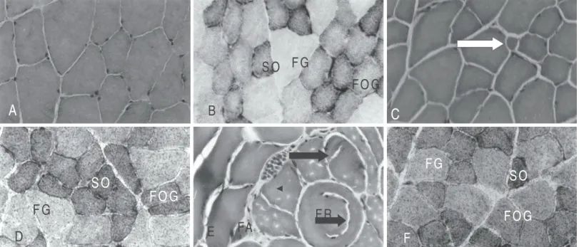
As revealed through the HE stained preparations, in the untrained group fed with normal diet and in the untrained group fed with hypercaloric diet, the morphology of the anterior tibial muscle re-vealed fibers with different diameters, polygonal shapes and some presented hypertrophy (figures 1A and 2A). In group T2XN, the fibers revealed to be smaller, with rounded shape and higher diam-eter variations (figure 1C).
Fig. 1 – Transversal sections of the anterior tibial muscle. A) normal untrained group (NU) HE. 128x; B) normal untrained group (NU), types of fibers (SO, FOG, FG) NADH-TR. 128x; C) normal group trained 2 days/week (T2XN) small fiber (arrow) HE. 128x; D) normal group trained 2 days/week (T2XN), types of fibers (SO, FOG, FG) Sudan Black. 128x; E) normal group trained 5 days/week (T5XN), rounded fiber (RF), atrophic fiber (AF), splitting (arrows), HE. 128x;F) normal group trained 5 days/week (T5XN), Sudan Black, 128x.
A
B
S O
F G
F O G
C
D
S O
F O G
F G
E
F R
FA
F
S O
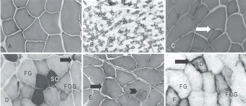
In group T2XH, some angular and atrophic and some hypertrophic fibers were observed and some of them presented segmented ap-pearance (splitting), fact also observed in groups T5XN and T5XH (fig-ures 1E; 2C and 2E). In group T5XN, besides the splitting with internal myonuclei, the presence of atrophic fibers occurred (figure 1E).
In group T5XH, besides the splitting (arrow head), cytoplasm areas with hypercontraction were also observed (arrow) (figure 2E). For both trained and untrained groups, the presence of three types of fibers was observed in the NADH-TR reaction, character-izing the mosaic distribution pattern (figures 1B and 2B). In trained groups, the SO and FOG fibers showed more intense reactivity pattern with shapeless formazan aggregates in the subsarcolem-mal region (figure 1F).
The myofibrillar ATPase reaction showed to be intense in SO fibers, moderate in FOG fibers and weak in FG fibers in all groups. In trained groups, the FOG fibers presented diameters a little smaller and more rounded, and the FG fibers, hypertrophy. In group T2XH, some SO fibers presented atrophic appearance (arrow) with ac-centuated polymorphism (figure 2D).
In normal and hypercaloric untrained groups, the Sudan Black method revealed high lipids concentration in SO fibers, moderated in FOG fibers and very weak in FG fibers. On the other hand, in trained groups, the lipids content in SO and FOG fibers was more intense. In FG fibers, the lipids concentration showed to be higher in normal groups and slightly lower in hypercaloric groups (figures 1D, 1F, and 2F).
The statistical treatment of the variable initial and final body weight of the rats studied is shown in table 1.
The statistical treatment of the muscular fibers average diame-ters of groups SN, SH, T2XN, T2XH, T5XN, T5XH is shown in table 2.
Fig. 2 – Transversal sections of the anterior tibial muscle. A) hypercaloric untrained group (HU) HE. 128x; B) hypercaloric untrained group (HU) NADH-TR. 34x;C) hypercaloric group trained 2 days/week (T2XH), splitting (arrow) HE. 128x; D) hypercaloric group trained 2 days/week (T2XH), types of fibers (SO, FOG, FG) m-ATPase. pH4,4. 128x; E) hypercaloric group trained 5 days/week (T5XH), splitting (arrow head), hypercontraction area (arrow). HE. 128x; F)
hypercaloric group trained 5 days/week formazan aggregate (arrow). NADH-TR, 128x.
A
B
C
D
S O
F O G
F G
E
F
S O
F O G
F G
TABLE 1
Comparison of initial body weight (IW) and final body weight (FW) of groups NU, NH, T2XN, T2XH, T5XN and T5XH. The significance
of differences is indicated by the value of P
Groups IW FW P
NU 437.5 ± 59.5 497.7 ± 56.2 0.19152 NH 437.5 ± 59.5 523.4 ± 60.5 0.0602 T2XN 431.2 ± 32.46 518.1 ± 14.8 0.00245 T2XH 443.7 ± 37.50 551,0 ± 50.7 0.0145 T5XN 368.7 ± 12.5 469.6 ± 14.1 0.000038 T5XH 418.7 ± 37.5 539.1 ± 40.2 0.00467
TABLE 2
Statistical treatment of the average diameter (µm) of fibers of rats from groups NU, NH, T2XN, T2XH, T5XN, T5XH
Groups NU NH T2XN T2XH T5XN
NH 0.48A
T2XN 2 x 10⁻⁵
B 5 x 10⁻⁴B
T2XH 0.06A 0.2A 0.007B
T5XN 0.03B 0.1A 0.02B0 0.7A T5XH 7 x 10⁻⁹
B 1 x 10⁻⁶B 0.32A0 5 x 10⁻⁵B 4 x 10⁻⁴B
A: averages of column with letter A are not statistically significant. B: averages of column with letter B are statistically significant. (P < 0.05)
TABLE 3
Number and percentage of fibers in function of the diameter of the anterior tibial muscle (intervals of 20 µm) of animals
from groups NU, NH, TX2N, TX2H, T5XN and T5XH
Diameter/fibers < 20 21-40 41-60 > 61 N Groups/rats
NU 174 (40.7%) 214 (50.1%) 39 0(9.1%) 400 NH 3 (0.7%) 146 (35.7%) 204 (49.8%) 57 (13.9%) 400 T2XN 151 (37.5%) 150 (37.5%) 99 (24.7%) 400 T2XH 1 (0.2%) 139 (34.7%) 217 (54.2%) 43 (10.7%) 400 T5XN 118 (29.5%) 221 (55.2%) 60 (15%)0, 400 T5XH 1 (0.2%) 116 (29%),0 189 (47.2%) 95 (23.7%) 400
The number and the frequencies (%) of fibers in function of di-ameters in intervals of 20 µm of the groups of rats studied are shown in table 3.
DISCUSSION
SN and T5XH, SH and T2XN, SH and T5XH, T2XN and T2XH, T2XH and T5XN, T5XN and T5XH, significant hypertrophy of fibers was observed.
The short pre-training period seems to have contributed, at least in part, for the fibers hypertrophy. Furthermore, the training itself seems to have been intense and the duration of 10 weeks, not long enough. These facts may have contributed for the hypertro-phy of some fibers.
Another aspect to be considered, according to Dall Pai et al.(16,17),
is the post-birth growth pattern of muscular fibers of rats, being linear from 12 to 45 days. In the subsequent ages, the growth is differential between the types of fibers, being more intense and continuous in the white fibers.
Besides the hypertrophy, the trained rats, especially from groups T2XH, T5XN and T5XH, presented cytoplasm longitudinal segmen-tation (splitting), rounded hypertrophic fibers, and fibers with smaller diameters suggestive of denervation and under phagocytosis pro-cess. These alterations showed to be more intense in groups trained five days/week. The internal or lateral segmentation process of fibers composes a morphophysiological adaptation aiming at re-ducing the distance between the capillary bed and the center of the cytoplasm, thus favoring metabolic changes and the occurrence of lesions.
On the other hand, the presence of 0.2 to 0.7% of fibers with diameter below 20 µm in both trained and untrained groups seems to show that the protocol used presented no significant effect on the number of muscular fibers, once its occurrence may be a re-sult of atrophic process and/or formation of new fibers.
The presence of different lesion degrees in the muscular fibers was also observed by Camargo et al.(18), who exercised rats during
60 days in treadmill, in daily sessions of 60 minutes, five days/ week. Elapsed 30, 45 and 60 days, all active groups revealed atro-phic fibers under phagocytosis process and other fibers with round-ed shape and centralizround-ed nucleuses. The lesions showround-ed to be more severe in animals belonging to active group of 60 days.
With regard to the oxidative metabolism, the exercised rats of this research showed reactivity slightly increased in SO and FOG fibers, especially in groups trained five days/week, demonstrating increase on the oxidative capacity. This aspect presented under the form of higher formazan aggregates in relation to the other animals studied.
Despite not being quantified, this result is in agreement with results obtained by Fitts et al.(19), when they submitted university
students to swimming training during 20 days with one daily ses-sions of 90 minutes in the first 10 days and two daily sesses-sions of 90 minutes in the last 10 days. According to the authors, the oxida-tive activity almost doubled in the first phase, with no significant alterations in the second phase.
Melichnaet al.(9) also observed elevation on the oxidative
capac-ity of fibers of the long extensor muscles of fingers and soleus in a dynamic training (treadmill) in young rats after four weeks in ses-sions of five days/week with duration from 20 minutes to two hours a session. Similar results were also obtained by Misumi et al.(11)
training horses in running and swimming. The animals were divid-ed into three groups: running, swimming plus progressive running, and running plus progressive swimming. Five months later, the authors observed an increase on the FOG fibers and a decrease on the FG fibers in groups that associated running and swimming train-ing.
A significant increase on the percentage of oxidative fibers and a decrease on the glycolytic fibers was also observed by Lopez-Rivero et al.(10) in horses submitted to a four-week training with
different velocities and session time variations.
In the same way, Saad et al.(20) observed significant increase on
the aerobic capacity in the straight muscles of the abdomen and in the paraexternal intercostalis muscles of rats submitted to swim-ming exercise as result of the increase on the percentile of SO
fibers and decrease on the FOG fibers in rats that swam 60 days, five days/week in sessions of 60 minutes. On the other hand, the FG fibers increased in rats that swam 15 days, five days/week in sessions of 60 minutes. A significant increase on the SO fibers in the paraexternal intercostalis muscles was observed in rats that swam 60 days, increase on the FOG fibers in rats that swam 45 and 60 days and decrease on the FG fibers in rats that swam dur-ing 30, 45 and 60 days.
Likewise, the results of the present research revealed intense lipids concentration in SO fibers, moderate in FOG fibers, and weak in FG fibers in group NU, being slightly higher in SO and FG fibers of group NH. With regard to the trained groups, in group T2XN, the lipids concentration was higher in SO and FOG fibers and lower in FG fibers when compared to group T2XH, where the lipids con-centration was lower in SO and FOG fibers and higher in FG fibers. In group T5XN, the lipid content showed to be very intense in SO fibers and more intense in the other fibers when compared with previous groups. In group T5XH, a higher lipids accumulation was observed in SO and FOG fibers and lower in the FG fibers, when compared with T5XN.
One may conclude that diets presented significant effect on the body weight of the trained groups. However, no statistically signif-icant difference was observed between the different diets (normal and hypercaloric). Furthermore, fibers distribution presented no alterations in both untrained and trained groups, keeping a mosaic distribution pattern. The reactivity pattern for slow and fast con-traction was similar in the different groups of rats studied. The swimming exercise, according to the protocol used here, promot-ed cytoplasm longitudinal segmentation (splitting), and the forma-tion of denervated fibers, and rounded and hypertrophic fibers, and this hypertrophy was statistically significant in some groups.
ACKNOWLEDGMENTS
To Professor Dr. Jair Rodrigues Garcia Júnior for the help in formatting this article.
All the authors declared there is not any potential conflict of inter-ests regarding this article.
REFERENCES
1. Pescatello L, VanHeest JL. Physical activity mediates a healthier body weight in the presence of obesity. Br J Sports Med 2000;34:86-93.
2. Marx J. Obesity – What is to be done? Science 2003;299:845-9.
3. Guerra RLF, Cunha CT, Montes RS, Santos Júnior JA, Dias A, Dâmaso AR. Efei-tos do exercício crônico com orientação nutricional sobre parâmetros lipídicos de mulheres obesas. Rev Bras Fisiot 2002;6:1-7.
4. Ross R, Dagnone D, Jones PJH, Smith H, Paddags A, Hudson R, et al. Reduc-tion in obesity and related comorbid after diet-induced weigh loss or exercise-induced weight loss in men American. Ann Intern Med 2000;133:92-103. 5. Stefanick MI. Exercise and weight control. In: Holloszy JO, editor. Reviews of
exercise & sport sciences, 1993;21:363-96.
6. Anderson DA, Wadden TA. Tratando o paciente obeso: sugestões para a prática de atendimento primário. JAMA 2000;4:3172-87.
7. Dioguardi GS, Faludi AA, Bertolami MC. Lípides nas atividades física e esporti-va. In: Ghorayeb N, Barros TL, editores. O exercício. São Paulo: Atheneu, 1999. 8. Chen KT, Yang RS. Effects of exercise in lipid metabolism and musculoskeletal
fitness in female athletes. World J Gastroenterol 2004;10:122-6.
9. Melichna J, Mackova EV, Semiginovsky B, Tolar M, Stichova J, Slavicek A, et al. Effect of exercise on muscle fiber composition and enzyme activities of skeletal muscles in young rats. Physiol Bohemoslov 1987;36:321-8.
10. Lopez-Rivero JL, Moralez-Lopez JL, Galisteo AM, Aguerra E. Muscle fiber type composition in untrained and endurance-trained Andalusian and Arab horses. Equine Vet J 1991;23:2191-3.
11. Misumi K, Sakamoto H, Shimizu R. Changes in skeletal muscle composition in response to swimming training for young horses. J Vet Med Sci 1995;57:959-61.
dieta normocalórica e hipercalórica [Dissertação de Mestrado]. UFSCar. São Paulo, 2001.
13. Rothwell NJ, Sotck MJ. A role for insulin in the diet-induced thermogenesis of cafeteria-fed rats. Metabolism 1981;30:673-8.
14. Prats E, Monfar M, Castella J, Iglesias R, Alemany M. Energy intake of rats fed a cafeteria diet. Physiol Behav 1989;45:263-72.
15. Dubowitz V. Muscle biopsy: modern approach. 2ed. London: Bailliere Tindall, 1985.
16. Dall Pai V, Macha N, Curi PR. On the cross sectional area of skeletal muscle fibres of the rat. Anat Anz 1983;154:337-41.
17. Dall Pai V, Thomaz E, Curi PR. Postnatal growth of skeletal muscle fibers of the rat. Gegenbaurs Morphol Jahrb 1984;130:827-34.
18. Camargo RCT, Camargo Filho JCS, Vanderlei LCM, Oliveira Júnior AS, Oliveira DAR. Efeitos do treinamento físico através da esteira rolante sobre o músculo sóleo de rato. Anais da XVIII Reunião Anual da FeSBE, Curitiba, 2003. 19. Fitts RH, Costill DL, Gardetto PR. Effect of swim exercise training on human
muscle fiber function. J Appl Physiol 1989;465-75.