Aldino Puppin Filho**
Angle
Class I malocclusion with bimaxillary dental
protrusion and missing mandibular first molars*
This case report describes the orthodontic treatment of a 24-year-old patient presenting with Angle Class I malocclusion, bimaxillary dental protrusion and recent loss of man-dibular molars. Treatment involved extraction of the maxillary first premolars and closing of mandibular first molar spaces. Treatment outcomes demonstrate the need for individu-alized treatment planning and highlight the key role played by biomechanical concepts in achieving proper orthodontic tooth movement. This case was presented to the Brazilian Board of Orthodontics and Facial Orthopedics (BBO) as representative of the free choice category in partial fulfillment of the requirements for obtaining the BBO Diploma. Abstract
Keywords: Angle Class I malocclusion. Corrective orthodontics. Biomechanics. Orthodontic space closure.
** MSc in Orthodontics, State University of Rio de Janeiro (UERJ). Specialist in Orthodontics and Facial Orthopedics, UERJ. Diplomate of the Brazilian Board of Orthodontics and Dentofacial Orthopedics.
* Case report, free choice category, approved by the Brazilian Board of Orthodontics and Facial Orthopedics (BBO).
HISTORY AND ETIOLOGY
Female Caucasian patient aged 24 years showed good overall health, moderate history of caries, adequate restorations, periodontal health and recent loss of lower molars.
Her chief complaint concerned the possibil-ity of closing the spaces left by the missing teeth. When asked about her facial appearance, she re-ported that she looked “toothy”.
DIAGNOSIS
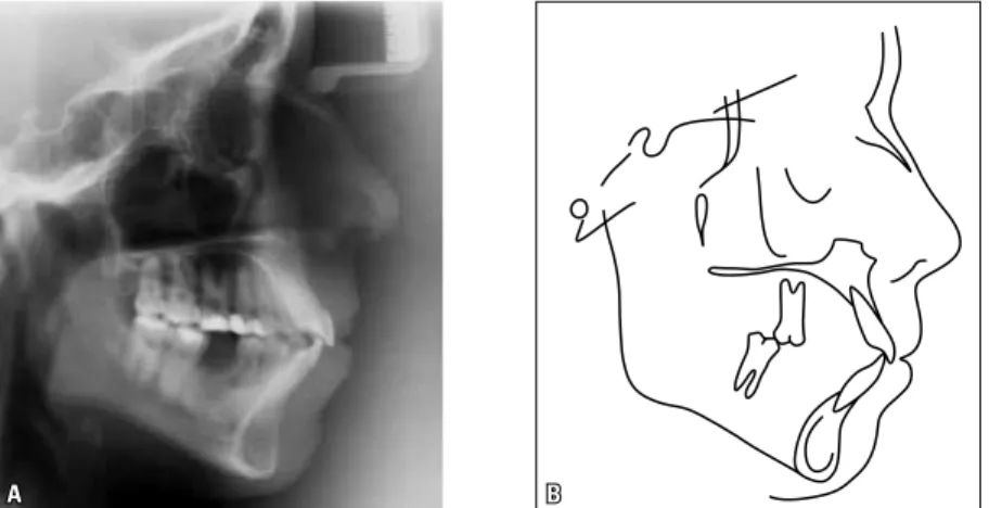
Analysis and evaluation of the data obtained in the clinical examination, with the aid of other diagnostic elements (extra- and intraoral photos; panoramic and periapical lateral cephalometric radiographs; and plaster models), revealed that the patient had a convex profile, lip incompe-tence, and protrusion of upper and lower lips (UL-S line=2 mm and LL-S line=3 mm).
How to cite this article : Puppin Filho A. Angle Class I malocclusion with bimaxillary dental protrusion and missing mandibular irst molars. Dental Press
J Orthod. 2011 Nov-Dec;16(6):119-29.
» The author reports no commercial, proprietary, or inancial interest in the
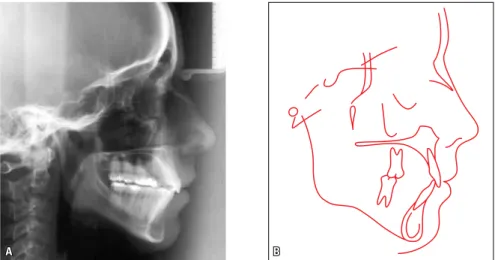
The anteroinferior face height appeared slightly increased and, based on height, the smile was clas-sified as average.¹ In the transverse plane, the face was considered symmetrical (Fig 1 and Table 1).
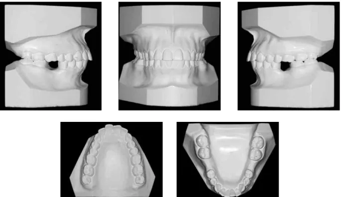
From a dental point of view, as can also be seen in Figure 1, given that the lower first mo-lars were missing, the patient was classified as presenting with an Angle Class I malocclusion, in view of the relationship between canines. The anterior region featured normal overjet and
overbite with upper and lower midlines that co-incided with each other and with the sagittal plane. As can be seen in Table 1, the upper and lower incisors were sharply inclined and pro-truded (1-NA= 30° and 7.5 mm; 1-NB= 37° and 12 mm; IMPA= 103.5°).
She had a Class II skeletal pattern (ANB=5.5°) with the mandible showing a slight tendency toward clockwise rotation (SN-GoGn=34.5°, FMA=28° and Y axis=61°).
FIGURE 2 - Initial models.
A B
TREATmENT GOALS
Treatment goals were set based on the charac-teristics of the case and the desires of the patient in seeking orthodontic treatment. Since there was no significant skeletal compromise, the treat-ment goals were focused on reducing upper and lower incisor protrusion and closing the spaces created by the missing first molars. Thus, a nor-mal canine occlusion would be preserved while molars would assume a Class II relationship.
As regards facial appearance, the goals would be to reduce lip protrusion, imparting balance and harmony to the relationship between nose, lips and chin.
TREATmENT pLANNING
To attain the set goals, all teeth in the upper and lower arches — including third molars — would have to be aligned and leveled using the Straight-Wire technique.
To allow the retraction of the upper teeth, extraction of the first upper premolars (teeth #14 and #24) was indicated, whereas for man-dibular retraction the spaces created by the missing first molar would be utilized.
The use of mini-implants was originally planned for anchorage control in the upper arch.
However, given that this hypothesis was reject-ed by the patient, a removable transpalatal arch was indicated in combination with headgear at-tached to the first molars. Additionally, plan-ning comprised canine distalization followed by incisor retraction. In the lower arch, no an-chorage control strategy was adopted.
In order to avert the tendency of lower mo-lars to tip mesially during space closure, the force action line was designed to operate as close as possible to the center of resistance of the teeth with the aid of a power arm seated on the auxil-iary tube of the second molars, along with large
Gurin® auxiliaries (Morelli, Sorocaba/SP, Brazil)
attached to a rectangular 0.019 x 0.025-in stain-less steel archwire, where elastomeric chains would be attached to close the space.
In like manner, two large Gurin®
auxilia-ries attached to a rectangular 0.019 x 0.025-in stainless steel archwire on the distal side of the incisors — where the elastomeric chains would be inserted — would be used for torque control of the upper incisors during retraction.
TREATmENT pROGRESS
A Straight-Wire, slot 0.022 x 0.030-in appli-ance was used, with the exception of the first upper molars, which were banded and had aux-iliary attachments welded to the buccal and pal-atal surfaces. All other auxiliaries were bonded directly to the buccal surfaces of the teeth.
The upper and lower arches were then aligned and leveled using straight 0.012-in, 0.014-in and 0.016-in nickel-titanium wires and 0.018-in, 0.020-in and 0.019 x 0.025-in stainless steel wires.
In the upper arch, subsequently to the use of 0.020-in archwires, the patient had teeth #14 and #24 extracted. As a result, a transpala-tal arch fabricated with 0.9 mm stainless steel wire was placed and the use of headgear began with parietal traction and a force of approxi-mately 150 to 200 g on each side.
After the extractions had been completed, distal movement of the upper canines was be-gun along with the en masse retraction of the lower teeth. In both the upper and lower arch forces were applied by means of elastomeric chains, which were replaced every four to six weeks. The initial force delivered to each ca-nine by the elastomeric chains reached 100 g in the upper arch and approximately 200 g on each side of the lower arch.
It is noteworthy that in the upper arch, while the canines were undergoing distal movement, omega loops were placed close to the first mo-lar tubes. After this phase, 0.019 x 0.025-in stainless steel archwires were inserted with
large Gurin® auxiliaries attached to the distal
region of the lateral incisors to retract the up-per incisors. The elastomeric chains, which were the actual source of orthodontic forces, were
attached to the first molars and the Gurin®
auxiliaries. The force applied to each side was around 150 g. The elastomeric chains were also replaced every four to six weeks.
In the finishing and detailing phase, after all
spaces had been closed, the transpalatal arch was removed and headgear use suspended to allow proper dental arch coordination. A pan-oramic radiograph was requested to evaluate the angulation of all teeth, which determined the need to rebond some of the brackets. To at-tain the final results, first, second and third or-der bends were placed and customized accord-ing to individual need. The use of intermaxil-lary elastics was also indicated with a view to improving intercuspation.
After removing the fixed appliance, retain-ers were installed. A straight wire wraparound Hawley retainer was used in the upper arch, and a fixed canine-to-canine stainless steel lin-gual retainer was bonded to the lower arch.
TREATmENT RESuLTS
Comparison between the initial and final pa-tient examinations demonstrates that the results were consistent with the proposed objectives.
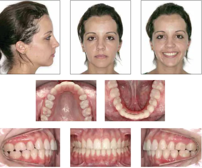
Facial appearance improved as well as lip posture, as viewed in frontal and lateral extra-oral photographs. A reduction of lip protrusion determined an excellent nose-lip-menton rela-tionship. As a result, the patient now features effortless lip competence with a pleasant and balanced smile (Figs 1 and 5).
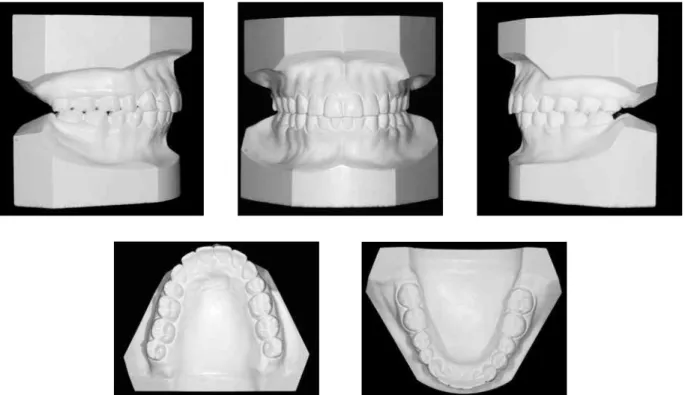
Retraction of the anterior teeth reduced the protrusion of upper and lower teeth (Table 1). Thus, a normal canine occlusion was preserved while molars assumed a Class II relationship. In the transverse plane, despite small midline deviations, the arches were well coordinated. Overbite and overjet remained appropriate (Figs 5 and 6).
increase in treatment time (which lasted a total of two years and seven months).
The spaces created by lower molar extractions performed prior to treatment were completely closed, with good root parallelism (Fig 7). As a re-sult of having been moved mesially, the lower first molars seem to have assumed a lower position in
the occlusal plane and this determined a slight mandibular counterclockwise rotation, which contributed to reducing facial convexity. The use of parietal traction headgear may also have con-tributed to preventing upper molar extrusion. The results achieved in this case corroborate some re-ports in the literature² (Fig 9).
FIGURE 6 - Final models.
A B
A B
FIGURE 8 - Final lateral cephalogram (A) and cephalometric tracing (B).
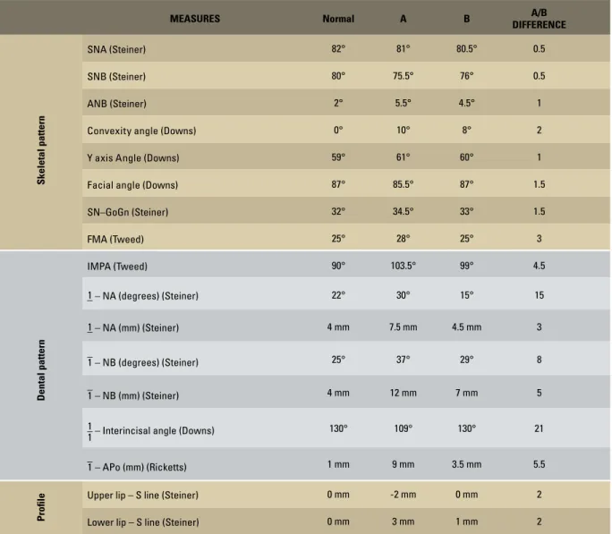
TABLE 1 - Summary of cephalometric measurements.
FINAL CONSIDERATIONS
A classic treatment strategy is adopted by most graduate orthodontics courses to tackle Angle Class I malocclusion with dental bimaxillary pro-trusion. This approach is taught since the initial stages of orthodontic training. It consists in indi-cating the extraction of the four first premolars.3
Choice of these teeth is usually due to their position close to the problem site. Importantly, however, common sense might suggest that the
amount of space available for retraction of ante-rior teeth varies according to the tooth to be ex-tracted, i.e., as a rule of thumb, the more distal the tooth, the greater the loss of anchorage and the lower the retraction.
The literature comprises estimates of anchor-age loss, which is quantified as one third of the
space in the case of first premolar extraction4,
and half the space when mandibular second
pre-molars are extracted.5 The maximum potential
MEASURES Normal A B A/B
DIFFERENCE
Skeletal pattern
SNA(Steiner) 82° 81° 80.5° 0.5
SNB(Steiner) 80° 75.5° 76° 0.5
ANB(Steiner) 2° 5.5° 4.5° 1
Convexity angle(Downs) 0° 10° 8° 2
Y axis Angle(Downs) 59° 61° 60° 1
Facial angle(Downs) 87° 85.5° 87° 1.5
SN–GoGn(Steiner) 32° 34.5° 33° 1.5
FMA(Tweed) 25° 28° 25° 3
Dental pattern
IMPA(Tweed) 90° 103.5° 99° 4.5
–
1 – NA (degrees)(Steiner) 22° 30° 15° 15
–
1 – NA (mm)(Steiner) 4 mm 7.5 mm 4.5 mm 3
–
1 – NB (degrees)(Steiner) 25° 37° 29° 8
–
1 – NB (mm)(Steiner) 4 mm 12 mm 7 mm 5
– 1
1 – Interincisal angle(Downs) 130° 109° 130° 21
–
1 – APo (mm)(Ricketts) 1 mm 9 mm 3.5 mm 5.5
Proile
Upper lip – S line(Steiner) 0 mm -2 mm 0 mm 2
retraction achieved comparing first and second premolar, and molar extraction, has been
esti-mated at 5 mm, 3 mm and 2 mm6, respectively.
In this clinical case, one particular factor in-terfered with the classic treatment plan: The patient had recently lost the first lower molars, thereby rendering impossible the extraction of two more teeth in the lower arch.
Once again, common sense might suggest that closing the spaces created by first molars would not be effective in reducing the protrusion of anterior teeth. However, the results clashed this hypothesis as they clearly demonstrate that the spaces created by extracting the first molars al-lowed proper retraction of anterior teeth with convenient protrusion correction. This is clearly depicted in the patient’s photographs and ceph-alometric superimpositions. It also emerges from a comparison between the initial and final ceph-alometric measurements, with particular focus on the reduction of 8° and 5 mm in measure 1-NB (Figs 1, 5 and 9, and Table 1).
It is noteworthy that the upper dental arch seems to behave according to common sense, i.e., the more distally the extracted tooth is po-sitioned, the less possibility of retraction, there-by underscoring the importance of implement-ing procedures for anchorage control.
The results obtained in this clinical case set the stage for new strategies in the treatment of cases that involve indication of extraction of per-manent teeth. Should the first premolars always be the first option? Shouldn’t the general condi-tion of the tooth (extent of injury, malformacondi-tion, endodontic treatment, etc.) be a major assessment factor? In addition, one should consider the use of mini-implants, which further expand the ability to indicate tooth extractions.
In cases where molar extractions are indi-cated the potential uses of third molars should
be explored. However, many patients who seek treatment have missing third molars even without clinical indication. A change in this approach would be welcome. Moreover, that information should be passed on to dentists and other dental specialists.
Another important point to consider is the mechanics used in closing first molar spaces. Forces applied directly to orthodontic auxil-iaries bonded to second molar crowns would unfailingly determine the mesial angulation of these teeth since the force action line would pass away from the centers of resistance of these teeth, located approximately in the fur-cation area. It is an often overlooked or
ne-glected basic mechanical concept7 because this
area is inaccessible to the placement of orth-odontic attachments. The indication of a power arm on the second molar auxiliary tube along
with the use of large Gurin® auxiliaries helped
to overcome this limitation and allowed the bodily movement of molars during space clo-sure. Power arms are not a new concept as they
were recommended by Andrews8 when he first
introduced his Straight-Wire appliance, for cases involving extractions. It should be noted, however, that it is not necessary to purchase ready-made power arms. Certain orthodontic auxiliaries are available on the market — such
as large Gurins®, crossed tubes and auxiliary
tubes — which when properly indicated allow movements consistent with the biomechanics.
1. Puppin Filho A. Avaliação quantitativa de medidas dento-faciais relacionadas à altura da linha do sorriso [dissertação]. Rio de Janeiro (RJ): Universidade do Estado do Rio de Janeiro; 2002.
2. Aras A. Vertical changes following orthodontic extraction treatment in skeletal open bite subjects. Eur J Orthod. 2002;24(4):407-16.
3. Tweed CH. Clinical orthodontics. St. Louis: C.V. Mosby Company; 1966.
4. Steiner C. Cephalometrics as a clinical tool. In: Riedel RA. Vistas in orthodontics. Philadelphia: Lea & Febiger; 1962. 397 p.
REFERENCES
Contact address Aldino Puppin Filho
Av. Américo Buaiz, 501, sala 1002, Torre Norte, Enseada do Suá Zip code: 29.050-911 – Vitória/ES, Brazil
E-mail: [email protected]
Submitted: August 2, 2011
Revised and accepted: August 26, 2011
5. Zachrisson B. JCO interviews: Dr. Bjorn Zachrisson on excellence
in inishing: Part 1. J Clin Orthod. 1986;20(7):460-82.
6. Profit WR, Fields HW Jr. Contemporary orthodontics. 2nd ed.
St Louis: Mosby Year Book; 1993.
7. Isaacson RJ, Lindauer SJ, Davidovitch M. The ground rules for arch wire design. Semin Orthod. 2001;7(1):34-41.