.
Mean platelet volume is associated with disease
severity in patients with obstructive sleep apnea
syndrome
Selahattin Akyol,IMustafa C¸o¨rtu¨k,IIAhmet Oytun Baykan,IKemal Kiraz,IIIAbdurrezzak Bo¨rekc¸i,IV Taner S¸eker,I,*Mustafa Gu¨r,IVMurat C¸ayliI
IAdana Numune Training and Research Hospital, Department of Cardiology, C¸ukurova/Adana, Turkey.IIAdana Numune Training and Research Hospital,
Department of Pulmonology, C¸ukurova/Adana, Turkey.IIIAntalya Numune Training and Research Hospital, Department of Pulmonology, Antalya, Turkey. IVKafkas University School of Medicine, Department of Cardiology, Kars, Turkey.
OBJECTIVE:Obstructive sleep apnea syndrome is associated with cardiovascular diseases and thromboembolic events. The mean platelet volume (MPV) is a predictor of cardiovascular thromboembolic events. The aim of the present study is to investigate the association between the MPV and disease severity in patients with obstructive sleep apnea syndrome.
METHODS:We prospectively included 194 obstructive sleep apnea syndrome patients without cardiovascular disease (mean age 56.5±12.5 years) who were undergoing sleep tests. An overnight full laboratory
polisomnography examination was conducted on each patient. The patients were divided into 3 groups according to the apnea-hypopnea index (AHI): (1) AHIlow group: 5pAHIo15, (2) AHImidgroup: 15oAHIp30, and (3) AHIhighgroup: AHI430.
RESULTS:The highest MPV values were found in the AHIhigh group compared with other groups (po0.05 for all). Multiple linear regression analysis indicated that the MPV was associated with the AHI (b=0.500,po0.001)
and the high sensitivity C-reactive protein (hs-CRP) level (b=0.194,p=0.010).
CONCLUSION:The MPV is independently associated with both disease severity and inflammation in patients with obstructive sleep apnea syndrome.
KEYWORDS: Obstructive sleep apnea; Apnea-hypopnea index; Mean platelet volume; High sensitivity C-reactive protein.
Akyol S, C¸o¨rtu¨k M, Baykan AO, Kiraz K, Bo¨rekc¸i A, S¸eker T, et al. Mean platelet volume is associated with disease severity in patients with
obstructive sleep apnea syndrome. Clinics. 2015;70(7):481-485
Received for publication onFebruary 3, 2015;First review completed onMarch 20, 2015;Accepted for publication onApril 20, 2015
E-mail: atanerseker@hotmail
*Corresponding author
’ INTRODUCTION
Obstructive sleep apnea syndrome (OSA) is a common sleep-related respiratory disorder that is characterized by repeated episodes of apnea and hypopnea due to intermittent upper airway obstruction. OSA, which is associated with repetitive nocturnal arterial oxygen desaturation and hyper-capnia as well as with alterations in systemic and pulmonary arterial pressure (1) is a highly prevalent illness that affects 4% of middle-aged men and 2% of middle-aged women (2). Furthermore, OSA is well known as an independent risk factor for cardiovascular diseases (CVDs) and hypertension
(HT) (1,3). Several possible mechanisms such as sympathetic nervous system activation, endothelial dysfunction, intermit-tent hypoxia, oxidative stress and inflammation explain the increased CVD prevalence in OSA patients (4). Although augmented hypercoagulability has been demonstrated in OSA patients who are not receiving continuous positive airway pressure (CPAP) therapy (5), the exact mechanism that drives the association between OSA and hypercoagulability is unknown. Moreover, few studies have investigated the asso-ciation between OSA severity and hypercoagulability (6). In a previous study, OSA was found to be associated with both arterial and venous thromboembolism (7).
The mean platelet volume (MPV) is a marker of thrombocyte activation and plays a pivotal role in the pathogenesis of CVDs (8,9). Larger platelets contain more granules and thromboxane A2 and express more glycopro-tein Ib and IIb/IIIa receptors; these platelets thus aggregate more quickly and strongly to collagen, possibly leading to increased thromboembolic events (10-12).
DOI:10.6061/clinics/2015(07)04
Increased MPVs have been found in hypertension, hypercholesterolemia, diabetes mellitus, acute myocardial infarction and acute ischemic stroke (11). Although a few studies have reported a relationship between OSA and increased platelet activation (15,16), the number of studies investigating the association between OSA and MPV is limited (17,18). The main purpose of the present study is to investigate the association between the MPV and the OSA severity in patients without CVD or hypertension and who do not take any medications that may affect platelet functions.
’ METHODS
Subjects who were clinically suspected of having sleep-related disorders (severe snoring, daytime sleepiness, and witnessed apnea) and who underwent a sleep test between March 2012 and July 2014 were prospectively enrolled in our study. A total of 194 patients (148 males; mean age 56.5±12.5
years) with an AHIX5 were included. All of the data were
collected prior to the administration of any treatment for OSA. After collecting a detailed medical history and performing a complete physical examination, each partici-pant was questioned regarding major cardiovascular risk factors, including age, sex, diabetes mellitus (DM), smoking status and hypertension (HT). Additionally, systolic blood pressure (SBP), diastolic blood pressure (DBP) and initial heart rate were recorded. Each of the patients underwent electrocardiography (ECG) and comprehensive transthoracic echocardiography. Patients with atherosclerotic heart disease such as coronary artery disease, cerebrovascular accident and peripheral vascular disease, heart failure, diabetes, hypertension, and hyperlipidemia and patients who were taking medications associated with these conditions were excluded. Patients with central sleep apnea syndrome, upper airway resistant syndrome, narcolepsy, or movement dis-order were excluded. Patients using any drug (such as aspirin, clopidogrel, dipyridamole, heparin, aminophylline, verapamil, nonsteroidal anti-inflammatory drugs, corticos-teroid, furosemide, antibiotics, and alcohol) that could affect platelet function were also excluded. Informed consent was obtained for each participant, and the local ethics committee approved the study protocol.
OSA diagnosis of and sleep testing
An overnight full laboratory polysomnography examination was conducted on each subject. All sleep recordings (E-Series, Compumedics, Melbourne, Australia) included electroencepha-lography, electroocuelectroencepha-lography, submental electromyography, and oxygen saturation (pulse oximetry), respiratory movement (inductance plethysmography), and nasal and oral airflow measurements. Sleep staging and sleep-disordered breathing were subsequently scored using standard techniques (19) but with all hypopneas including a mandatory minimum 4% oxygen desaturation. The average numbers of apnea and hypopnea episodes per hour of sleep were measured as the AHI. Patients were classified into 3 separate groups to determine the OSA severity according to their AHI scores, as follows: AHImild group (5oAHIo15), AHImoderate group
(15pAHIo30), and AHIseveregroup (AHIX30).
Echocardiography
All echocardiographic examinations were performed using commercially available equipment (Vivid-7; GE Vingmed
Sound, Horten, Norway) with a 2.5–3.5 MHz transducer. A single echocardiographer who was blinded to the patients’ clinical and laboratory data interpreted each echocardio-graphic examination independently. Simultaneous ECG recordings were also obtained. All patients were examined at rest in the left lateral decubitus position. Echocardio-graphic techniques and calculations of different cardiac dimensions were performed in accordance with the recom-mendations of the American Society of Echocardiography. The left ventricular ejection fraction (EF) was calculated using a modified Simpson’s rule technique (20).
Laboratory Analysis
Fasting venous blood samples on admission were obtained from all patients to determine their plasma fasting blood glucose, total cholesterol, high-density lipoprotein cholesterol (HDL-C), low-density lipoprotein cholesterol (LDL-C), triglycer-ide, creatinine, and high-sensitivity CRP (hs-CRP) levels and blood counts. Blood samples were collected through the brachial vein into tubes containing dipotassium EDTA. Serum creatinine, fasting glucose, alanine aminotransferase (ALT), aspartate aminotransferase (AST), and lipid profiles were measured using an autoanalyzer (Roche Diagnostics Modular Systems, Tokyo, Japan). To measure hematologic parameters, platelet counts and MPVs, samples were analyzed within 20 minutes after collection using a Sysmex XT 1800i automated hematology analyzer (Roche Diagnostics, Shanghai, China). For the MPV, the cut-off value was 9-13 fL, and the intra- and inter-assay coefficients of variation (CVs) were below 4.1% and 7.1%, respectively.
Statistical Analysis
All analyses were conducted using SPSS 17.0 (SPSS for Windows 17.0, Chicago, IL, USA). Data were expressed as the mean±SD. A comparison of categorical variables between
groups was performed using a chi-square test. Analysis of variance (ANOVA) was applied to analyze continuous variables between the AHI groups. Normality analysis was performed using a Kolmogorov–Smirnov test. Associations between other variables and the AHI were assessed by Pearson’s correlation coefficient. Multiple linear regression analysis was performed to identify independent associations of the AHI by including the parameters that were correlated with the AHI in the bivariate analysis. Standardized b
regression coefficients and their significance according to multiple linear regression analysis were reported. Ap-value of
o0.05 was considered statistically significant.
’ RESULTS
A total of 194 patients who fulfilled the selection criteria were included in the analysis. The patients were divided into three groups according to their corresponding AHI values: (a) AHImildgroup, 62 patients (5oAHIo15); (b) AHImoderate
group, 61 patients (15pAHIo30); and (c) AHIseveregroup,
71 patients (30pAHI). The mean AHI values were 9.0±2.7,
22.9±4.0 and 56.6±20.1 for the mild, moderate and severe
groups, respectively. Of the 194 participants, 148 (76%) were men, and 46 (24%) were women; the mean age was 56.5±
12.5 years. No significant differences were found between the groups in terms of age and sex.
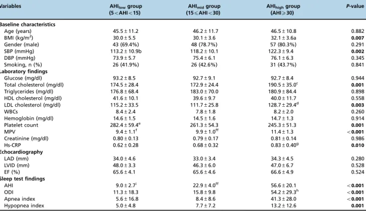
AHImild groups (po0.05 for both). Additionally, systolic
blood pressure was higher in the AHImoderateand AHIsevere
groups compared with the AHImildgroup (po0.05 for both).
Laboratory findings. Total cholesterol, LDL cholesterol and hs-CRP levels were higher in the AHIsevere group
compared with the AHImoderate and AHImild groups
(po0.05 for all). Platelet counts were lower in the AHImild
group than in the AHImoderateand AHIseveregroups (po0.05
for both). The highest MPVs were observed in the AHIsevere
group compared with the AHImoderate and AHImild groups
(po0.05 for all). Moreover, the MPV was higher in the
AHImoderate group than in the AHImild group (po0.05).
MPVs according to the AHI groups are shown in Table 1.
Sleep test findings. Higher oxygen desaturation index (ODI) values were observed in the AHIseveregroup than in
the AHImoderateand AHImildgroups (po0.05 for both).
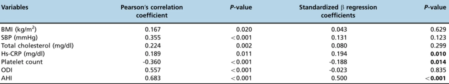
Bivariate and multivariate relationships of the mean platelet volume. According to the results of bivariate analysis, the MPV was associated with the BMI (r=0.167, p=0.020), SBP (r=0.355, po0.001), total cholesterol levels
(r=0.224, p=0.002), hs-CRP levels (r=0.189, p=0.011), the platelet count (r=-0.360, po0.001), the ODI (r=0.557,
po0.001) and the AHI (r=0.683,po0.001). The relationship
between the AHI and MPV is shown in Table 2.
Multiple linear regression analysis showed that the MPV was associated with hs-CRP levels (b=0.194, p=0.010), the platelet count (b=-0.188, p=0.014) and the AHI (b=0.500, po0.001).
’ DISCUSSION
The present study shows that MPV is independently associated with both the disease severity, as indicated by the AHI, and inflammation in OSA patients. Our study also suggests that MPV might play a role in thromboembolic events in OSA patients with severe AHI.
Although several previous studies have demonstrated that OSA is associated with CVD, the mechanism behind this association remains controversial (1,3,21). Of all of the health consequences of OSA, those that affect the cardiovascular system are the most undesirable, and platelet activation plays a pivotal role in CVD (18). Individual characteristics such as advanced age, increased blood pressure and obesity are the primary risk factors for both CVD and OSA (22).
Table 1 -Baseline characteristics and laboratory, echocardiographic and sleep test findings.
Variables AHIlowgroup (5oAHIo15)
AHImidgroup (15pAHIo30)
AHIhighgroup (AHIX30)
P-value
Baseline characteristics
Age (years) 45.5±11.2 46.2±11.7 46.5±10.8 0.882
BMI (kg/m2) 30.0±5.5 30.1±3.6 32.1±3.6a 0.007
Gender (male) 43 (69.4%) 48 (78.7%) 57 (80.3%) 0.291
SBP (mmHg) 113.2±10.9b 118.2±10.1 122.3±9.4 0.002
DBP (mmHg) 73.9±5.7 75.4±6.1 76.1±6.3 0.345
Smoking, n (%) 26 (41.9%) 26 (42.6%) 31 (43.7%) 0.841
Laboratory findings
Glucose (mg/dl) 93.2±8.5 92.7±9.1 92.7±8.4 0.944
Total cholesterol (mg/dl) 174.5±28.4 172.9±24.4 190.5±35.0c 0.001
Triglycerides (mg/dl) 176.8±68.4 183.0±70.0 180.9±84.4 0.898
HDL cholesterol (mg/dl) 41.6±10.1 39.6±9.7 40.0±11.7 0.558
LDL cholesterol (mg/dl) 115.2±33.5 111.7±25.8 128.7±29.4d
0.003
WBCs 8.4±2.4 7.8±1.8 8.2±2.0 0.260
Hemoglobin (mg/dl) 14.6±1.5 14.5±1.6 14.7±1.3 0.914
Platelet count 282.4±59.4e 261.3±54.3 245.3±51.3 0.001
MPV 9.4±1.1f 9.9±1.0ff 11.4±1.3 o0.001
Creatinine (mg/dl) 0.80±0.13 0.79±0.17 0.81±0.14 0.986
Hs-CRP 0.62±0.28 0.68±0.32 0.83±0.40g 0.010
Echocardiography
LAD (mm) 34.0±4.6 33.0±3.4 34.3±4.5 0.280
LVID (mm) 48.0±3.3 46.3±6.0 47.0±6.7 0.528
EF (%) 65.6±4.1 65.6±4.6 66.6±4.9 0.524
Sleep test findings
AHI 9.0±2.7i 22.9±4.0ff 56.6±20.1 o0.001
ODI 11.3±18.3 15.8±9.8 54.2±29.3h o
0.001
Apnea index 5.6±16.8 8.4±8.6 41.3±28.0 o0.001
Hypopnea index 5.0±4.8 7.7±7.2 13.2±12.6 0.001
ap=0.006 vs. AHI
lowgroup,p=0.008 vs. AHImidgroup bP=0.05 vs. AHI
midgroup,po0.001 vs. AHIhighgroup cp=0.003 vs. AHI
lowgroup,p=0.001 vs. AHImidgroup dp=0.010 vs. AHI
lowgroup,p=0.001 vs. AHImidgroup eP=0.034 vs. AHI
midgroup,po0.001 vs. AHIhighgroup fP=0.010 vs. AHI
midgroup,po0.001 vs. AHIhighgroup ffpo0.001 vs. AHI
highgroup gp=0.005 vs. AHI
lowgroup,p=0.026 vs. AHImidgroup iPo0.001 vs. AHI
midgroup and AHIhighgroup hpo0.001 vs. AHI
Likewise, markers of inflammation such as Hs-CRP are elevated in patients with OSA and CVD (17,22,23).
The present study shows that MPV is associated with disease severity in OSA patients. The relationship between OSA and the MPV has been previously investigated in a limited number of studies (17,18). A recent study assessed the relationship between OSA severity and the MPV in 205 OSA patients and found that MPV was higher in patients with moderate and severe OSA (17). However, the patients with moderate or severe OSA in that study were more likely to be hypertensive and active smokers, and in contrast to our study, the authors did not exclude patients with these cardiovascular risk factors, which may have influenced the MPV. In a similar study, Varol et al. investigated the effects of OSA on the MPV in hypertensive patients and active smokers (18); however, there were several differences compared with our study. First, we included a larger population of study subjects. Second, we excluded patients who had disorders or received medications that could affect the MPV. The exact mechanism for the association between OSA and the MPV is not known. Several possible mechan-isms might be responsible for this relationship, including sympathetic nervous system activation due to repetitive nocturnal hypoxemia, increased inflammation and oxidative stress and increased cardiovascular risk factors such as hypertension and obesity (4,24). In the present study, patients with cardiovascular risk factors such as hyperten-sion and diabetes were excluded from the study. It is well known that the MVP increases with hypoxemia (24). Additionally, the relationship between the MPV and hs-CRP in the present study confirms the effect of inflammation on platelet activation in OSA patients. However, the relation-ship between the MPV and oxidative stress was not investigated in the present study. Therefore, repetitive nocturnal hypoxemia and increased inflammation may be responsible for the increased MPV in patients with severe OSA.
Previous studies have demonstrated that MPV is a marker of inflammation in various clinical conditions (25,26). CRP is also a well-known marker of inflammation, and increased CRP levels are associated with atherothrombic events (27). Moreover, low-grade inflammation exists in patients with non-dipper HT, and the MPV is correlated with CRP levels (28). In a previous study, Panautsoulos et al. observed a significant decrease in CRP levels after nasal CPAP treatment in OSA patients (29). In the present study, we demonstrated that both hs-CRP and MPV are correlated with the OSA severity, as determined by the AHI. Using bivariate and
multivariate analyses, we confirmed the association between the MPV and hs-CRP levels. Because we excluded patients with clinical conditions that may alter the MPV, the relationship between the MPV and AHI identified in this study is more reliable.
There are some notable limitations to our current study, the most prominent of which is the absence of a control group without OSA. However, it has been previously demonstrated that OSA patients exhibit increased platelet activation and higher MPVs compared with controls. Because the main purpose of our study was to investigate the association between the MPV and the OSA severity, we did not include a control group. Additionally, we did not prospectively follow the patients and did not investigate the effects of various treatments such as nasal CPAP treatment on MPVs. However, this study was designed to demonstrate a relationship between the MPV and OSA severity.
In conclusion, the findings of our current study demon-strated that the MPV, a marker of cardiovascular disease, and hs-CRP, a marker of systemic inflammation, are both significantly associated with the OSA severity. Additional large-scale studies should be designed to explore the possible mechanisms of this relationship and to assess alterations in platelet activation and the MPV in response to adequate OSA treatment.
’ AUTHOR CONTRIBUTION
Baykan AO participated in the study design. Çörtük M obtained the
funding. Börekçi A, S¸eker T and Kiraz K collected the data and performed
the literature review. Gür M analyzed the data. Akyol S wrote the manuscript. Çayli M critically revised the manuscript.
’ REFERENCES
1. McNicholas WT, Bonsigore MR. Sleep apnoea as an independent risk factor for cardiovascular disease: current evidence basic and research priorities. Eur Respir J. 2007;29(1):156–78, http://dx.doi.org/10.1183/ 09031936.00027406.
2. Young T, Palta M, Dempsey J, Skatrud J, Weber S, Badr Set. The occur-rence of sleep-disordered breathing among middle aged adults. N Engl J Med. 1993;328(17):1230–5, http://dx.doi.org/10.1056/NEJM19930429328 1704.
3. Dincer HE, O’Neill W. Deleterious effects of sleep-disordered breathing on the heart and vascular system. Respiration. 2006; 73(1):124–30, http:// dx.doi.org/10.1159/000089814.
4. Garvey JF, Taylor CT, McNicholas WT. Cardiovascular disease in obstructive sleep apnoea syndrome: the role of intermittent hypoxia and inflammation. Eur Respir J. 2009;33(5):1195–205, http://dx.doi.org/ 10.1183/09031936.00111208.
Table 2 -Bivariate and multivariate relationships of the MPV in patients with obstructive sleep apnea syndrome.
Variables Pearson’s correlation coefficient
P-value Standardizedbregression coefficients
P-value
BMI (kg/m2) 0.167 0.020 0.043 0.629
SBP (mmHg) 0.355 o0.001 0.131 0.123
Total cholesterol (mg/dl) 0.224 0.002 0.080 0.299
Hs-CRP (mg/dl) 0.189 0.011 0.194 0.010
Platelet count -0.360 o0.001 -0.188 0.014
ODI 0.557 o0.001 -0.023 0.835
AHI 0.683 o0.001 0.500 o0.001
aMultiple linear regression analysis
5. Guardiola JJ, Matheson PJ, Clavijo LC, Wilson MA, Fletcher EC. Hyper-coagulability in patients with obstructive sleep apnea. Sleep Med. 2001;2 (6):517–23, http://dx.doi.org/10.1016/S1389-9457(01)00088-0.
6. Shitrit D, Peled N, Shitrit AB, Meidan S, Bendayan D, Sahar G, et al. An association between oxygen desaturation and D-dimer in patients with ostructive sleep apnea syndrome. Thromb Haemost. 2005; 94(3):544–7. 7. Bosanquet JP, Bade BC, Zia MF, Karo A, Hassan O, Hess BT, et al. Patients
with venous thromboembolism appear to have higher prevalence of obstructive sleep apnea than the general population. Clin Applied Thromb Hemost. 2011;17(6):119–24, http://dx.doi.org/10.1177/1076029610389023. 8. Tsiara S, Elisaf M, Jagroop IA, Mikhailidis DP. Platelets as predictors of
vascular risk: is there a practical index of platelet activity? Clin Appl Thromb Hemost. 2003;9(3):177–90, http://dx.doi.org/10.1177/10760296 0300900301.
9. Park Y, Schoene N, Haris W. Mean platelet volume as an indicator of platelet activation: methodological issues. Platelets. 2002;13(5-6):301–6, http://dx.doi.org/10.1080/095371002220148332.
10. Martin JF, Trowbridge EA, Salmon GL, Plumb J. The biological sig-nificance of platelet volume: its relationship to bleeding time, platelet thromboxane B2 production and megakaryocyte nuclear DNA con-centration. Thromb Res. 1983;32(5):443–60, http://dx.doi.org/10.1016/ 0049-3848(83)90255-4.
11. Giles H, Smith REA, Martin JF. Platelet glycoprotein Iib–IIIa and size are increased in acute myocardial infarction. Eur J Clin Invest. 1994;24(1): 69–72, http://dx.doi.org/10.1111/j.1365-2362.1994.tb02062.x.
12. Jakubowski JA, Thompson CB, Vaillancourt R, Valeri CR, Deykin D. Ara-chidonic acid metabolism by platelets of differing size. Br J Haematol. 1983;53 (3):503–11, http://dx.doi.org/10.1111/j.1365-2141.1983.tb02052.x.
13. Vizioli L, Muscari S, Muscari A. The relationship of mean platelet volume with the risk and prognosis of cardiovascular diseases. Int J Clin Pract. 2009;63(10):1509–15, http://dx.doi.org/10.1111/ijcp.2009.63.issue-10. 14. Minoguchi K, Yokoe T, Tazaki T, Minoguchi H, Oda N, Tanaka A, et al.
Silent brain infarction and platelet activation in obstructive sleep apnea. Am J Respir Crit Care Med. 2007;175(6):612–7, http://dx.doi.org/ 10.1164/rccm.200608-1141OC.
15. Bokinsky G, Miller M, Ault K, Husband P, Mitchell J. Spontaneous platelet activation and aggregation during obstructive sleep apnea and its response to therapy with nasal continuous positive airway pressure. A preliminary investigation. Chest. 1995; 108(3):625–30, http://dx.doi.org/ 10.1378/chest.108.3.625.
16. Akinnusi ME, Paasch LL, Szarpa KR, Wallace PK, El Solh AA. Impact of nasal continuous positive airway pressure therapy on markers of platelet activation in patients with obstructive sleep apnea. Respiration. 2009;77 (1):25–31, http://dx.doi.org/10.1159/000158488.
17. Kanbay A, Tutar N, Kaya E, Buyukoglan H, Ozdogan N, Oymak FS, et al. Mean platelet volume in patients with obstructive sleep apnea syndrome
and its relationship with cardiovascular diseases. Blood Coagul Fibrino-lysis. 2013;24(5);532–6.
18. Varol E, Ozturk O, Gonca T, Has M, Ozaydin M, Erdogan D, et al. Mean platelet volume is increased in patients with severe obstructive sleep apnea. Scand J Clin Invest. 2010;70(7):497–502, http://dx.doi.org/ 10.3109/00365513.2010.520733.
19. Berry RB, Budhiraja R, Gottlieb DJ, Gozal D, Iber C, Kapur VK, et al. The AASM manual for the scoring of sleep and associated events: rules, ter-minology, and technical specification, 1st ed. Westchester, IL:American Academy of Sleep Medicine, J Clin Sleep Med. 2012;8(5):597–619. 20. Schiller NB, Shah PM, Crawford M, DeMaria A, Devereux R, Feigenbaum
H, et al. Recommendations for quantitation of the left ventricle by two-dimensional echocardiography. American Society of Echocardiography Committee on Standards, Subcommittee on Quantitation of Two-Dimensional Echocardiograms. J Am Soc Echocardiogr. 1989;2(5):358–67, http://dx.doi.org/10.1016/S0894-7317(89)80013-6.
21. Pack AI, Gislason T. Obstructive sleep apnea and cardiovascular disease: a perspective and future directions. Prog Cardiovasc Dis. 2009;51(5): 434–51, http://dx.doi.org/10.1016/j.pcad.2009.01.002.
22. Phillips BG, Somers VK. Sleep disordered breathing and risk factors for cardiovascular disease. Curr Opin Pulm Med. 2002;8(6):516–20, http://dx.doi.org/10.1097/00063198-200211000-00006.
23. Lin QC, Xie HS, Liu XJ, Zhou JL, Zhao JM. Relationship between obstructive sleep apnea-hypopnea syndrome and high sensitivity C-reactive protein in non-obese subjects. Zhonghua Yi Xue Za Zhi. 2013;93(30):2355–8. 24. Parish JM, Somers VK. Obstructive sleep apnea and cardiovascular
disease. Mayo Clin Proc. 2004;79(8):1036–46, http://dx.doi.org/10.4065/ 79.8.1036.
25. Shah T, Casas JP, Cooper JA, Tzoulaki I, Sofat R, McCormack V, et al. Critical appraisal of CRP measurement for the prediction of coronary heart disease events: new data and systematic review of 31 prospective cohorts. Int J Epidemiol. 2008;38(1):217–31, http://dx.doi.org/10.1093/ ije/dyn217.
26. Gasparyan AY, Ayvazyan L, Mikhailidis DP, Kitas GD. Mean platelet volume: a link between thrombosis and inflammation? Curr Pharm Des. 2011;17(1):47–58, http://dx.doi.org/10.2174/138161211795049804. 27. Danesh J, Wheeler JG, Hirshfield GM, Eda S, Eiriksdottir G, Rumley A,
et al. C-reactive protein and other circulating markers of inflammation in the prediction of coronary heart disease. N Engl J Med. 2004;350 (14):1387–97, http://dx.doi.org/10.1056/NEJMoa032804.
28. Kaya MG, YarliogluesM, Gunebakmaz O, Gunturk E, Inanc T, Dogan A, et al. Platelet activation and inflammatory response in patients with non-dipper hypertension. Atherosclerosis. 2010; 209(1):278–82, http://dx. doi.org/10.1016/j.atherosclerosis.2009.09.010.