CLINICAL SCIENCE
Preoperative nodal staging of non-small cell lung
cancer using
99m
Tc-sestamibi spect/ct imaging
Juliana Muniz Miziara,IEuclides Timo´teo da Rocha,IJose´ Elias Abra˜o Miziara,IGustavo Fabene Garcia,I Maria Izilda Previato Simo˜es,IMarco Antoˆnio Lopes,ILı´gia Maria Kerr,ICarlos Alberto BuchpiguelII IHospital de Caˆncer de Barretos, Barretos/SP, Brazil.IIFaculdade de Medicina da Universidade da Sa˜o Paulo, Hospital das Clı´nicas da Faculdade de
Medicina da Universidade de Sa˜o Paulo, Radiologia, Sa˜o Paulo/SP, Brazil.
OBJECTIVES:The proper nodal staging of non-small cell lung cancer is important for choosing the best treatment modality. Although computed tomography remains the first-line imaging test for the primary staging of lung cancer, its limitations for mediastinum nodal staging are well known. The aim of this study is to evaluate the accuracy of hybrid single-photon emission computed tomography and computed tomography using99mTc-sestamibi
in the nodal staging of patients with non-small cell lung cancer and to identify potential candidates for surgical treatment.
METHODS:Prospective data were collected for 41 patients from December 2006 to February 2009. The patients underwent chest computed tomography and single-photon emission computed tomography/computed tomogra-phy examinations with 99mTc-sestamibi within a 30-day time period before surgery. Single-photon emission
computed tomography/computed tomography was considered positive when there was focal uptake of sestamibi in the mediastinum, and computed tomography scan when there was lymph nodes larger than 10 mm in short axis. The results of single-photon emission computed tomography and computed tomography were correlated with pathology findings after surgery.
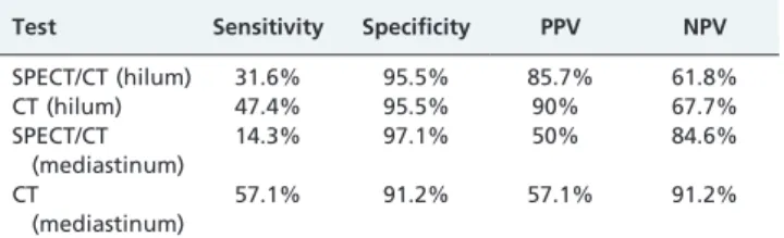
RESULTS:Single-photon emission computed tomography/computed tomography correctly identified six out of 19 cases involving hilar lymph nodes and one out of seven cases involving nodal metastases in the mediastinum. The sensitivity, specificity, positive predictive value, and negative predictive value for 99mTc-sestamibi single-photon emission computed tomography/computed tomography in the hilum assessment were 31.6%, 95.5%, 85.7%, and 61.8%, respectively. The same values for the mediastinum were 14.3%, 97.1%, 50%, and 84.6%, respectively. For the hilar and mediastinal lymph nodes, chest tomography showed sensitivity values of 47.4% and 57.1%, specificity values of 95.5% and 91.2%, positive predictive values of 90% and 57.1% and negative predictive values of 67.7% and 91.2%, respectively.
CONCLUSION:Single-photon emission computed tomography/computed tomography with99mTc-sestamibi showed very low sensitivity and accuracy for the nodal staging of patients with non-small cell lung cancer, despite its high level of specificity. In addition, the performance of single-photon emission computed tomography/computed tomography added no relevant information compared to computed tomography that would justify its use in the routine preoperative staging of non-small cell lung carcinoma.
KEYWORDS: Lung cancer; Lymph nodes; MIBI; Single-photon emission computed tomography; Functional imaging.
Miziara JM, Rocha ET, Miziara JEA, Garcia GF, Simo˜es MIP, Lopes MA, et al. Preoperative nodal staging of non-small cell lung cancer using99m
Tc-sestamibi spect/ct imaging. Clinics. 2011;66(11):1901-1909.
Received for publication onMay 28, 2011;First review completed onJune 16, 2011;Accepted for publication onJuly 18, 2011 E-mail: [email protected]
Tel.: 55 17 3321-6600
INTRODUCTION
The survival of lung cancer patients is related to the extent of their disease at the time of diagnosis. In the absence of distant metastases, the spread of tumors to the
mediastinal lymph nodes is a major determinant of both the prognosis and the therapeutic approach. Proper staging is important for selecting patients who may benefit from surgical resection and for defining the treatment modalities of patients who will undergo radiotherapy.
The histopathologic evaluation of lymph nodes is consid-ered the gold standard in assessing the presence or absence of metastases in the mediastinum. There are several invasive methods that can be used for this purpose: mediastinoscopy, anterior mediastinotomy, transthoracic needle aspiration, endobronchial or esophageal ultrasound with needle aspira-tion, and thorachoscopy.1,2 Mediastinoscopy is the most
Copyrightß2011CLINICS– This is an Open Access article distributed under the terms of the Creative Commons Attribution Non-Commercial License (http:// creativecommons.org/licenses/by-nc/3.0/) which permits unrestricted non-commercial use, distribution, and reproduction in any medium, provided the original work is properly cited.
common invasive test. It has a mean sensitivity of up to 80%, with a range of 44% to 97%. With this method, only high and low paratracheal, pre-tracheal, and subcarinal lymph nodes are accessible; there is also a low but real risk of morbidity and mortality.1
In an attempt to reduce the frequency of invasive methods or to guide the most appropriate procedures for lymph node biopsies, noninvasive imaging tests are used when applic-able. Computed tomography (CT) is the imaging method of choice in the evaluation and staging of primary cancers. The diagnostic CT criteria for the involvement of lymph nodes are based on their sizes, especially when their minor axes are longer than ten millimeters. However, small lymph nodes that are considered normal according to such criteria may contain tumor cells, while inflammatory and infectious diseases may be responsible for enlarged lymph nodes, limiting the overall effectiveness of this diagnostic test. In a meta-analysis by Toloza that evaluated 20 studies, chest CT examinations showed a sensitivity of 57%, specificity of 82%, and positive and negative predictive values of 56% and 83%, respectively.3
Tomographic imaging in nuclear medicine is based on the metabolic activity of tissues and may be useful for identifying pathological changes before they are detected by radiological examinations such as CTs. PET (positron emission tomography) scans with18F-FDG (18F
-fluorodeox-yglucose) have superior sensitivity and specificity com-pared to chest CTs and are considered the most accurate imaging method for staging patients with lung cancers.3-5 However, there are limitations related to positive predictive value of this method because there may be FDG uptake in inflammatory cells.6 The sensitivity can also be decreased when lymph node metastasis is microscopic or below the spatial resolution threshold of current, state-of-the-art scanners.7 In Brazil, the availability of PET is restricted to a few institutions because of equipment costs and avail-ability of comercial doses of FDG regarding the number of cyclotrons installed in Brazil.
Alternatively, single-photon emission computed tomo-graphy (SPECT) is widely available, has lower costs than PET, and does not require the presence of a cyclotron adjacent to the hospital. Sestamibi (hexakis-2-methoxyiso-butyl-isonitrile) labeled with technetium (99mTc-sestamibi) is a lipophilic cation that is routinely used in myocardial perfusion imaging and has been used as a tumor-seeking agent.8
Encouraging results have been obtained with SPECT scanning using sestamibi to detect primary lung malignan-cies9-11 and to perform mediastinal staging with a higher diagnostic accuracy higher than chest CTs.12,13
Despite the positive results obtained with sestamibi, particularly regarding its specificity, there are difficulties in the analysis of mediastinal images. The main difficulty is related to the limited spatial resolution of SPECT. Furthermore, the vascular structures in the mediastinum and heart, where sestamibi is taken up, interfere with the correct interpretation of the images and could cause false positive results and decrease the diagnostic capability of the method.
The association of functional images and anatomical information from CTs may be useful in interpreting SPECT by providing more accurate data regarding the location and extent of tumor lesions. Hybrid devices usually have dual detectors, with scintillation cameras and low-dose
CT scanners. Sequentially, data from both CT and SPECT are acquired. The two images are merged, creating SPECT images that are superimposed on corresponding anatomical planes. This image fusion may help to differentiate between tumors and other areas of physiological activity.14,15
The aim of this study is to evaluate the accuracy of SPECT/CT using the radiotracer 99mTc-sestamibi in the mediastinal lymph node staging of patients with non-small cell lung cancer and candidates to surgical treatment.
MATERIALS AND METHODS
Patients
A cross-sectional study with prospective data collection was conducted from December 2006 to February 2009 at the Hospital de Caˆncer de Barretos-SP. The study was approved by the Institutional Ethics Committee. The inclusion criteria were as follows: 1) patients of either sex, 2) patients who were at least 18 years old, 3) patients with histological diagnoses of non-small cell lung cancer (e.g., squamous cell carcinoma, adenocarcinoma or large cell carcinoma) or pulmonary lesions that were strongly suspicious for neoplasia, 4) patients with clinical stages I, II, or III, as classified by the sixth edition of TNM16 with performance
status that allowed surgery enrollment (ECOG PS zero or 1), and 5) patients who agreed to participate in the study and signed the informed consent form. Patients were excluded if their diagnosis of non-small cell lung cancer was not confirmed after surgical resection. Patients with bulky lymph node metastases that were considered unresectable and pregnant patients were also excluded.
The clinical evaluation included: physical examination, hematologic and biochemic screening, cardiologic evalua-tion, bronchoscopy when the pulmonary lesions were considered accessible for this method, bone scan, chest and upper abdomen CT, brain MRI or CT. Forty one patients were enrolled in the study. All of the patients were submitted to surgical procedures for diagnosis and treat-ment which were performed within 30 days after chest CT and SPECT/CT. The type of resection performed on each patient was defined by the thoracic surgery team in accordance with the extent of the primary tumor. Mediastinal systematic lymph node dissection was per-formed for adequate pathological staging according to the tumor location. For tumors of the right lung, a mediastinal dissection included the ipsilateral hilar region as well as the upper and lower paratracheal, subcarinal and paraesopha-geal lymph nodes. For left lung tumors, the left hilar region, lower paratracheal area, aortopulmonary window, and para-aortic, subcarinal, and paraesophageal lymph nodes were sampled. Lymph nodes were identified according to the Mountain nodal station classification system16and were
sent for histopathological analysis, which was performed by an experienced pathologist with expertise in lung cancer. The largest diameter of the metastatic foci in the lymph nodes was divided into two groups: those ,10 mm and those$10 mm.
99mTc-sestamibi SPECT/CT
USA). Images were acquired 10 minutes after the adminis-tration of sestamibi with patients placed in supine positions with their arms elevated.
SPECT: Emission images were acquired using a dual-head, large field-of-view scintillation camera equipped with a low-energy, high-resolution collimator (VPC-45). The images were acquired every 20 seconds, at a 3˚angle, in a circular orbit of 180˚per detector array, and using a 1286128 matrix. The equipment was calibrated for a photopeak of 140 keV with a symmetric 20% window.
CT images were acquired sequentially in a non-dedicated 3rd-generation scanner installed in the SPECT camera gantry, with a 10 mm slice thickness (maximum of 40 slices), a maximum current of 2.5 mA and a 140 kV potential.
The raw data from SPECT and CT were transferred from the acquisition equipment to an Entegra workstation (General Electric Healthcare), and the tools provided by the manufacturer were used to process the data. The following iterative processing protocol in standard 2-D was used. The image reconstruction was made after automatic pre-processing, which included the reconstruc-tion of the SPECT plane imaging, the reformatting of the anatomical and functional sections according to the type of organ and the creation of maximum-intensity projection imaging. The Butterworth filter was used with a cutoff frequency of 0.28 Nyquist and an order of 10. After this initial step, the images were reoriented to obtain transaxial, coronal, and sagittal views.
All images were independently interpreted by two nuclear physicians who were blinded to the chest CT and pathology findings. Disagreements in the analysis were resolved by consensus.
The following aspects were analyzed regarding the primary pulmonary lesion: 1) presence or absence of sestamibi uptake; 2) qualitative visual assessment of sestamibi uptake, which was classified as mild, moderate or intense. As a parameter, the uptake in primary tumor was compared with uptake in physiological thoracic structures as follows: more than soft tissues (mild) and less than hepatic uptake; similar to hepatic uptake (moderate); similar to cardiac uptake (intense). The intensity of uptake was correlated with histology and tumor size. 3) a semi-quantitative analysis obtained by outlining the regions of interest (ROI) over the tumor (T) and in the contralateral normal lung (L). ROI was defined manually on coronal images that showed the tumors’ highest degree of uptake. The maximum ROI values were measured, and the TL/T index was calculated. This analysis was correlated with the histology, tumor size and tumor size was analyzed as a continuous variable and also divided into three categories: 1) less than or equal to 3.0 cm, 2) greater than 3.0 cm and less than or equal to 7.0 cm and 3) greater than 7.0 cm.
With regard to the lymph nodes, the following aspects were analyzed: 1) the presence or absence of focal uptake in the mediastinum or hilum; 2) a qualitative visual assess-ment of the sestamibi uptake, which was classified as mild, moderate or intense; 3) a semi-quantitative analysis that was performed in the same manner as the analysis conducted for the primary lesion, comparing radiotracer uptake in the lymph nodes and the lung; and 4) the identification of uptake lymph node chains in accordance with the interna-tional system adopted by the American Joint Committee on Cancer (AJCC) and the International Union Against Cancer
(UICC), which is the classification proposed by the American Thoracic Society and modified by Mountain-Dresler (MD-ATS).17
Chest CT
All CT scans were performed on a Hispeed CT (GE, Milwaukee) after intravenous administration of an iodi-nated contrast agent at a dose of 1.0 ml/kg. Helical CT scans were performed with a slice thickness of 7 mm, pitch of 1.5 and reconstruction thickness of 5 mm. The scans had a window for the lung parenchyma and a window for the mediastinum and were acquired from the lower cervical region to the upper lumbar region, including the adrenal glands.
The images were read independently by two radiologists who were blinded to the SPECT and pathological findings. Disagreements in the assessments were resolved by con-sensus. In the axial plane, those lymph nodes larger than 10 mm on their smallest axis were considered suspicious for metastasis. The lymph node stations were identified according to the international system adopted by the AJCC and the UICC.17
Statistical analysis
To calculate the diagnostic efficacy as determined by the sensitivity, specificity, positive predictive value and nega-tive predicnega-tive value, the SPECT/CT results were compared to pathology analyses, which were defined as the reference test. The same assessment was conducted to evaluate the accuracy of chest CTs. A descriptive analysis was performed by measuring the central tendency, dispersion, and relative frequencies of patient and tumor characteristics.
The inter-observer agreement was calculated by the Kappa index. In the analysis of the correlation between numerical variables, a Spearman’s correlation coefficient test was used. The chi-square test was used to assess the relationships between categorical variables. Kruskal-Wallis and Mann-Whitney nonparametric tests were used to evaluate the relationships between continuous and catego-rical variables. The level of significance was set at 0.05.
All statistical analyses were performed with the SPSS (version 18.0) software package.
RESULTS
From December 2006 to February 2009, 46 patients with potentially resectable lung lesions were included in our study. Among these patients, five were excluded for the following reasons. In one patient, the pathological results revealed a carcinoid tumor. For another patient, a chest CT was not performed at our institution. In three other cases, SPECT evaluations were not performed.
The characteristics of the 41 patients that were included in this study are summarized in Table 1. Two lesions, a squamous cell carcinoma, and an adenocarcinoma, were diagnosed in one patient. Therefore, we analyzed a total of 42 primary lung lesions in 41 patients.
Primary tumor
concentrate sestamibi. The mean size of these lesions was 1.9 cm. For those tumors that concentrated sestamibi, the mean size was 5.0 cm. Although the mean size of those lesions that showed sestamibi uptake was significantly higher than the mean size of those lesions that did not (p =0.009), no statistically significant differences between
the intensity of uptake (mild or moderate) and the tumor size (p =0.8) or between the degree of uptake and the tumor
histological type (p =0.2) were found.
Regarding the quantitative analysis, the rate of uptake in primary tumors ranged from 0.17 to 0.76 (average of 0.50). There was no correlation between the rate of uptake and the tumor size (correlation coefficient = 0.081, p =0.6).
Further-more, there was no statistically significant difference between the rate of uptake and primary tumor lesions that were smaller than or equal to 3.0 cm, greater than 3.0 cm and less than or equal to 7.0 cm or greater than 7.0 cm (p =0.8). The histological
type did not influence the rate of uptake (p =0.55).
Lymph Nodes
The mean number of resected hilar and mediastinal lymph nodes and the number of patients with hilar and mediastinal metastases by pathology examination, which was considered to be the reference method, are described in Table 1. Of the seven patients with metastases in the mediastinal lymph nodes, six also had metastases in the hilum. There was only one case of metastasis in the mediastinum with no involvement of the hilar lymph nodes (skip metastasis). The metastatic foci were smaller than 10 mm in 12 out of 19 patients (63.1%) with metastases of the hilar lymph nodes and in three out of the seven patients (42.9%) with mediastinal lymph node metastases.
With regard to the pathological staging of the lymph nodes, 21 patients (51.3%) had no regional metastases and were therefore classified by the TNM system as pN0. Thirteen (31.7%) had metastases only in the hilar lymph nodes and were staged as pN1, and seven patients (17%) were classified as pN2 because they had metastases in the mediastinal lymph nodes.
Lymph nodes: SPECT/CT and CT
SPECT/CT with sestamibi correctly identified six out of 19 cases with involvement of the hilar lymph nodes (Figure 1). In four of these cases, the metastatic foci were larger than or equal to 10 mm. In two cases, the foci were smaller than 10 mm. In ten out of 13 cases in which SPECT was falsely negative, the metastases were smaller than 10 mm (i.e., below the spatial resolution threshold of SPECT).
In 22 cases, there was no involvement of the hilar lymph nodes by pathology examination, and sestamibi correctly staged 21 of these cases. Therefore, there was one false positive result where the analysis of this lymph node showed a reactive process, with the presence of sinus histiocytosis and follicular hyperplasia.
Regarding mediastinum staging by SPECT/CT, the method correctly evaluated only one out of seven patients with nodal metastases, as confirmed by pathology examina-tions. The positive node in this case was located on the aorto-pulmonary window, and the metastasis was larger than 10 mm (Figure 2). In the six false negative cases, the sizes of the lymph node metastases were smaller than 10 mm in three cases and larger than 10 mm in three other cases. SPECT/CT made a correct diagnosis in 33 out of 34 patients without lymph node involvement, as confirmed by pathology examinations. Therefore, there was one false positive result, which occurred in the same patient in which there was a false positive finding in the hilum.
The degree of uptake was considered to be mild in all cases. The mediastinal lymph node uptake index was 0.14 and ranged from 0.2 to 0.47 for the hilar lymph nodes.
CT provided the correct diagnosis in nine out of 19 cases with metastasis of the hilum. Of the 22 patients without lymph node involvement, CT was negative in 21 patients. Regarding the mediastinum, CT correctly assessed four out of seven cases with nodal metastases and 31 out of 34 patients without lymph node involvement.
The sensitivity, specificity, positive predictive value, and negative predictive value of SPECT/CT and CT in the analyses of the hilar and mediastinal lymph nodes are described in Table 2.
Table 3 describes the locations and sizes of the primary tumors, histology, chains with positive nodes, sizes of lymph node metastases, and diagnoses of the two imaging methods (SPECT/CT and CT) for the seven patients with positive lymph nodes in the mediastinum.
The reproducibility analysis of the imaging method in relation to the pathology results revealed regular kappa values for CT in the analysis of the mediastinum (0.5) and the hilum (0.4) and a weak agreement for SPECT/CT for the mediastinum (Kappa 0.1) and the hilum (Kappa 0.3).
Between the two observers who evaluated the SPECT/CT results, there was complete diagnostic agreement in all 41 cases for the evaluation of the mediastinum (Kappa 1) and disagreement in two cases for the hilum analysis (kappa 0.8). For the CT images, the diagnosis differed in only one Table 1 -Patient characteristics (n = 41).
Age (years), range (mean) 45-78 (62.6)
Gender (male/female) 28 (68.3%)/13 (31.7%)
Histopathology diagnosis Thoracotomy Fine-needle aspiration Bronchoscopy 24 (58.5%) 11 (26.8%) 6 (14.6%) Histology Adenocarcinoma Squamous cell carcinoma Broncholoalveolar Adenosquamous Non-small cell carcinoma
22 (52.2%) 13 (31%)
5 (12%) 1 (2.4%) 1 (2.4%)
Primary tumor size (cm), range (mean) 1.5-11.5 (4.7)
Tumor classification (TNM)
T1 T2 T3 T4 9 (21.4%) 23 (54.8%) 9 (21.4%) 1 (2.4%) Lesion size
Range (mean) without sestamibi uptake (cm) Range (mean) with sestamibi uptake (cm)
1.5-2.5 (1.9) 2.0-11.5 (5.0)
Number of resected hilar lymph nodes 2-28 (10.3)
Number of resected mediastinal lymph 4-44 (22.3)
Hilar metastasis 19 (46.3%)
Mediastinal metastasis 7 (17.7%)
Size of metastatic foci
Hilar lymph nodes ,10 mm $10 mm
Mediastinal lymph nodes ,10 mm
$10 mm
12 (63.1%) 7 (36.9%)
case in the assessment of the hilum (Kappa 0.9) and in two cases in the assessment of the mediastinum (Kappa 0.8).
DISCUSSION
Chest CT is the most commonly performed imaging test for the primary staging of lung cancers. However, it has low sensitivity and may compromise the selection of treatment options for patients. Currently, the sizes of the lymph nodes
are the main radiological criteria used to classify lymph nodes as suspicious for metastatic disease; they are usually considered positive when the short axis diameter is larger than 10 mm. However, lymph nodes of a normal size may contain tumor cells and thus lead to false negative results and low sensitivity. With chest CTs, our study found a sensitivity of 57% in the evaluation of the mediastinum, which was similar to the sensitivity described in other studies.3,18For the hilar lymph nodes, the sensitivity of CT
was 47.4%, which was lower than the mediastinal CT sensitivity.
In a prospective study that assessed the role of chest CT in the preoperative staging of 49 patients with non-small cell lung cancer, the hilar CT sensitivity was only 11%, which was lower than the sensitivity of 67% found for mediastinal lymph nodes.19Perhaps this difference is due to the greater
difficulty in evaluating the hilar region using this method
than in evaluating other regions. In our study, CT specificity was higher than 90% for both the pulmonary hilum and the mediastinum. Such a high specificity might be related to the study population, which included only patients who were being considered for surgical treatment and who therefore had no bulky adenopathies. Toloza et al. found a mean specificity of 82% (ranging from 57% to 93%) in their meta-analysis. Moreover, the accuracy of chest CTs in the staging
of the mediastinum was not superior to that found in earlier meta-analyses, suggesting that despite the superior resolu-tion of most modern scanners, CTs did not lead to better accuracy for nodal staging.3
However, in a recent retrospective study that evaluated the accuracy of multi-slice chest CTs in mediastinum nodal staging in 86 patients with non-small cell lung cancer, the sensitivity, specificity, positive and negative predictive values, and accuracy were 100%, 98.5%, 94.4%, 100%, and 98.8%, respectively. For its diagnostic criteria, this study used not only lymph node size but also the locations of the lymph nodes in relation to the position of the primary tumor and the lymph node structure. For example, the presence of central calcification and fat density were used as common signs of negative or normal nodes. Peripheral calcifications and necrosis were also considered as positive findings. When only the sizes of the lymph nodes (short axis larger than 10 mm) were considered, the effectiveness of the test was decreased, with sensitivity, specificity, positive and negative predictive values and accuracy values of 64%, 61%, 87%, 40%, and 62%, respectively. These findings suggest that when factors other than size are also considered, the diagnostic efficacy of multi-slice CTs can be enhanced.20
Although PET results have been shown to be superior to chest CTs in the evaluation of regional lymph nodes, and although the association between PET and CT (PET/CT) seems to be more effective in staging the mediastinum than simply relying on PET imaging,4-6 PET/CT was not available in our institution. The rationale for performing this study was based on the scarce and controversial evidence regarding the value of SPECT/CT with sestamibi
in staging lung cancers, especially considering its higher availability and lower costs compared to PET.9-13
SPECT/CT showed mild or moderate sestamibi uptake in 39 out of 42 pulmonary lesions (92.85%). Only three lesions did not concentrate sestamibi, and these lesions were the smallest tumors in the sample. However, the mean size of the lesions that showed no uptake was 1.9 cm, which is considered to be within the diagnostic power of modern SPECT scanners.
In 31 cases, the degree of uptake was mild according to the criteria adopted in the study (i.e., lower than the degree of uptake in the liver and higher than the degree of uptake in soft tissues). In eight cases, the degree of uptake was similar to the degree of liver uptake and was therefore considered moderate. Although the degree of uptake was considered mild for most lesions, it was much higher than the degree of uptake seen in soft tissues. It is possible that the comparison criteria that were selected in an attempt to make the interpretation more objective were not adequate because we observed that the degree of liver uptake was very similar to the degree of heart uptake, which can be considered intense in these two organs. In contrast to the findings reported with FDG-PET in lung cancer, our study did not show any direct correlation between the degree of sestamibi uptake in primary lesions and lymph nodes and the size and histology classification of tumors. That means, larger lesions or more agressive histology types of tumors were not associated with higher sestamibi uptake. One potential explanation is a difference in the transmembrane electrical potential, while another is the degree of neovas-cularity. Although aggressive histologies should show higher levels of sestamibi uptake, this phenomenon was not observed in our study sample. The size of the tumor is important because larger tumors are more easily detected by SPECT imaging. However, larger tumors have larger necrotic areas. In addition, necrotic, fibrotic and/or inflam-matory tissues were not quantified by the pathology examinations in our study, and no corrective measurements were taken according to the histology findings.
In a study evaluating 25 patients with indeterminate pulmonary nodules, Minai et al. found a sensitivity of 85.7% and a specificity of 100% for sestamibi SPECT. Two of the three false negative cases occurred with the smallest nodules in the study, which had sizes ranging from 1.2 to Table 2 -SPECT/CT and CT results for the analyses of the
hilar and mediastinal lymph nodes.
Test Sensitivity Specificity PPV NPV
SPECT/CT (hilum) 31.6% 95.5% 85.7% 61.8%
CT (hilum) 47.4% 95.5% 90% 67.7%
SPECT/CT (mediastinum)
14.3% 97.1% 50% 84.6%
CT
(mediastinum)
57.1% 91.2% 57.1% 91.2%
Note: PPV: positive predictive value; NPV: negative predictive value.
Table 3 -Patients with mediastinal lymph node metastasis (n = 7).
Patient
Primary tumor localization
Size of primary
tumor (cm) Histology
Positive lymph
node stations Metastasis size SPECT TC
2 SRL 2.7 Adenocarcinoma 2
3 4R
,10 mm ,10 mm $10 mm
-3 ILL 4.5 Adenocarcinoma 7 ,10 mm - +1.7 cm
8 ILL 11.5 Adenocarcinoma 5
8
$10 mm ,10 mm
+
-+1.2 cm
-11 ILL 7.0 Adenocarcinoma 7
8
$10 mm $10 mm
-13 IRL 2.4 Adenocarcinoma 4R
7 8
,10 mm ,10 mm $10 mm
-22 SLL 11.0 Adenocarcinoma 4L
5 6
,10 mm ,10 mm ,10 mm
-+1.1 cm
-31 SLL 7.5 SCC 5 ,10 mm - +1.2 cm
1.3 cm.9Santini et al. also studied the role of sestamibi in the
diagnosis of pulmonary lesions. There was sestamibi uptake in 91.6% of the malignant lesions, while only 8.3% of the identified tumors showed no sestamibi uptake.11 These results are consistent with the results of our study.
SPECT/CT with sestamibi correctly identified six of 19 cases with involvement of the hilar lymph nodes, with a sensitivity of 31.6%. Only one out of seven patients with mediastinal nodal metastases, identified by pathology examinations, were correctly identified, leading to a sensitivity of only 14.3%. SPECT/CT sensitivity values for both the hilum and the mediastinum were inferior to the sensitivity of chest CT; these values were also lower than the sensitivity values described in previous studies. One hypothesis is that the sizes of the metastases in the lymph nodes were smaller than the sizes reported in previous studies. In the 13 cases in which SPECT have a false negative result for the hilum, the majority of metastases were smaller than 10 mm (n = 10; 76.9%). In the mediastinum, 50% of the lymph nodes were smaller than 10 mm.
Regarding mediastinal nodal staging by sestamibi SPECT, Chiti et al. evaluated 36 patients with pulmonary lesions and showed a higher accuracy for SPECT than for chest CT. SPECT correctly staged 10 out of 11 patients with mediastinal metastases and 21 out of 25 without mediastinal metastases, with sensitivity and specificity values of 91% and 84%, respectively. However, node enlargement was seen by CT in the majority of the patients with confirmed nodal involvement (80%), in contrast to our findings.12
Nosotti et al. studied 87 patients with non-small cell lung cancer and found a sensitivity of 54.5% and a specificity of 100% for sestamibi SPECT in the analysis of nodal staging. The lymph nodes were evaluated by mediastinoscopy or thoracotomy; however, the numbers and sizes of the lymph nodes that were resected were not described in detail.13
In a prospective study evaluating the value of PET/CT in the nodal staging of 51 patients with lung cancers who were scheduled for surgery, the sensitivity was as low as 40%, mainly because of the small sizes of the lymph nodes (,10 mm).7
The sensitivity of PET/CT in the preoperative staging of regional thoracic lymph nodes was 54.2% in a study involving 159 patients with resectable lung cancers. The specificity was 91.9%. The mean size of the lymph nodes that were falsely negative on PET/CT was 7.8 mm, with a minimum size of 3 mm and a maximum size of 15 mm. The method correctly identified 85.3% of the lymph nodes that were larger than 10 mm and 32.4% of the patients with lymph node metastases that were smaller than 10 mm. The authors concluded that the spatial resolution of PET/CT appears to be inadequate to detect lymph node metastases that are smaller than 10 mm.21 This finding is also more relevant for SPECT that has lower spatial resolution than PET.
Although the hybrid system used in the present study had an attached CT with a much lower resolution than a multidetector CT, the method was effective in excluding the areas of physiological uptake and possibly decreasing the rate of false positive results. There was only one false positive result, which was represented by an inflammatory reaction in the affected lymph node and found through a pathology examination.
Previous studies have shown that the fusion of SPECT and CT can help to differentiate tumor activity and
physiological activity.15The main advantage of SPECT/CT
in our study was the ability to distinguish areas of tumor uptake from those of cardiac and/or vascular uptake, helping to reduce the frequency of false positive results while simultaneously providing higher specificity (.95%).
Despite the limited resolution of SPECT, which made it difficult to interpret the results due to low sensitivity and many false negative results, the strong agreement between observers in the analysis of the mediastinum and the hilum should be noted. This agreement suggests that image interpretation was consistent between experts and that the poor results were likely due to the limitations of the method. In conclusion, SPECT/CT using99mTc-sestamibi seems to play no role in the nodal staging of patients with non-small cell lung carcinoma because the technique is associated with a much lower sensitivity than CT or pathology analyses. Moreover, no incremental diagnostic value of SPECT could be observed in the present study, especially when compared to the staging information provided by chest CT. However, more studies are needed that use devices equipped with multidetector CT scanners, which could provide more accurate registration and simultaneously offer more ade-quate attenuation corrections for emission scans.
AUTHOR CONTRIBUTIONS
Miziara JM was responsible for the design and coordination of the project, and writing of the manuscript. Buchpiguel CA was responsible for guiding the project’s implementation and writing of the manuscript. Rocha ET was responsible for guiding the project’s implementation and performing nuclear medicine image analyses. Miziara JEA was responsible for the design of the project and surgical procedures. Garcia GF and Pinheiro MAL were responsible for the analysis of the radiological images. Simo˜es MIP was responsible for the nuclear medicine image analyses. Kerr LM was responsible for all the histopathology analyses.
REFERENCES
1. Detterbeck FC, Jantz MA, Wallace M, Vansteenkiste J, Silvestri GA. Invasive mediastinal staging of lung cancer: ACCP evidence-based clinical practice guidelines (2nd edition). Chest. 2007;132(3 Suppl):202S-20S, doi: 10.1378/chest.07-1362.
2. Guimara˜es MD, Chojniak R, Gross JL, Bitencourt AG. Predictive success factors for CT-guided fine needle aspiration biopsy of pulmonary lesions. Clinics (Sao Paulo). 2009;64:1139-44.
3. Toloza EM, Harpole L, McCrory DC. Noninvasive staging of non-small cell lung cancer: a review of the current evidence. Chest. 2003;123(1 Suppl):137S-46S, doi: 10.1378/chest.123.1_suppl.137S.
4. Konishi J, Yamazaki K, Tsukamoto E, Tamaki N, Onodera Y, Otake T, et al. Mediastinal lymph node staging by FDG-PET in patients with non-small cell lung cancer: analysis of false-positive FDG-PET findings. Respiration. 2003;70:500-6, doi: 10.1159/000074207.
5. Vansteenkiste JF, Stroobants SG, De Leyn PR, Dupont PJ, Verschakelen JA, Nackaerts KL, et al. Mediastinal lymph node staging with FDG-PET scan in patients with potentially operable non-small cell lung cancer: a prospective analysis of 50 cases. Leuven Lung Cancer Group. Chest. 1997;112:1480-6.
6. Vansteenkiste JF, Stroobants SS. PET scan in lung cancer: current recommendations and innovation. J Thorac Oncol. 2006;1:71-3, doi: 10. 1097/01243894-200601000-00014.
7. Perigaud C, Bridji B, Roussel JC, Sagan C, Mugniot A, Duveau D, et al. Prospective preoperative mediastinal lymph node staging by integrated positron emission tomography-computerised tomography in patients with non-small-cell lung cancer. Eur J Cardiothorac Surg. 2009;36:731-6, doi: 10.1016/j.ejcts.2009.05.044.
8. Piwnica-Worms D, Holman BL. Noncardiac applications of hexakis(alk-ylisonitrile) technetium-99m complexes. J Nucl Med. 1990;31:1166-7. 9. Minai OA, Raja S, Mehta AC, Sullivan EJ, Khan SU, Dasgupta A, et al.
Role of Tc-99m MIBI in the evaluation of single pulmonary nodules: a preliminary report. Thorax. 2000;55:60-2, doi: 10.1136/thorax.55.1.60. 10. Sergiacomi G, Schillaci O, Leporace M, Laviani F, Carlani M, Manni C,
11. Santini M, Fiorello A, Mansi L, Rambaldi PF, Vicidomini G, Busiello L, et al. The role of technetium-99m hexakis-2-methoxyisobutyl isonitrile in the detection of neoplastic lung lesions. Eur J Cardiothorac Surg. 2009; 35:325-31, doi: 10.1016/j.ejcts.2008.09.033.
12. Chiti A, Maffioli LS, Infante M, Grasselli G, Incarbone M, Gasparini MD, et al. Assessment of mediastinal involvement in lung cancer with technetium-99m-sestamibi SPECT. J Nucl Med. 1996;37:938-42. 13. Nosotti M, Santambrogio L, Gasparini M, Baisi A, Bellaviti N, Rosso L.
Role of (99m)tc-hexakis-2-methoxy-isobutylisonitrile in the diagnosis and staging of lung cancer. Chest. 2002;122:1361-4, doi: 10.1378/chest. 122.4.1361.
14. Chowdhury FU, Scarsbrook AF. The role of hybrid SPECT-CT in oncology: current and emerging clinical applications. Clin Radiol. 2008;63:241-51, doi: 10.1016/j.crad.2007.11.008.
15. Katyal S, Kramer EL, Noz ME, McCauley D, Chachoua A, Steinfeld A. Fusion of immunoscintigraphy single-photon emission computed tomography (SPECT) with CT of the chest in patients with non-small cell lung cancer. Cancer Res. 1995;55(23 Suppl):5759s-63s.
16. Mountain CF. Revisions in the International System for Staging Lung Cancer. Chest. 1997;111:1710-7, doi: 10.1378/chest.111.6.1710.
17. Mountain CF, Dresler CM. Regional lymph node classification for lung cancer staging. Chest. 1997;111:1718-23, doi: 10.1378/chest.111.6.1718. 18. Webb WR, Gatsonis C, Zerhouni EA, Heelan RT, Glazer GM, Francis IR,
et al. CT and MR imaging in staging non-small cell bronchogenic carcinoma: report of the Radiologic Diagnostic Oncology Group. Radiology. 1991;178:705-13.
19. Sioris T, Jarvenpaa R, Kuukasjarvi P, Helin H, Saarelainen S, Tarkka M. Comparison of computed tomography and systematic lymph node dissection in determining TNM and stage in non-small cell lung cancer. Eur J Cardiothorac Surg. 2003;23:403-8, doi: 10.1016/s1010-7940(02) 00806-0.
20. Volterrani L, Mazzei MA, Banchi B, Voltolini L, La Sala F, Carbone SF, et al. MSCT multi-criteria: A novel approach in assessment of mediastinal lymph node metastases in non-small cell lung cancer. Eur J Radiol. 11.