Frontal presentation of Alzheimer’s disease
A series of patients with biological
evidence by CSF biomarkers
Leonardo Cruz de Souza1, Maxime Bertoux1, Aurélie Funkiewiez2, Dalila Samri2, Carole Azuar1, Marie-Odile Habert3, Aurélie Kas3, Foudil Lamari4, Marie Sarazin1, Bruno Dubois1
ABSTRACT. Besides its typical amnesic presentation, focal atypical presentations of Alzheimer’s disease (AD) have been described in neuropathological studies. These phenotypical variants of AD (so-called “atypical AD”) do not follow the typical amnestic pattern and include non-amnestic focal cortical syndromes, such as posterior cortical atrophy and frontal variant AD. These variants exhibit characteristic histological lesions of Alzheimer pathology at post-mortem exam. By using physiopathological markers, such as cerebrospinal fluid markers, it is now possible to establish in vivo a biological diagnosis of AD in these focal cortical syndromes. We report a series of eight patients who were diagnosed with behavioural variant frontotemporal dementia based on their clinical, neuropsychological and neuroimaging findings, while CSF biomarkers showed an AD biological profile, thus supporting a diagnosis of frontal variant of AD.
Key words: Alzheimer’s disease, frontotemporal dementia, CSF biomarkers.
APRESENTAÇÃO FRONTAL DA DOENÇA DE ALZHEIMER: UMA SÉRIE DE PACIENTES COM EVIDÊNCIA BIOLÓGICA POR BIOMARCADORES NO LCR
RESUMO. Além da típica forma amnésica, apresentações focais atípicas da doença de Alzheimer (DA) foram descritas em estudos anatomopatológicos. Essas variantes fenotípicas da DA (“DA atípica”) não seguem o padrão amnésico convencional e incluem síndromes corticais focais não amnésicas, tais como a atrofia cortical posterior e a variante frontal da DA. Essas variantes apresentam lesões histológicas características da DA ao exame patológico post-mortem. O uso de marcadores fisiopatológicos da DA, como os biomarcadores do líquido cefalorraquidiano, permite estabelecer in vivo um diagnóstico biológico de DA nessas síndromes corticais focais. Reportamos uma série de oito pacientes que foram clinicamente diagnosticados como portadores da variante comportamental da demência frontotemporal (de acordo com critérios clínicos, neuropsicológicos e de neuroimagem), mas nos quais a investigação dos biomarcadores do líquor mostrou um perfil biológico de DA, de modo que o diagnóstico da variante frontal de DA foi finalmente estabelecido.
Palavras-chave: doença de Alzheimer, demência frontotemporal, biomarcadores do líquor.
INTRODUCTION
A
lzheimer’s disease (AD) has been classi-cally deined as a progressive amnestic neurodegenerative disorder with subsequent emergence of other cognitive and neuropsy-chiatric changes that impair activities of daily living.1 In typical presentations, patients withAD manifest early episodic memory deicit
followed by various associations with execu-tive, language and visuospatial deicits. he identiication of this speciic clinical and cognitive proile has been the core of the clinical diagnosis of AD, as established by the NINCDS–ADRDA criteria.2
In contrast to this typical amnestic proile, focal atypical presentations of AD have been
1Université Pierre et Marie Curie Paris 6, Centre de Recherche de l’Institut du Cerveau et de la Moelle Epinière, UMR-S975, 47-83 bd de l’Hôpital, 75013 Paris,
France. Inserm, U975, 47-83 bd de l’Hôpital, 75013 Paris, France. CNRS, UMR 7225, 47-83 bd de l’Hôpital, 75013 Paris, France 4 Institut du Cerveau et de la Moelle Epinière, ICM, 47-83 bd de l’Hôpital, 75013 Paris, France. Alzheimer Institute; Research and Resource Memory Centre; Centre de Référence des Démences Rares, Centre de Référence Maladie d’Alzheimer jeune, AP-HP, Pitié-Salpêtrière Hospital, 47-83 boulevard de l’Hôpital, 75013 Paris, France. 2Université Pierre et
Marie Curie Paris 6, Centre de Recherche de l’Institut du Cerveau et de la Moelle Epinière, UMR-S975, 47-83 bd de l’Hôpital, 75013 Paris, France. Alzheimer Insti-tute; Research and Resource Memory Centre; Centre de Référence des Démences Rares, Centre de Référence Maladie d’Alzheimer jeune, AP-HP, Pitié-Salpêtrière Hospital, 47-83 boulevard de l’Hôpital, 75013 Paris, France. 3Service de Médecine Nucléaire, AP-HP, Groupe hospitalier Pitié-Salpêtrière, F-75013, Paris, France. 4Department of Metabolic Biochemistry, Pitié-Salpêtrière Hospital, Paris, France.
Leonardo Cruz de Souza. Alzheimer Institute; Research and Resource Memory Centre; Centre de Référence des Démences Rares, Centre de Référence Maladie d’Alzheimer jeune, AP-HP, Pitié-Salpêtrière Hospital, 47-83 boulevard de l’Hôpital – 75013 Paris – France. E-mail : [email protected] Disclosure: The authors report no conflicts of interest.
described in neuropathological studies.3-6 hese
pheno-typical variants of AD (so-called “apheno-typical AD”)7 do not
follow the typical amnestic pattern and include non-amnestic focal cortical syndromes, such as posterior cortical atrophy and frontal variant AD. hese variants exhibit characteristic histological lesions of Alzheimer pathology at post-mortem exam. Alzheimer pathology is indeed the most frequent pathological diagnosis associ-ated with posterior cortical atrophy. By contrast, it is less frequently reported in patients presenting prominent behavioural deicits3,5,8,9 such as those observed in the
behavioural variant frontotemporal dementia (bvFTD). With the recent advances in physiopathological markers of AD, the underlying pathological process of AD may be identiied in vivo in patients who present with an atypical clinical presentation. By using physio-pathological markers, such as amyloid markers on neu-roimaging and cerebrospinal luid (CSF) markers, it is now possible to establish in vivo a biological diagnosis of AD in these focal cortical syndromes.10-12
Here we report a series of eight patients who were diagnosed with bvFTD based on their clinical, neuro-psychological and neuroimaging indings, while CSF biomarkers showed an AD biological proile, thus sup-porting a diagnosis of frontal variant of AD.
METHODS
We searched the database at the Memory and Alzheim-er Institute of the Pitié-Salpêtrière Hospital for patients for whom a diagnosis of bvFTD had been established according to clinical criteria. From this series, we se-lected those patients with a “CSF AD biomarker proile”. A “CSF AD biomarker proile” was deined as a P-Tau/ Ab42 ratio higher than 0.21, as this distinguishes AD
from bvFTD with a high sensitivity (91.2%) and speci-icity (92.6%).11 All selected patients in this “frontal AD
group” fulilled the revised Lund-Manchester consensus criteria for bvFTD13-15 including: [1] a corroborated
his-tory of initial progressive decline in social interpersonal conduct and behavioral symptoms such as emotional blunting, apathy, reduced empathy, disinhibition, ste-reotypic behaviors, alterations in food preference and poor self-care; [2] the presence of dysexecutive dii-culties at the neuropsychological exam; [3] anatomical magnetic resonance imaging (MRI) and/or single Pho-ton Emission Computed Tomography (SPECT) disclos-ing frontal atrophy and/or blood hypoperfusion.
We did not include subjects who presented with the following: [1] clinical or neuroimaging evidence of focal lesions; [2] severe depression; [3] early impairment of praxis and spatial skills; [4] patients with language
dis-orders characteristic either of progressive non-luent aphasia or semantic aphasia; [5] severe cortical or sub-cortical vascular lesions, and [6] inlammatory, infec-tious or vascular diseases that could account for cogni-tive/behavioral impairment.
Clinical and neuropsychological data from patients with frontal AD were compared with three groups of subjects selected from the database of the Memory and Alzheimer Institute of the Pitié-Salpêtrière Hospital: [i] patients with typical amnestic AD (n=18), with CSF AD biological proile (P-Tau/Ab42 ratio higher than 0.21);
[ii] patients with bvFTD (n=18) that fulilled the last re-vised diagnostic criteria for bvFTD15 and who had
nor-mal CSF biomarker proile (P-Tau/Ab42 ratio lower than
0.21); and [iii] normal controls (n=18) selected accord-ing to the followaccord-ing criteria: Mini-Mental State Exam (MMSE) ≥27 and normal neuropsychological testing.16
Subjects from frontal AD, typical amnestic AD and bvFTD groups were matched for educational level and disease duration.
Statistical behavioral analysis. All statistical analyses were performed with the STATISTICA 5.5A software (© Stat-Soft, Tulsa, Oklahoma, USA). Descriptive statistics were used to characterize each group. he Mann-Whitney test was employed to compare diferences in distribu-tions between the “frontal AD” group and each of the other three groups (healthy controls, typical AD and bvFTD groups).
Measurement of CSF biomarkers. CSF samples were col-lected by lumbar puncture (LP) and analyzed for total Tau, Tau phosphorylated at threonine 181 (P-Tau) and Ab42 with a double-sandwich enzyme-linked
immuno-sorbent assay (ELISA) method (Innogenetics, Gent, Bel-gium) at the Metabolic Biochemistry Department of the Pitié-Salpêtrière Hospital, as previously described.11
For all patients, the biological and clinical data were generated during a routine clinical work-up and were retrospectively extracted for this study. Accord-ing to French legislation, explicit informed consent was waived, as patients and their relatives had been in-formed that individual data might be used in retrospec-tive clinical research studies.
RESULTS
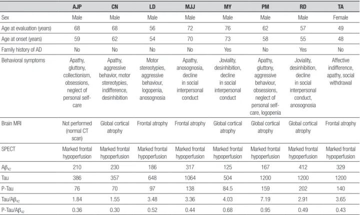
Table 1. Demographic, clinical, neuroimaging and CSF data for each patient.
AJP CN LD MJJ MY PM RD TA
Sex Male Male Male Male Male Male Male Female
Age at evaluation (years) 68 68 56 72 76 62 57 49
Age at onset (years) 59 62 54 70 73 58 55 48
Family history of AD No No No No Yes No Yes No
Behavioral symptoms Apathy,
gluttony, collectionism,
obsessions, neglect of personal
self-care
Apathy, aggressive behavior, motor
stereotypies, indifference, desinhibition
Motor stereotypies,
aggressive behaviour, logopenia, anosognosia
Apathy, anosognosia,
decline in social interpersonal
conduct
Joviality, desinhibition,
decline in social interpersonal
conduct
Apathy, gluttony, aggressive
behaviour, obsessions,
neglect of personal self-care, logopenia
Joviality, desinhibition,
decline in social interpersonal
conduct, anosognosia
Affective indifference, apathy, social
withdrawal
Brain MRI Not performed
(normal CT scan)
Global cortical atrophy
Frontal atrophy Frontal atrophy Global cortical atrophy
Global cortical atrophy
Global cortical atrophy
Frontal atrophy
SPECT Marked frontal
hypoperfusion
Marked frontal hypoperfusion
Marked frontal hypoperfusion
Marked frontal hypoperfusion
Marked frontal hypoperfusion
Marked frontal hypoperfusion
Marked frontal hypoperfusion
Marked frontal hypoperfusion
Ab42 210 230 186 317 125 167 412 329
Tau 386 357 648 1064 504 1200 1200 1200
P-Tau 76 70 97 138 84.5 159 202 140
Tau/Ab42 1.84 1.55 3.48 3.36 4.03 7.19 2.91 3.65
P-Tau/Ab42 0.36 0.30 0.52 0.44 0.68 0.95 0.49 0.43
Table 2. Neuropsychological data for each patient.
AJP CN LD MJJ MY PM RD TA
MMSE ( /30) 16 22 10 19 26 10 17 21
Orientation time/space ( /10) 8 10 4 8 10 2 4 8
MATTIS ( /144) 121 126 108 77 NA 101 111 121
MATTIS – Attention ( /37) 35 36 33 29 NA 36 36 37
MATTIS – Initiation ( /37) 35 27 28 13 NA 17 23 28
MATTIS – Construction ( /6) 5 6 4 4 NA 5 5 6
MATTIS – Concepts ( /39) 30 36 26 12 NA 36 35 31
MATTIS – Memory ( /25) 16 21 39 19 NA 7 12 19
Memory: Encoding (FCSRT) ( /16) 10 10 3 NA 14 2 9 8
Total Free Recall (FCSRT) ) ( /48) 8 9 NC NA 13 NC 0 8
Total (Free + Cued) Recall (FCSRT) ( /48) 27 18 NC NA 41 NC 12 14
Verbal Span (Direct – Indirect) 5-3 5-4 4-2 4-2 5-4 4-3 5-4 6-4
Phonemic Fluency in 2 minutes 14 10 3 1 4 3 14 7
Category Fluency in 2 minutes 18 13 3 2 21 5 15 6
FAB ( /18) 13 14 5 2 13 7 8 13
Wisconsin ( /20) 3 6 NA 3 9 3 NA 9
Mini-SEA 11.7 21.7 NA NA 18.8 13.5 NA 12.1
Gestural Apraxia Absent Absent Absent Absent Absent Absent Absent Absent
the time of clinical evaluation varied between 49 and 76 years. he age at onset of symptoms varied between 48 and 73 years. No patient had familial antecedents of bvFTD, but there was a family history of AD for two patients (patients “MY” and “RD”). According to inclu-sion criteria, all frontal AD patients had abnormal P-Tau/Ab42 ratio. Moreover, all frontal AD patients had
reduced Ab42 (<450 pg/mL) and high P-Tau (>60 pg/mL)
levels.
he most frequent behavioral signs among patients with frontal AD were apathy (5 out of 8 patients), obses-sive stereotypies (4 out of 8), decline in social interper-sonal conduct (3 out of 8), irritability/aggressive behav-ior (3 out of 8), binge eating (2 out of 8), and neglect of personal self-care (2 out of 8). At the onset of the disease, two patients had predominantly inert behavior presentation; three patients had a disinhibited proile, and three others had a mixed proile.
Scores on the MMSE difered signiicantly between
frontal AD patients and healthy controls, with lower scores for frontal AD patients (Table 3). All scores on frontal tests were signiicantly lower in frontal AD pa-tients as compared to healthy controls (Table 3). More speciically, four frontal AD patients had time-space disorientation at neuropsychological exam, while four had good orientation. All frontal AD patients had poor performance on working memory tests (verbal spans). Dysexecutive deicits were present in all patients. Five out of eight frontal AD patients were evaluated with the short version of the Social Cognition and Emotional As-sessment (Mini-SEA);17 all these patients had severe
def-icits on theory of mind and facial emotion recognition tests. Five out of eight frontal AD patients had episodic memory impairment on the Free and Cued Selective Reminding Test (FCSRT),18 with the so-called “amnestic
syndrome of the medial temporal type” (low free recall not normalized with cueing). Two frontal AD patients had severe encoding deicits that limited the evaluation
Table 3. Clinical and neuropsychological data between groups.
Frontal AD (n=8) Controls (n=18) AD (n=18) bvFTD (n=18)
Age at evaluation (years) 63.5 (±8.9) 68.6 (±7)NS 64.8 (±9.6)NS 65.7 (±7.9)NS
Disease duration (years) 3.5 (±2.4) NA 3.4 (±1.7)NS 3.2 (±1.6)NS
Sex (male/female) 7/1 10/8 9/9 9/9
Educational level (in years) 10.4 (±3.9) 12.2 (±2.3)NS 10.5 (±3.9)NS 9.8 (±4.3)NS
MMSE ( /30) 17.6 (±5.6) 29.6 (±0.6)* 22.2 (±2.9)** 23.3 (±3.6)**
Orientation time/space ( /10) 6.3 (±2.9) 9.4 (±2.3)* 8 (±1.9)NS 8 (±1.8)NS
MATTIS ( /144) 109.3 (±16.7) 143.3 (±1.5)* NA 119.2 (±12.3)NS
MATTIS – Attention ( /37) 34.6 (±2.8) 37 (±0)** NA 33.9 (±3.6)NS
MATTIS – Initiation ( /37) 24.4 (±7.4) 36.5 (±1.0)** NA 27.4 (±5.6)NS
MATTIS – Construction ( /6) 5 (±0.8) 6 (±0)** NA 5.6 (±0.7)NS
MATTIS – Concepts ( /39) 29.4 (±8.5) 39 (±0)* NA 32 (±4.9)NS
MATTIS – Memory ( /25) 19 (±10) 24.8 (±0.5)NS NA 19 (±3.7)NS
Memory: Encoding (FCSRT) ( /16) 8 (±4.2) 15.7 (±0.6)* 12.4 (±1.9)* 13.8 (±3)**
Total Free Recall (FCSRT) ( /48) 5.4 (±5.3) 33.8 (±7.9)* 12 (±5.3)** 16.9 (±5.2)*
Total (Free + Cued) Recall (FCSRT) ( /48) 16 (±14.6) 46 (±1.8)* 30.8 (±8.6)** 39.9 (±7.6)**
Verbal Span (Direct) 4.8 (±0.7) 5.5 (±0.7)** 4.8 (±1.3)NS 5.2 (±1.4)NS
Verbal Span (Indirect) 3.3 (±0.09) 4 (±0.6)NS 3.4 (±1.2)NS 3.1 (±0.9)NS
Phonemic Fluency in 2 minutes 7 (±5.1) 13.7 (±3.2)** 8.2 (±6.3)NS 6.6 (±3.8)NS
Category Fluency in 2 minutes 10.4 (±7.3) 19.1 (±4.5)** 13.8 (±4.8)NS 10.6 (±3.9)NS
FAB ( /18) 9.4 (±4.5) 16.9 (±0.9)* 13 (±2.3)** 11.6 (±3.3)NS
Wisconsin ( /20) 6 (±3) 19 (±1.1)* NA 2.5 (±1.7)NS
Mini-SEA 24 (±16) 41.2 (±6.9)* NA 26.2 (±3.4)**
FAB: Frontal Assessment Battery; FCSRT: Free and Cued Selective Reminding Test; Mini-SEA: Mini version of the Social Cognition and Emotional Assessment; MMSE: Mini-Mental State Exam; NA: Data not available. Comparison between frontal variant AD patients and other groups was performed using a non-parametric Mann-Whitney U-test with the following annotations: NSNon significant vs frontal variant
of episodic memory by the FCSRT and one patient did not undergo episodic memory evaluation with the FC-SRT. No frontal AD patients presented gestural apraxia.
here was no signiicant diference between frontal AD and bvFTD groups for MATTIS scores, verbal spans, verbal luencies (phonemic and category) and for Wis-consin score (Table 3).
As an illustrative example, we report the clinical vignette of one of the patients included in this study. Mrs. TA, a medical nurse aged 48 years, was referred to the Behavioral Unit of the Pitié-Salpêtrière Hospital in April 2010 for marked apathy, afective indiference and social withdrawal which had been evolving for ap-proximately one year. he patient also had a history of reduced verbal output. Her husband did not report memory diiculties or spatial disorientation. he activi-ties of daily living were globally preserved. A previous psychiatric referral led to a diagnosis of depression, and the patient was in use of antidepressants (venlafaxine). Her preceding medical history was unremarkable. here was no history of hallucinations, head trauma, neuro-leptic medications, or alcohol/drug abuse. here was no family history of neurological diseases or dementia. he standard neurological examination was normal, with-out abnormalities of eye movement. She had no motor signs, no extrapyramidal syndrome, and no myoclonus.
Neuropsychological tests showed an impairment in global cognitive eiciency (MMSE 21/30 and MAT-TIS scale 121/144), without disorientation in time and space. he patient presented a severe dysexecutive syn-drome, with attentional and working memory deicits. Mental lexibility and the abilities of conceptualizing and programming were severely impaired on the Trail Making Test (TMT),19 on the modiied Wisconsin Card
Sorting Test20 and on the copy of Rey complex igure.21
he patient had impaired performance on tests of theo-ry of mind and of facial emotion recognition (mini-SEA). here was an episodic memory deicit characterized by a low free recall (free recall score=8/48) not normalized with cueing (total recall=14/48). he patient had no def-icits on the execution of gestures from the limb apraxia battery.22 here were no signs of Bálint or Gertsmann
syndromes.
Language assessment demonstrated that there was no reading or writing impairment, with preserved writ-ten language comprehension. Verbal luencies were re-duced, both in phonemic (only four “p” words in two minutes) and categorical modalities (only nine fruits in two minutes), with slight diiculty on the denomina-tion task (74/80). here was no speech apraxia and no semantic deicit.
Brain MRI (Figure 1) revealed mild cortical frontal atrophy, without medial temporal atrophy. Brain SPECT (Figure 2A) showed moderate hypoperfusion in medial and dorsolateral prefrontal cortex, with left predomi-nance. here was very mild hypoperfusion in the left parietal cortex. Cerebral perfusion was preserved in me-dial temporal regions.
he patient underwent a complete blood and CSF exam in order exclude other causes of non-neurodegen-erative dementia in young patients (autoimmune dis-eases, paraneoplastic pathology, CNS infection, meta-bolic diseases, and so on). hese exams were all negative. he diagnosis of bvFTD was initially established on a clinical basis by the neurologist (BD), as the clinical picture fulilled the criteria for the disease: [1] a cor-roborated history of initial progressive decline in social interpersonal conduct, with apathy, afective indifer-ence and loss of empathy; [2] the presindifer-ence of severe diiculties in executive and social-emotional abilities; [3] atrophy of frontal lobes on brain MRI and marked hypoperfusion of frontal lobes on SPECT, with preser-vation of medial temporal and parietal regions. Taking into account these clinical data, the presence of signii-cant amnesia was not considered incompatible with the diagnosis of bvFTD.
However, some weeks after hospitalization, data on CSF biomarkers were available and showed low Ab42
(329 pg/mL), high Tau (1200 pg/mL) and high P-Tau (140 pg/mL), in favor of an AD diagnosis. All derived ra-tios (Tau/Ab42 and P-Tau/Ab42) were also in favor of AD.
Considering these results, a diagnosis of frontal variant of AD was proposed.
On the clinical follow-up over 30 months, there was a marked deterioration in cognitive abilities and the pa-tient manifested disorientation in time and space as well as limb apraxia and presented an aggravation of both
A B
amnesia and dysexecutive deicits. he patient clini-cally progressed to multi-domain cognitive impairment, with loss of autonomy, thus deining the dementia stage of the disease. he patient underwent another brain SPECT exam (Figure 2B) which showed severe hypoper-fusion in prefrontal cortex (with left predominance), se-vere hypoperfusion in the left parieto-temporal cortex and mild hypoperfusion in the right parieto-temporal cortex. here was very mild hypoperfusion in the medial temporal regions. he patient has been treated with an-tidepressants and with an anticholinesterasic. She was also included in a clinical immunotherapy trial.
DISCUSSION
We reported a series of eight patients which fulilled clinical consensual criteria for bvFTD, but for whom a diagnosis of frontal variant AD was inally proposed on the basis of CSF biomarkers. Previous studies with either biological,23 genetic24 or pathological
conirma-Figure 2. Brain scintigraphy (SPECT) from patient TA, 48 year-old woman. [A] Brain SPECT after approximately one year since symptoms onset (May/2010): moderate hypoperfusion in medial and dorsolateral prefrontal cortex, with left predominance; very mild hypoperfusion in the left parietal cortex; no hypoperfusion in medial temporal regions. [B] Brain SPECT approximately two years after symptoms onset (March/2011): severe hypoperfusion in prefrontal cortex (with left predominance); severe hypoperfusion in the left parieto-temporal cortex; mild hypoperfusion in the right parieto-temporal cortex; very mild hypoperfusion in the medial temporal regions.
A B
tion3,5,8,25,26 have also reported focal atypical
presenta-tions of AD mimicking bvFTD.
FTD is the second most frequent cause of degen-erative dementia in patients below 65 years old27 and
includes three clinical subtypes: the behavioral variant (bvFTD) and the language variants, progressive non-luent aphasia and semantic aphasia.27,28
bvFTD is the most common presentation of FTD29
and is clinically characterized by an insidious and gradu-ally progressive behavioral syndrome deined by a de-cline in social interpersonal conduct, impairment in regulation of personal conduct, emotional blunting and a loss of insight.14 bvFTD is typically associated with
frontal and anterior temporal atrophy, in particular in the mesial and orbital prefrontal cortex, anterior insula and anterior cingulate cortices.30
frontal and temporal cortex.28 FTLD have two major
histopathological subtypes: FTLD with tau-positive in-clusions (FTLD-tau), and FTLD with ubiquitin-positive and TAR DNA-binding protein (TDP) inclusions, but Tau-negative inclusions (FTLD-TDP).28 Alzheimer
pa-thology is less frequently identiied in patients clini-cally diagnosed as bvFTD.3,5,8,9 In agreement with these
pathological data, it has been demonstrated that CSF biomarkers, which are considered surrogate markers of Alzheimer’s pathophysiology, eiciently discriminate AD from bvFTD.31 In a previous study, we reported that
only one out of 27 bvFTD patients presented a CSF AD biomarker proile.11
hese pathological observations of AD presenting with symptoms that mimic bvFTD have led to the con-cept of “frontal AD”.8,25,26 Besides this behavioral variant,
AD may also present as other non-amnesic atypical focal variants, such as posterior cortical atrophy and logope-nic aphasia. Taken together, these observations empha-size that not all patients with AD manifest a “typical” clinical pattern and that patients sharing a common pa-thology may be clinically heterogeneous.6
Conversely, pathologically diferent neurological disorders may share common symptomatology. his is the case for typical AD and bvFTD. For instance, apathy, a common feature of bvFTD, is also frequently observed in AD, even at initial stages of the disease.32
On the other hand, recent evidence has shown that marked amnesia, a hallmark of AD, is not uncommon in bvFTD patients. Episodic memory performance has been traditionally considered relatively preserved in bvFTD and amnesia was considered an exclusion crite-rion for the clinical diagnosis of bvFTD.15 However, it
is increasingly recognized that bvFTD patients exhibit amnesia,33-36 even at early stages of the disease as up to
10% of pathologically-proven cases of bvFTD reported memory deicits.37 In a recent study, Hornberger, et al.38
analyzed the structural integrity of the memory circuit in AD and FTD in vivo and at post-mortem. Patients with FTD and AD patients did not difer on memory mea-sures (visual recall with the Rey-Osterrieth Complex Figure Test, visual recognition with the Doors and Peo-ple Test, and immediate recall from the Rey Auditory Verbal Learning Test). Moreover, they found that FTD and AD patients had similar degrees of hippocampal at-rophy in vivo. More interestingly, they showed that FTD had more severe hippocampal atrophy at post-mortem. In line with these observations, neuroimaging studies have previously demonstrated that measures of hippo-campal volumes do not accurately distinguish AD and bvFTD patients.39-42
Taken together, for diagnostic purposes, the reli-ance on exclusively phenotypical features assessed by “topographical markers”7, such as episodic memory
deicits and hippocampal atrophy, may lead to misdiag-nosis between AD and bvFTD. Until the development of biomarkers, the in vivo diagnosis of neurodegenerative dementias had been largely based on the identiication of the presenting cognitive proile supported by neuro-imaging. However, the diagnosis established according only to clinical “phenotypical criteria”, without reference to an accurate biomarker, may lack conidence, as not all patients with dementia syndromes manifest a “typical” clinical pattern. Moreover, patients sharing a common pathology may be clinically heterogeneous and, con-versely, pathologically diferent diseases may share com-mon symptoms. he correspondence between clinical phenotype and underlying pathology is not always opti-mal.5 Accordingly, new proposals for diagnostic criteria
of AD7,43,44 include “pathophysiological markers” such
as CSF biomarkers for increased diagnostic eiciency. he CSF is the optimal source of biological physio-pathological markers, as it is in direct contact with the cerebral extracellular space.45 he neuropathological
studies that analysed correlations of the levels of in vivo
CSF biomarkers (total Tau [T-tau], phosphorylated Tau [P-Tau] and beta-amyloid peptide 1-42 [Ab42]) with the
intensity of the post-mortem cerebral lesions showed that CSF biomarkers predicted the presence of AD pathologic features with high accuracy.46-50 Considering
these data, CSF biomarkers can be considered surrogate markers of AD-associated pathologic changes in the brain.45,47-49
he CSF levels of T-tau, P-Tau and Ab42 or, even more
speciically, the combination of low Ab42 and high
lev-els of T-tau and P-Tau, provide optimal sensitivity and speciicity in the diagnosis of AD patients (even at MCI stage) against normal controls.51,52 he combined
analy-sis of the CSF biomarkers, especially P-Tau/Ab42 ratio,
is also useful for the diferential diagnosis between AD and frontotemporal lobar degeneration, regardless of its behavioural (bvFTD) or semantic presentation.11,31
CSF biomarkers or amyloid imaging may also iden-tify patients with Alzheimer underlying pathology in atypical focal cortical presentations of AD, and thus may identify eligible patients for emerging anti-amyloid therapies.10-12,23,53-57 Including pathophysiological
In this present series, all patients manifested typi-cal bvFTD presentation and the diagnosis of atypitypi-cal AD was possible only with CSF biomarker investiga-tion. It is essential to use pathophysiological markers, especially in young subjects, in order to identify patients with atypical AD presentations and to propose a speciic treatment for them.
While pathological data may be important for es-tablishing diagnosis for patients, no autopsies were available in our cohort. It should be noted, however, that clinical diagnosis was established using accepted consensus criteria; all patients were extensively evalu-ated with clinical, biological, neuropsychological and
neuroimaging exams at a center with expertise in the ield of dementias. Furthermore, we selected patients with strict biological inclusion criteria based on CSF biomarker results, such as reduced Ab42 level, high
P-Tau and abnormal P-P-Tau/Ab42 ratio, which have been
demonstrated to be highly correlated with Alzheimer pathology at post-mortem exam.47
It would also be of value to compare the clinical and neuroimaging features across patients with frontal AD and bvFTD. Further studies with a greater number of patients are needed to investigate whether clinical, neu-roimaging and neuropsychological parameters difer during disease progression of frontal AD and bvFTD.
REFERENCES
1. Querfurth HW, LaFerla FM. Alzheimer’s disease. N Engl J Med 2010;362: 329-344.
2. McKhann G, Drachman D, Folstein M, Katzman R, Price D, Stadlan EM. Clinical diagnosis of Alzheimer’s disease: report of the NINCDS-ADRDA Work Group under the auspices of Department of Health and Human Services Task Force on Alzheimer’s Disease. Neurology 1984;34:939-944.
3. Alladi S, Xuereb J, Bak T, et al. Focal cortical presentations of Alzheim-er’s disease. Brain. 2007;130:2636-2645.
4. Galton CJ, Patterson K, Xuereb JH, Hodges JR. Atypical and typical presentations of Alzheimer’s disease: a clinical, neuropsychological, neuroimaging and pathological study of 13 cases. Brain 2000;123:484-498.
5. Snowden JS, Thompson JC, Stopford CL, et al. The clinical diagnosis of early-onset dementias: diagnostic accuracy and clinicopathological relationships. Brain 2011;134:2478-2492.
6. Murray ME, Graff-Radford NR, Ross OA, Petersen RC, Duara R, Dick-son DW. Neuropathologically defined subtypes of Alzheimer’s disease with distinct clinical characteristics: a retrospective study. Lancet Neurol 2011;10:785-796.
7. Dubois B, Feldman HH, Jacova C, et al. Revising the definition of Al-zheimer’s disease: a new lexicon. Lancet Neurol 2010;9:1118-1127. 8. Grossman M, Libon DJ, Forman MS, et al. Distinct antemortem profiles
in patients with pathologically defined frontotemporal dementia. Arch Neurol 2007;64:1601-1609.
9. Mendez MF, Joshi A, Tassniyom K, Teng E, Shapira JS. Clinicopatho-logic differences among patients with behavioral variant frontotemporal dementia. Neurology 2013;80:561-568.
10. de Souza LC, Corlier F, Habert MO, Uspenskaya O, Maroy R, Lamari F, et al. Similar amyloid-{beta} burden in posterior cortical atrophy and Alzheimer’s disease. Brain 2011;134:2036-2043.
11. de Souza LC, Lamari F, Belliard S, et al. Cerebrospinal fluid biomarkers in the differential diagnosis of Alzheimer’s disease from other cortical dementias. J Neurol Neurosurg Psychiatry 2011;82:240-246. 12. Rosenbloom MH, Alkalay A, Agarwal N, et al. Distinct clinical and
meta-bolic deficits in PCA and AD are not related to amyloid distribution. Neu-rology 2011;76:1789-1796.
13. Neary D, Snowden JS, Gustafson L, et al. Frontotemporal lobar de-generation: a consensus on clinical diagnostic criteria. Neurology 1998; 51:1546-1554.
14. McKhann GM, Albert MS, Grossman M, Miller B, Dickson D, Trojanows-ki JQ. Clinical and pathological diagnosis of frontotemporal dementia: report of the Work Group on Frontotemporal Dementia and Pick’s Dis-ease. Arch Neurol 2001;58:1803-1809.
15. Rascovsky K, Hodges JR, Knopman D, et al. Sensitivity of revised di-agnostic criteria for the behavioural variant of frontotemporal dementia. Brain 2011;134:2456-2477.
16. de Souza LC, Volle E, Bertoux M, et al. Poor creativity in frontotemporal dementia: a window into the neural bases of the creative mind. Neuro-psychologia 2010;48:3733-3742.
17. Bertoux M, Delavest M, de Souza LC, et al. Social Cognition and Emo-tional Assessment differentiates frontotemporal dementia from depres-sion. J Neurol Neurosurg Psychiatry 2012;83:411-416.
18. Grober E, Buschke H, Crystal H, Bang S, Dresner R. Screening for de-mentia by memory testing. Neurology 1988;38:900-903.
19. Reitan R. Validity of the Trail Making test as an indicator of organic brain damage. Percept Mot Skills 1958;8:271-276.
20. Nelson HE. A modified card sorting test sensitive to frontal lobe defects. Cortex 1976;12:313-324.
21. Liberman J, Stewart W, Seines O, Gordon B. Rater agreement for the Rey-Osterrieth Complex Figure Test. J Clin Psychol 1994;50:615-624. 22. Peigneux P, Van der Linden M. Présentation d’une batterie
neuropsy-chologique et cognitive pour l’évaluation de l’apraxie gestuelle. Rev Neuropsychol 2000;10:311-362.
23. Richard-Mornas A, Dirson S, Perret-Liaudet A, Decousus M, Thomas-Anterion C. Diagnosis contribution of biomarkers in Alzheimer disease: A case of frontotemporal dementia. Rev Neurol (Paris) 2011;167:160-163.
24. Raux G, Gantier R, Thomas-Anterion C, et al. Dementia with prominent frontotemporal features associated with L113P presenilin 1 mutation. Neurology 2000;55:1577-1578.
25. Johnson JK, Head E, Kim R, Starr A, Cotman CW. Clinical and patho-logical evidence for a frontal variant of Alzheimer disease. Arch Neurol 1999;56:1233-1239.
26. Taylor KI, Probst A, Miserez AR, Monsch AU, Tolnay M. Clinical course of neuropathologically confirmed frontal-variant Alzheimer’s disease. Nat Clin Pract Neurol 2008;4:226-232.
27. Piguet O, Hornberger M, Mioshi E, Hodges JR. Behavioural-variant frontotemporal dementia: diagnosis, clinical staging, and management. Lancet Neurol 2011;10:162-172.
28. Seelaar H, Rohrer JD, Pijnenburg YA, Fox NC, van Swieten JC. Clinical, genetic and pathological heterogeneity of frontotemporal dementia: a review. J Neurol Neurosurg Psychiatry 2011;82:476-486.
29. Kertesz A, McMonagle P, Blair M, Davidson W, Munoz DG. The evolu-tion and pathology of frontotemporal dementia. Brain 2005;128:1996-2005.
30. de Souza LC, Lehericy S, Dubois B, Stella F, Sarazin M. Neuroimaging in dementias. Curr Opin Psychiatry 2012;25:473-479.
31. Bian H, Van Swieten JC, Leight S, et al. CSF biomarkers in frontotempo-ral lobar degeneration with known pathology. Neurology 2008;70:1827-1835.
32. Weiner MF, Hynan LS, Bret ME, White C 3rd. Early behavioral symp-toms and course of Alzheimer’s disease. Acta Psychiatr Scand 2005; 111:367-371.
33. Yew B, Alladi S, Shailaja M, Hodges JR, Hornberger M. Lost and For-gotten? Orientation versus Memory in Alzheimer’s Disease and Fronto-temporal Dementia. J Alzheimers Dis 2013;33:473-481.
35. Hornberger M, Piguet O. Episodic memory in frontotemporal dementia: a critical review. Brain 2012;135:678-692.
36. Hornberger M, Piguet O, Graham AJ, Nestor PJ, Hodges JR. How pre-served is episodic memory in behavioral variant frontotemporal demen-tia? Neurology 2010;74:472-479.
37. Hodges JR, Davies RR, Xuereb JH, et al. Clinicopathological correlates in frontotemporal dementia. Ann Neurol 2004;56:399-406.
38. Hornberger M, Wong S, Tan R, et al. In vivo and post-mortem memory circuit integrity in frontotemporal dementia and Alzheimer’s disease. Brain 2012;135:3015-3025.
39. Frisoni GB, Laakso MP, Beltramello A, Geroldi C, Bianchetti A, Soininen H, et al. Hippocampal and entorhinal cortex atrophy in frontotemporal dementia and Alzheimer’s disease. Neurology 1999;52:91-100. 40. Laakso MP, Frisoni GB, Kononen M, et al. Hippocampus and entorhinal
cortex in frontotemporal dementia and Alzheimer’s disease: a morpho-metric MRI study. Biol Psychiatry 2000;47:1056-1063.
41. Lindberg O, Walterfang M, Looi JC, et al. Hippocampal shape analysis in Alzheimer’s disease and frontotemporal lobar degeneration subtypes. J Alzheimers Dis 2012;30:355-365.
42. Davatzikos C, Resnick SM, Wu X, Parmpi P, Clark CM. Individual patient diagnosis of AD and FTD via high-dimensional pattern classification of MRI. Neuroimage 2008;41:1220-1227.
43. Albert MS, Dekosky ST, Dickson D, et al. The diagnosis of mild cog-nitive impairment due to Alzheimer’s disease: Recommendations from the National Institute on Aging-Alzheimer’s Association workgroups on diagnostic guidelines for Alzheimer’s disease. Alzheimers Dement 2011;7:270-279.
44. Jack CR, Jr., Albert MS, Knopman DS, et al. Introduction to the recom-mendations from the National Institute on Aging-Alzheimer’s Associa-tion workgroups on diagnostic guidelines for Alzheimer’s disease. Al-zheimers Dement 2011;7:257-262.
45. Blennow K, Hampel H, Weiner M, Zetterberg H. Cerebrospinal fluid and plasma biomarkers in Alzheimer disease. Nat Rev Neurol 2010;6: 131-144.
46. Tapiola T, Overmyer M, Lehtovirta M, et al. The level of cerebrospinal fluid tau correlates with neurofibrillary tangles in Alzheimer’s disease. Neuroreport 1997;8:3961-3963.
47. Tapiola T, Alafuzoff I, Herukka SK, et al. Cerebrospinal fluid {beta}-am-yloid 42 and tau proteins as biomarkers of Alzheimer-type pathologic changes in the brain. Arch Neurol 2009;66:382-389.
48. Seppala TT, Nerg O, Koivisto AM, et al. CSF biomarkers for Alzheim-er disease correlate with cortical brain biopsy findings. Neurology 2012;78:1568-1575.
49. Buerger K, Ewers M, Pirttila T, et al. CSF phosphorylated tau protein correlates with neocortical neurofibrillary pathology in Alzheimer’s dis-ease. Brain 2006;129:3035-3041.
50. Strozyk D, Blennow K, White LR, Launer LJ. CSF Abeta 42 levels corre-late with amyloid-neuropathology in a population-based autopsy study. Neurology 2003;60:652-656.
51. Hansson O, Zetterberg H, Buchhave P, Londos E, Blennow K, Mint-hon L. Association between CSF biomarkers and incipient Alzheimer’s disease in patients with mild cognitive impairment: a follow-up study. Lancet Neurol 2006;5:228-2234.
52. Mattsson N, Zetterberg H, Hansson O, et al. CSF biomarkers and in-cipient Alzheimer disease in patients with mild cognitive impairment. JAMA 2009;302:385-393.
53. Leyton CE, Villemagne VL, Savage S, et al. Subtypes of progressive aphasia: application of the international consensus criteria and valida-tion using {beta}-amyloid imaging. Brain 2011;134:3030-3043. 54. Baumann TP, Duyar H, Sollberger M, et al. CSF-tau and
CSF-Abe-ta(1-42) in posterior cortical atrophy. Dement Geriatr Cogn Disord 2010; 29:530-533.
55. Rabinovici GD, Jagust WJ, Furst AJ, et al. Abeta amyloid and glucose metabolism in three variants of primary progressive aphasia. Ann Neurol 2008;64:388-401.
56. Seguin J, Formaglio M, Perret-Liaudet A, et al. CSF biomarkers in pos-terior cortical atrophy. Neurology 2011;76:1782-1788.
57. Kas A, Uspenskaya O, Lamari F, et al. Distinct brain perfusion pattern associated with CSF biomarkers profile in progressive aphasia J Neurol Neurosurg Psychiatry 2012;83:695-698.
58. Rabinovici GD, Rosen HJ, Alkalay A, et al. Amyloid vs FDG-PET in the differential diagnosis of AD and FTLD. Neurology 2011;77:2034-2042. 59. Rowe CC, Ng S, Ackermann U, et al. Imaging beta-amyloid burden in
![Figure 1. Brain MRI from patient TA, 48 year-old woman. [A] Axial slice: mild cortical frontal atrophy, predominant in medial regions](https://thumb-eu.123doks.com/thumbv2/123dok_br/15188374.527030/5.892.440.788.105.283/figure-brain-patient-cortical-frontal-atrophy-predominant-regions.webp)
![Figure 2. Brain scintigraphy (SPECT) from patient TA, 48 year-old woman. [A] Brain SPECT after approximately one year since symptoms onset (May/2010):](https://thumb-eu.123doks.com/thumbv2/123dok_br/15188374.527030/6.892.110.831.102.591/figure-brain-scintigraphy-spect-patient-brain-approximately-symptoms.webp)
