Behavioural-variant
frontotemporal dementia
An update
Olivier Piguet1, John R. Hodges1
ABSTRACT. Behavioural-variant frontotemporal dementia (bvFTD) is characterised by insidious changes in personality and interpersonal conduct that reflect progressive disintegration of the neural circuits involved in social cognition, emotion regulation, motivation and decision making. The underlying pathology is heterogeneous and classified according to the presence of intraneuronal inclusions of tau, TDP-43 or occasionally FUS. Biomarkers to detect these histopathological changes in life are increasingly important with the development of disease-modifying drugs. Gene mutations have been found which collectively account for around 10-20% of cases including a novel hexanucleotide repeat on chromosome 9 (C9orf72). The recently reviewed International Consensus Criteria for bvFTD propose three levels of diagnostic certainly: possible, probable and definite. Detailed history taking from family members to elicit behavioural features underpins the diagnostic process with support from neuropsychological testing designed to detect impairment in decision-making, emotion processing and social cognition. Brain imaging is important for increasing the level of diagnosis certainty. Carer education and support remain of paramount importance.
Key words: frontotemporal dementia, genetics, cognition, social cognition, neuroimaging. DEMÊNCIA FRONTOTEMPORAL-VARIANTE COMPORTAMENTAL: UMA REVISÃO
RESUMO. A demência frontotemporal-variante comportamental (DFTvc) é caracterizada por mudanças insidiosas de personalidade e conduta interpessoal, que refletem a desintegração progressiva de circuitos neurais envolvidos em cognição social, regulação emocional, motivação e tomada de decisão. O substrato patológico é heterogêneo e classificado de acordo com a presença de inclusões intraneuronais de proteína tau, TDP-43 ou, ocasionalmente, de FUS. Biomarcadores capazes de detectar estas alterações histopatológicas durante a vida vêm ganhando importância com o desenvolvimento de drogas específicas modificadoras da doença. Algumas mutações genéticas já foram encontradas, sendo em conjunto responsáveis por 10-20% dos casos, incluindo a recentemente descrita repetição de hexanucleotídeo no cromossomo 9 (C9orf72). A versão revisada dos Critérios Internacionais do Consenso em DFTvc propõe três níveis de certeza diagnóstica: possível, provável e definida. História clínica detalhada obtida com familiares, para identificar as alterações de comportamento características, auxilia no diagnóstico, juntamente com o apoio de avaliação neuropsicológica dirigida à detecção de comprometimento em tarefas de tomada de decisão, processamento emocional e cognição social. A neuroimagem é importante para aumentar o grau de certeza diagnóstica. Educação e suporte dos cuidadores continuam sendo medidas de extrema relevância.
Palavras-chave: demência frontotemporal, genética, cognição, cognição social, neuroimagem.
INTRODUCTION
F
rontotemporal dementia (FTD) is the clinical diagnostic label now preferred to describe patients with a range of progressive dementia syndromes associated with focal atrophy of the orbitomesial frontal and ante-rior temporal lobes. Epidemiological studies suggest that FTD is the second mostcom-mon cause of young onset dementia after Alzheimer’s disease (AD).1,2 Two independent studies from the UK revealed a prevalence of around 15 per cases per 100,000 population aged 45 to 64 with broad conidence intervals (8 to 27).1 Although regarded as a rare cause of dementia after 65 years, FTD may be more common than assumed because older adults
1Neuroscience Research Australia, Barker St, Randwick NSW 2031, Australia. School of Medical Sciences, the University of New South Wales, Sydney, Australia.
ARC Centre of Excellence in Cognition and its Disorders, the University of New South Wales, Sydney, Australia.
John Hodges. Neuroscience Research Australia. Barker St,Randwick NSW 2031, Australia. E-mail: [email protected] Disclosure: The authors report no conflicts of interest.
rarely undergo the types of investigation needed to es-tablish a conident in vivo diagnosis and are not followed to autopsy.
Unlike AD, both the clinical proile and underlying pathology are heterogeneous in FTD. Two broad pre-sentations are recognised: progressive deterioration in social function and personality, known as bvFTD (or sometimes simply FTD) and insidious decline in language skills, known as primary progressive aphasia which can, in turn, be subdivided according the pre-dominant pattern of language breakdown into progres-sive nonluent aphasia and semantic dementia.3-5 he syndrome of FTD overlaps with motor neurone disease (MND) both clinically and pathologically, and with a number of the extrapyramidal motor disorders. Around 10% of patients with FTD develop clinical and neuro-physiological evidence of MND.6,7 and likewise patients with MND show behavioural and/or language changes which, in some instances, are severe enough to qualify for a diagnosis of FTD.8 Of the extrapyramidal disorders, corticobasal degeneration and progressive supranuclear palsy show substantial overlap with FTD and share the inding of abnormal tau pathology.9
his review cannot cover every facet of this rapid-ly evolving ield and focuses on the clinical aspects of bvFTD within the context of recent pathological and ge-netic discoveries. Readers are referred to recent authori-tative reviews on the language presentations of FTD.3,4
PATHOLOGY
he subtypes of underlying pathology in patients with FTD are classiied on the basis of the pattern of protein accumulation and are referred to collectively as fron-totemporal lobar degeneration (FTLD).10 At
postmor-tem, cases share, by deinition, the inding of bilateral frontotemporal atrophy with neuronal loss, microvacu-olation and a variable degree of astrocytic gliosis. he progression of this atrophy has been examined by map-ping the pattern in patients with diferent disease dura-tion.11 Initially, mesial and orbital frontal regions bear the brunt of the atrophy followed by the temporal pole, hippocampal formation, dorsolateral frontal cortex and the basal ganglia. his pattern of progression of atrophy has been shown to relate to the volume of cortical and subcortical regions12 and underlying neuron loss.13
Inclusions of the microtubular binding protein tau are present in approximately 40% of cases (FTLD-tau).14 Tau positive cases include the subset with mutations of the microtubule associated phosphoprotein tau (MAPT) gene. Further sub-classiication is based on morpho-logic criteria and the predominance of either tau with
three microtubule binding repeats (3R tau) or four microtubule binding repeat (4R tau).10 he majority of the remaining cases are tau negative ubiquitin positive and have inclusions comprising the TAR DNA binding protein of 43-kDA or TDP-43 (FTLD-TDP). A minor-ity (around 5 to 10%), which are both tau and TDP-43 negative, harbours inclusions of FUS, fused in sarcoma protein (FTLD-FUS).14,15 A small proportion of cases has either no inclusions (FTLD-ni) or shows ubiquitin inclu-sions which TDP-43 and FUS negative (FTLD-UPS)14 suggesting that additional protein abnormalities will be found in FTLD.
In bvFTD, any of the histological variants can be found, with an approximately 50-50 split between FTLD-tau and FTLD-TDP,16,17 and a small proportion of FTLD-FUS cases.18 With the advent of potential disease-modifying therapies, ascertainment of a pathological diagnosis in vivo will be increasing important. As yet, no reliable method of determining pathology in life exists.
GENETICS
Up to 40% of patients with FTD are said to have a fam-ily history of dementia2 but the high community preva-lence of non-FTD dementia means that many of the el-derly family members included in such estimates almost certainly have other causes of dementia. Patients with an autosomal dominant pattern (afected irst degree relatives across two generations) account for only 10% of cases.19 he strength of family history is highly predic-tive in that mutations can now be demonstrated in the majority of patients with two or more irst degree rela-tives with a dementia syndrome compatible with FTD.19 Mutations of the MAPT, and the progranulin (GRN) genes each account for 5-11% of total FTD cases.19 Link-age studies of familial FTD-MND clusters indicated a common locus in the region of chromosome 9p13.2– 21.3.20 In 2011, the responsible mutation, a novel hex-amino acid expansion termed C9orf72 was identiied21,22 which appears to be the most common gene abnormal-ity in FTD.23 It is particularly associated with familial FTD-MND but is also found in patients with bvFTD, some of whom may have a family history of MND.23 Pa-tients with this mutation appear to have a particularly high rate of psychotic features which are otherwise rare in FTD.24,25 In our own experience, screening 89 patients with FTD syndromes revealed 10% with the C9orf72 mutation24 compared to an earlier similar study which found a prevalence of the GRN mutation of around 4%.26 he three most common mutations – MAPT, GRN and
C9orf72 together account around three-quarters of
encoding for TDP -43 (TARDBP) and FUS, recognised as a cause of familial ALS, have also been identiied in a small number of cases of FTD-ALS27,28 but seems rare in uncomplicated FTD.19 Rare genetic mutations caus-ing FTD include valosin-containcaus-ing protein (VCP) and charged multivesicular body protein 2B (CHMP2B). Mu-tation of the VCP gene causes FTD in association with inclusion body myopathy and Paget’s disease of bone,29 whereas the CHMP2B gene mutation is mostly conined to a large Danish cohort with FTD.30,31
From a practical perspective, a detailed family his-tory should be taken in all patients with suspected FTD bearing in mind the overlap between MND and FTD, that a diagnosis of FTD or Pick’s disease was rarely made in the past and the phenotypic variability within fami-lies with gene mutations: one member may present with bvFTD and others have a progressive aphasic syndrome or corticobasal syndrome. Based upon a comprehensive analysis of the frequency gene mutations according to strength of family history and clinical syndrome in a large clinical cohort19 and recent indings related to
the C9orf72 gene expansion, we recommend that
pa-tients with one or more irst -degree relatives with a disease within in the FTD spectrum, including MND, be screened for MAPT, GRN and C9orf72 gene mutations after appropriate counselling in a clinical genetics set-ting. If thepatient has FTD-MND, or a family history of MND, or features of psychosis, then screening for
C9orf72 should be conducted irst. hose with an
infor-mative family history that reveals no afected relatives can be conidently reassured and need not undergo gene screening. It should be noted that the age of onset in patients with MAPT gene mutations is almost always below 65 whereas those with GRN mutations are often older.19
BEHAVIOURAL FEATURES OF BVFTD
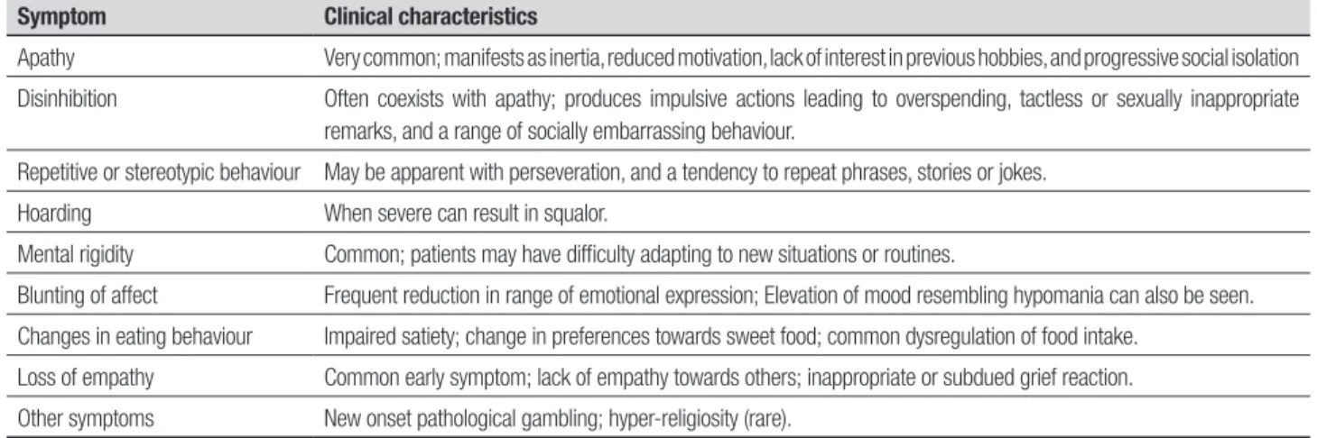
Insidious changes in personality, interpersonal conduct and emotional modulation characterise bvFTD and re-lect progressive disintegration of the neural circuits involved in social cognition, emotion regulation, moti-vation and decision making.32-34 he onset is typically diicult to pinpoint. Since insight is limited, or absent, an interview with a close family member to elicit the nature of the early symptoms and their progression is vital. he assessment and diagnosis has been greatly assisted by the development of carer based question-naires designed to document the range of symptoms found in the dementia, notably the Neuropsychiatric Inventory,35 Cambridge Behavioural Inventory36 and the Frontal Behavioural Inventory.37All of the features found in bvFTD can occur in other dementias but it is their predominance and early emergence that typify bvFTD (Table 1).
Psychotic symptoms such as delusions, paranoid ideation and hallucinations are relatively rare in FTD, except in patients with FTD or FTD-MND associated with the C9orf72 gene expansion in whom a prevalence of up to 40% of psychosis has been reported.24,25 Psycho-sis has also been reported in young-onset patients with FTLD-FUS6,38 who can present with lorid behavioural symptoms. In these patients, their age of onset is often exceptionally young with an average of 41 years and a positive family history appears rare in keeping with the absence of FUS gene mutations in this group.18
Social disinhibition, euphoria, stereotypical or aber-rant motor behaviour, and changes in eating preference are the features that most clearly discriminate bvFTD from AD.36.39 Increased behavioural changes have been associated with disease severity.40 Agitation, disinhibi-tion and irritability also seem to be more frequent in
Table 1. Symptoms characteristic of behavioural-variant frontotemporal dementia.
Symptom Clinical characteristics
Apathy Very common; manifests as inertia, reduced motivation, lack of interest in previous hobbies, and progressive social isolation
Disinhibition Often coexists with apathy; produces impulsive actions leading to overspending, tactless or sexually inappropriate remarks, and a range of socially embarrassing behaviour.
Repetitive or stereotypic behaviour May be apparent with perseveration, and a tendency to repeat phrases, stories or jokes.
Hoarding When severe can result in squalor.
Mental rigidity Common; patients may have difficulty adapting to new situations or routines.
Blunting of affect Frequent reduction in range of emotional expression; Elevation of mood resembling hypomania can also be seen.
Changes in eating behaviour Impaired satiety; change in preferences towards sweet food; common dysregulation of food intake.
Loss of empathy Common early symptom; lack of empathy towards others; inappropriate or subdued grief reaction.
the later stages,41 while restlessness and hyperorality are present throughout the disease.42
In summary, behavioural assessment is a central component of the examination of patients with poten-tial bvFTD and appears more sensitive in distinguish-ing bvFTD from AD than standard cognitive testdistinguish-ing. Despite a considerable increase of our knowledge of the behavioural changes in bvFTD, which are at the root of so much carer distress, much remains uncertain partic-ularly concerning their speciicity, neural basis and their relation to underlying pathology.
THE BVFTD PHENOCOPY SYNDROME AND
IMPLICATIONS FOR DIAGNOSTIC CRITERIA
he diagnosis of bvFTD is by no means an easy task in the early stages and many of the symptoms overlap with those seen in psychiatric disorders as well as other de-mentias.38 It is also increasingly apparent that a subset of patients who present with the clinical features of bvFTD do not progress to frank incapacitating dementia.43 Such patients are almost always men and they remain stable over many years or improve.44,45 he symptom proile as reported by family members is identical except that ac-tivities of daily living are less disrupted.45,46 A number of features distinguish these non-progressor or phenocopy cases from those with true FTD, notably normal or mar-ginal impairment on neuropsychological tests of execu-tive function, preserved memory and social cognition, a lack of overt atrophy on MRI, and normal metabolic (PET) imaging brain.43-45,47
To qualify for a diagnosis of the phenocopy syndrome we recommend that patients should remain stable with-out evidence of brain atrophy or decline on cognitive tasks with maintenance of ADLs over a period of three years. Diagnostic caution is advised in patients with a possible bvFTD diagnosis as some of these do progress over time. In our experience, those who eventually fall in the “phenocopy” category remain stable over many years. In Cambridge, some have been observed over a decade without progression to frank dementia.
he aetiology of the phenocopy syndrome is a mat-ter of debate. A proportion of patients appear to have a developmental personality disorder in the Asperger’s spectrum with decompensation due to altered life cir-cumstances (personal observation). Some may have a chronic low-grade mood disorder, but others remain a mystery, although a genetic aetiology may be a possi-bility.48 Within the international consensus criteria for bvFTD, phenocopy cases qualify for a diagnosis of pos-sible bvFTD, on the basis of the presence of three core behavioural or cognitive features (social disinhibition,
apathy, loss of empathy, stereotypic behaviours or al-terations in eating pattern, neuropsychological deicits indicative of frontal executive dysfunction). A diagno-sis of probable bvFTD, however, is not applicable, as it requires evidence of functional decline and unequivocal neuroimaging abnormalities.
NEUROPSYCHOLOGY OF
BEHAVIOURAL-VARIANT FTD
Cognition in bvFTD. Early in the disease process, bvFTD patients may perform relatively well on formal neu-ropsychological tests despite the presence of signii-cant personality and behavioural changes.49 he Mini-Mental State Examination (MMSE) is insensitive but the Addenbrooke’s Cognitive Examination (ACE and ACE-Revised) appears to detect around 90% of cases at presentation.50 he prototypical cognitive proile is one of relatively preserved language and visuospatial/con-structive abilities. Whether bvFTD patients exhibit ex-ecutive dysfunctions remains contentious,51,52 and has been complicated by the inclusion of phenocopy cases (see above). Such deicits, however, constitute a central diagnostic feature of the newly proposed clinical diag-nostic criteria.52 he combination of speciic tests (e.g., Digit Span backward, the Hayling test of response inhi-bition and the short version of the executive and social cognition battery) may help diferentiate these cases, as tests are typically abnormal in patients with true bvFTD and normal in phenocopy cases.51,53
he current international consensus criteria for bvFTD advocate a relative sparing of episodic memory.54 A proportion (10-15%) of patients with pathologically conirmed FTLD, however, present with severe amne-sia.16,55 Deicits in episodic memory appear more com-mon than previously reported,56 with deicits being, in some instances, as severe as in AD on tests of episodic memory, even after accounting for disease severity.57 hese deicits are found not only on tests of anterograde memory but also on tests of autobiographical memory and tests of future thinking.58,59
he existing evidence indicates that no speciic cog-nitive proile appears to be associated with bvFTD early in the disease, although careful cognitive evaluation will reveal deicits, generally in the domains of executive function and episodic memory. With disease progres-sion, the pattern of deicits becomes less distinct from other FTD subtypes, notably semantic dementia.9
the use of naturalistic tasks (i.e., tasks relecting cog-nitive and non cogcog-nitive demands more akin to daily activities). he orbitomesial frontal cortices are critical for performance in these domains and lesions within these brain regions have been shown to impact nega-tively on tests measuring these cognitive constructs.60 Not surprisingly, bvFTD patients have been found to be impaired on tasks such as the Iowa Gambling task, Go-NoGo, or Reversal learning.61,62
Emotion recognition and social cognition in bvFTD. A striking impairment in emotion detection and recognition is evident early in the course of bvFTD and appears to be most pronounced for negative emotions, such as fear, sadness, anger and disgust.63 Disorders of emotion de-tection and regulation are part of the clinical diagnos-tic criteria for the disease (i.e., early emotional blunt-ing; early decline in social interpersonal conduct). It is important to note, however, that such deicits are not limited to this subtype of FTD and are also present in semantic dementia.64 Diiculties in detecting and un-derstanding emotions are observed with static (photos) or dynamic (ilms) visual stimuli,65,66 voices,67 emotional words68 or even music.69 Importantly, physiological re-sponses (e.g., skin conductance) to some emotional stimuli appear preserved.70 Deicits have also been ob-served in detecting more complex emotions, such as embarrassment.71,72 Some of these deicits are modu-lated by coexisting cognitive deicits73,74 and might be amenable to retraining to enhance their recognition and improve interpersonal relationships.
Patients with bvFTD patients are also impaired on many aspects of social cognition. For example, the often reported feature of lack of empathy and coldness is con-irmed on formal testing.75 heory of mind is impaired in bvFTD, as exempliied by defective ability to infer intention and mental states in others, to take someone else’s point of view,62,76,77 detection of social faux pas,76 discrimination of sincere from sarcastic exchanges47,78 and understanding of situations requiring moral judg-ment.79 hese deicits have been recently described as a failure to process contextual information.80 While most of the tasks developed remain in the research arena, well-validated tests of emotion and sarcasm detection exist81,82 which will hopefully become part of the stan-dard cognitive evaluation in suspected bvFTD.
NEUROIMAGING IN BVFTD
In most cases, atrophy of the mesial frontal, orbitofron-tal and anterior insula cortices can be visually observed on magnetic resonance imaging (MRI) acquired in the
coronal plane.83-85 A normal MRI to visual inspection does not, however, completely exclude a diagnosis of bvFTD, as the changes may be subtle in the early stages.
Automated quantitative methods including voxel-based morphometry and cortical thickness mapping have revealed selective atrophy of the anterior cingu-late and frontal insula cortices early in the course of bvFTD.85,86 he anterior cingulate-frontal insula complex contains the von Economo cells, a unique population of neurons thought to be involved in the development and maintenance of social cognition, which are depleted in patients with bvFTD coming to autopsy.34,88 Signii-cant changes in structural and functional connectivity among the regions most sensitive to atrophy in bvFTD compared to healthy controls or patients with other de-mentia syndromes have also been reported.87,89 Patterns of grey matter atrophy may be predictive of the under-lying pathological process in bvFTD,18,90,91 although pat-terns of atrophy appear to relate more closely to clinical features than to speciic pathologies.92
Brain atrophy in bvFTD is also present in subcorti-cal brain regions, including amygdala, hippocampus, caudate, striatum, putamen, thalamus, and hypothala-mus,93,94 accompanied by reduction in functional and structural connectivity among a number of subcortical and cortical structures.89,95
In contrast to the well-documented cortical grey matter changes, presence and severity of white matter changes in bvFTD have only recently been investigated. Frontal lobe white matter volume reduction largely par-allels the atrophy in the adjacent grey matter in bvFTD with diferent subtypes of FTD showing speciic pat-terns of white matter atrophy.96-98 Using difusion ten-sor imaging (DTI), which is an index of changes in the microstructure organisation of the white matter, stud-ies have successfully diferentiated bvFTD from AD, as well as the diferent FTD subtypes.96,98,99 Patients with bvFTD appear to show a selective reduction in some white matter tracts (superior longitudinal fasciculus, uncinate fasciculus, cingulum tracts and genu and sple-nium of the corpus callosum), particularly those within the frontal lobe (e.g., genu of the corpus callosum) or those connecting frontal and temporal brain regions (e.g., uncinate fasciculus).96,99,100
predominant in the temporoparietal and posterior cin-gulate cortices.101 Although SPECT appears to be more sensitive than structural MRI in detecting early patho-logical changes in bvFTD, quantiication and speciicity of these changes are not established. Hypometabolism on FDG-PET is detected consistently and reliably in the frontal brain regions in bvFTD patients compared to AD patients who show posterior cingulate hypometabolism early in the disease process.102 hese changes are detect-ed before any changes are visible on structural MR imag-es making FDG-PET the most sensitive diagnostic tool currently available. It is also particularly useful to help identify phenocopy cases who will show preserved fron-tal metabolism. In patients showing clear brain atrophy on structural MR images, however, little additional di-agnostic beneit is gained by conducting a PET scan, as focal atrophy is a positive predictive marker of FTD.
he novel PET technique, employing the β–amyloid detecting [11C] Pittsburgh Compound B (PIB), shows promising results in discriminating AD and FTD cas-es,103,104 particularly those presenting with language deicits rather than behavioural changes. Its use as a routine test remains to be established but its clinical applicability is evident as therapeutic interventions are being developed that are likely to be pathology speciic.
In summary, neuroimaging investigations in the di-agnosis of bvFTD are powerful tools, which can reliably diferentiate bvFTD from other FTD subtypes and from other dementia syndromes, and can corroborate clinical diagnostics based on neuropsychiatric symptoms.
MANAGEMENT
No disease speciic treatment interventions for FTD currently exist. Treatment largely remains supportive and involves a combination of non-pharmacological and pharmacological measures, aimed at reducing the efect of distressing symptoms.105 he role of pharmacological interventions in FTD remains uncertain, and only small and often conlicting treatment trials have been con-ducted thus far that have also failed to consider impact on carer stress as a major outcome variable. Selective serotonin reuptake inhibitors (SSRIs) have been used to treat disinhibition and challenging behaviours, but evi-dence for their use remains contradictory.106,107 Atypical antipsychotics such as olanzapine have been used for patients with prominent agitation, aggressive behaviour or psychosis.108 Anticholinesterase inhibitors, the main-stay of AD therapy, do not have an established role in the treatment of FTD. One study reported improvement in measures of behavioural disturbance and carer stress with rivastigmine,109 however deterioration in
neuropsy-chiatric symptoms without cognitive improvement was demonstrated with donepezil.110 Two recently reported double-blind, placebo-controlled, trials of memantine, a non competitive inhibitor of NMDA receptors, failed to show any signiicant beneit in terms of symptom improvement with a suggestion of cognitive worsen-ing.111,112 A number of drugs under development at-tempt to reduce aggregation of tau or TPD-43 and hence slow the fundamental pathological process in FTD.105,113
Limited information is available on the efectiveness of systematic caregiver intervention, although a recent pilot study which employed the antecedent-behaviour-consequence (ABC) model114 produced an encouraging reduction in caregiver distress and improved coping strategies.115 Studies have conirmed the clinical im-pression that caregiver burden is much greater in FTD than in AD.116-118 Behavioural changes, rather than level of disability, appear to be correlated with caregiver dis-tress and burden in bvFTD,116 although a younger age at disease onset and disease severity also appear to impact on burden of care.119,120 Evidence indicates that care-giver health is a major contributor to carer stress, with depression accounting for 58% of the variance of stress scores on FTD caregivers.117 It seems the key for reduc-ing caregiver stress lies in increasreduc-ing their understand-ing of the symptoms and ways of dealunderstand-ing with challeng-ing behaviours.
CONCLUSIONS AND FUTURE DIRECTIONS
Knowledge of the clinical presentation in bvFTD and its pathological processes has improved dramatically over the past 20 years. Clinicians have become more aware of this disabling neurodegenerative condition afecting individuals who are not uncommonly still in the work-force or with young children. Careful medical history and information from family members, combined with clinical investigations, neuropsychological testing in-cluding investigations of social cognition have increased case identiication. Sensitivity has also improved with the use of advanced structural and functional neuro-imaging techniques. he major challenge that remains, however, is to improve the prediction of the underlying neuropathology in bvFTD patients during life. Eforts to identify potential disease biomarkers for the disease are promising but will require further investigations. his line of research will become particularly relevant as disease-modifying agents are being developed.
REFERENCES
1. Ratnavalli E, Brayne C, Dawson K, Hodges JR. The prevalence of fron-totemporal dementia. Neurology 2002;58:1615-1621.
2. Rosso SM, Donker Kaat L, Baks T, et al. Frontotemporal dementia in The Netherlands: patient characteristics and prevalence estimates from a population-based study. Brain 2003;126:2016-2022.
3. Grossman M. Primary progressive aphasia: clinicopathological correla-tions. Nat Rev Neurol 2010;6:88-97.
4. Hodges JR, Patterson K. Semantic dementia: a unique clinicopatho-logical syndrome.Lancet Neurol 2007;6:1004-1014.
5. Neary D, Snowden JS, Gustafson L, et al. Frontotemporal lobar de-generation: a consensus on clinical diagnostic criteria. Neurology 1998; 51:1546-1554.
6. Lillo P, Garcin B, Hornberger M, Bak TH, Hodges JR. Neurobehavioral features in frontotemporal dementia with amyotrophic lateral sclerosis. Arch Neurol 2010;67:826-830.
7. Lomen-Hoerth C, Anderson T, Miller B. The overlap of amyotrophic lateral sclerosis and frontotemporal dementia. Neurology 2002;59:1077-1079. 8. Lillo P, Mioshi E, Zoing MC, Kiernan MC, Hodges JR. How common
are behavioural changes in amyotrophic lateral sclerosis? Amyotroph Lateral Scler 2011;12:45-51.
9. Kertesz A, McMonagle P, Blair M, Davidson W, Munoz DG. The evolu-tion and pathology of frontotemporal dementia.Brain 2005;128:1996-2005.
10. Cairns NJ, Bigio EH, Mackenzie IR, et al. Neuropathologic diagnostic and nosologic criteria for frontotemporal lobar degeneration: consensus of the Consortium for Frontotemporal Lobar Degeneration Acta Neuro-pathol (Berlin) 20076;114:5-22.
11. Broe M, Hodges JR, Schofield E, Shepherd CE, Kril JJ, Halliday GM. Staging disease severity in pathologically confirmed cases of frontotem-poral dementia. Neurology 2003;60:1005-1011.
12. Kril JJ, Macdonald V, Patel S, Png F, Halliday GM. Distribution of brain atrophy in behavioral variant frontotemporal dementia J Neurol Sci 2005;232:83-90.
13. Kersaitis C, Halliday GM, Kril JJ. Regional and cellular pathology in fron-totemporal dementia: relationship to stage of disease in cases with and without Pick bodies Acta Neuropathol 2004;108:515-523.
14. Mackenzie IR, Neumann M, Bigio EH, et al. Nomenclature and nosology for neuropathologic subtypes of frontotemporal lobar degeneration: an update. Acta Neuropathol 2010;119:1-4.
15. Neumann M, Rademakers R, Roeber S, Baker M, Kretzschmar HA, Mackenzie IR. A new subtype of frontotemporal lobar degeneration with FUS pathology Brain 2009;132:2922-2931.
16. Hodges JR, Davies RR, Xuereb JH, et al. Clinicopathological correlates in frontotemporal dementia Annals of Neurology 2004;56:399-406. 17. Shi J, Shaw CL, Du Plessis D, et al. Histopathological changes
underly-ing frontotemporal lobar degeneration with clinicopathological correla-tion. Acta Neuropathol 2005;110:501-512.
18. Seelaar H, Klijnsma KY, de Koning I, et al. Frequency of ubiquitin and FUS-positive TDP-43-negative frontotemporal lobar degeneration. J Neurol 2010;257:747-753.
19. Rohrer JD, Guerreiro R, Vandrovcova J, et al. The heritability and genetics of frontotemporal lobar degeneration. Neurology 2009;73: 1451-1456.
20. Vance C, Al-Chalabi A, Ruddy D, et al. Familial amyotrophic lateral scle-rosis with frontotemporal dementia is linked to a locus on chromosome 9p132-213 Brain 2006;129:868-876.
21. DeJesus-Hernandez M, Mackenzie IR, Boeve BF, et al. Expanded GGGGCC hexanucleotide repeat in noncoding region of C9ORF72 causes chromosome 9p-linked FTD and ALS. Neuron 2011;72: 245-256
22. Renton AE, Majounie E, Waite A, et al.A hexanucleotide repeat expan-sion in C9ORF72 is the cause of chromosome 9p21-linked ALS-FTD. Neuron 2011;72:257-268.
23. Hodges J. Familial frontotemporal dementia and amyotrophic lateral sclerosis associated with the C9ORF72 hexanucleotide repeat.Brain 2012;135:652-655.
24. Dobson-Stone C, Hallupp M, Bartley L, et al.C9ORF72 repeat expan-sion in clinical and neuropathologic frontotemporal dementia cohorts. Neurology 2012;79:995-1001.
25. Snowden JS, Rollinson S, Thompson JC, et al. Distinct clinical and
pathological characteristics of frontotemporal dementia associated with C9ORF72 mutations. Brain 2012;135:693-708.
26. Schofield EC, Halliday GM, Kwok J, Loy C, Double KL,Hodges JR. Low serum progranulin predicts the presence of mutations: a prospective study. J Alzheimers Dis 2010;22:981-984.
27. Benajiba L, Le Ber I, Camuzat A, et al.TARDBP mutations in moto-neuron disease with frontotemporal lobar degeneration Ann Neurol 2009;65:470-473.
28. Blair IP, Williams KL, Warraich ST, et al. FUS mutations in amyotrophic lateral sclerosis: clinical pathological neurophysiological and genetic analysis. J Neurol Neurosurg Psychiatry 2010;81:639-645.
29. Watts GD, Wymer J, Kovach MJ, et al. Inclusion body myopathy as-sociated with Paget disease of bone and frontotemporal dementia is caused by mutant valosin-containing protein. Nat Genet 2004;36: 377-381.
30. Parkinson N, Ince PG, Smith MO, et al. ALS phenotypes with muta-tions in CHMP2B (charged multivesicular body protein 2B). Neurology 2006;67:1074-1077.
31. van der Zee J, Urwin H, Engelborghs S, et al.CHMP2B C-truncating mutations in frontotemporal lobar degeneration are associated with an aberrant endosomal phenotype in vitro. Hum Mol Genet 2008;17: 313-322.
32. Huey ED, Goveia EN, Paviol S.Executive dysfunction in frontotemporal dementia and corticobasal syndrome. Neurology 2009;72:453-459. 33. Kipps CM, Mioshi E,Hodges JR.Emotion social functioning and
ac-tivities of daily living in frontotemporal dementia. Neurocase 2009; 15: 182-189.
34. Seeley WW. Selective functional regional and neuronal vulnerability in frontotemporal dementia Curr Opin Neurol 2008;21:701-707. 35. Cummings JL, Mega M, Gray K, Rosenberg-Thompson S, Carusi
DAGornbein J. (1994) The Neuropsychiatric Inventory: comprehen-sive assessment of psychopathology in dementia. Neurology 1944;44: 2308-2314.
36. Bozeat S, Gregory CA, Ralph MA,Hodges JR.Which neuropsychiatric and behavioural features distinguish frontal and temporal variants of frontotemporal dementia from Alzheimer’s disease? J Neurol Neurosurg Psychiatry 2000;69:178-186.
37. Kertesz A, Davidson W,Fox H.Frontal behavioral inventory: diagnostic criteria for frontal lobe dementia. Can J Neurol Sci 1997;24:29-36. 38. Loy CT, Kril JJ, Trollor JN, et al.The case of a 48 year-old woman with
bizarre and complex delusions. Nat Rev Neurol 2010;6:175-179. 39. Liu W, Miller BL, Kramer JH.Behavioral disorders in the frontal and
tem-poral variants of frontotemtem-poral dementia Neurology 2004;62:742-748. 40. Diehl-Schmid J, Pohl C, Perneczky R, Forstl H,Kurz A.Behavioral distur-bances in the course of frontotemporal dementia. Dement Geriatr Cogn Disord 2006;22:352-357.
41. Srikanth S, Nagaraja AV,Ratnavalli E.Neuropsychiatric symptoms in dementia-frequency relationship to dementia severity and comparison in Alzheimer’s disease vascular dementia and frontotemporal dementia. J Neurol Sci 2005;236:43-48.
42. Pasquier F, Lebert F, Lavenu I,Guillaume B.The clinical picture of fron-totemporal dementia: diagnosis and follow-up. Dement Geriatr Cogn Disord 1999;10( Suppl 1):10-14.
43. Kipps CM, Nestor PJ, Fryer TD,Hodges JR.Behavioural variant fron-totemporal dementia: not all it seems? Neurocase 2007;13:237-247. 44. Davies RR, Kipps CM, Mitchell J, Kril JJ, Halliday GM,Hodges
JR.Progression in frontotemporal dementia: identifying a benign be-havioral variant by magnetic resonance imaging. Arch Neurol 2006;63: 1627-1631.
45. Hornberger M, Shelley BP, Kipps CM, Piguet O,Hodges JR.Can pro-gressive and non-propro-gressive behavioral variant frontotemporal demen-tia be distinguished at presentation? J Neurol Neurosurg Psychiatry 2009;80:591-593.
46. Piguet O, Hornberger M, Shelley BP, Kipps CMHodges JR.Sensitivity of current criteria for the diagnosis of behavioral variant frontotemporal dementia. Neurology 2009;72:732-737.
48. Khan BK, Yokoyama JS, Takada LT. Atypical slowly progressive behav-ioural variant frontotemporal dementia associated with C9ORF72 hexa-nucleotide expansion. J Neurol Neurosurg Psychiatry 2012;83:358-364. 49. Mioshi E, Kipps CM, Dawson K, Mitchell J, Graham A,Hodges JR. Ac-tivities of daily living in frontotemporal dementia and Alzheimer disease Neurology 2007;68:2077-2084
50. Kipps CM, Nestor PJ, Dawson CE, Mitchell J, Hodges JR. Measuring progression in frontotemporal dementia: Implications for therapeutic in-terventions. Neurology 2008;70:2046-2052.
51. Giovagnoli AR, Erbetta A, Reati F,Bugiani O. Differential neuropsycho-logical patterns of frontal variant frontotemporal dementia and Alzheim-er’s disease in a study of diagnostic concordance. Neuropsychologia 2008;46:1495-1504.
52. Hornberger M, Piguet O, Kipps C,Hodges JR. Executive function in progressive and nonprogressive behavioral variant frontotemporal de-mentia. Neurology 2008;71:1481-1488.
53. Gleichgerrcht E, Torralva T, Roca M,Manes F. Utility of an abbreviated version of the executive and social cognition battery in the detection of executive deficits in early behavioral variant frontotemporal dementia patients. J Int Neuropsychol Soc 2010;19:1-8.
54. Rascovsky K, Hodges JR, Knopman D, et al. Sensitivity of revised di-agnostic criteria for the behavioural variant of frontotemporal dementia. Brain 2011;34:2456-2477.
55. Graham A, Davies R, Xuereb J, et al. Pathologically proven frontotempo-ral dementia presenting with severe amnesia. Brain 2005;128:597-605. 56. Hornberger M, Piguet O. Episodic memory in frontotemporal dementia:
a critical review. Brain 2012;135:678-692.
57. Hornberger M, Piguet O, Graham AJ, Nestor PJ,Hodges JR. How pre-served is episodic memory in behavioral variant frontotemporal demen-tia? Neurology 2010;74:472-479.
58. Irish M, Hornberger M, Lah S, et al. Profiles of recent autobiographi-cal memory retrieval in semantic dementia behavioural-variant fronto-temporal dementia and Alzheimer’s disease. Neuropsychologia 2011; 49:2694-2702.
59. Irish M, Piguet O, Hodges JR. Self-projection and the default network in frontotemporal dementia. Nat Rev Neurol 2011;8:152-161.
60. Fellows LK, Farah MJ. Ventromedial frontal cortex mediates affective shifting in humans: evidence from a reversal learning paradigm. Brain 2003;126:1830-1837.
61. Bertoux M, Funkiewiez A, O’Callaghan C, Dubois B. Hornberger M. Sensitivity and specificity of ventromedial prefrontal cortex tests in be-havioral variant frontotemporal dementia. Alzheimers Dement 2012: S1552-5260(12)02499-5.
62. Torralva T, Roca M, Gleichgerrcht E, Bekinschtein T,Manes F. A neu-ropsychological battery to detect specific executive and social cogni-tive impairments in early frontotemporal dementia. Brain 2009;132: 1299-1309.
63. Kumfor F, Piguet O. Disturbance of emotion processing in frontotempo-ral dementia: a synthesis of cognitive and neuroimaging findings. Neu-ropsychol Rev 2012;22:280 -297.
64. Rankin KP, Gorno-Tempini ML, Allison SC, et al. Structural anatomy of empathy in neurodegenerative disease Brain 2006;129:2945-2956 65. Rankin KP, Salazar A, Gorno-Tempini ML, et al. Detecting sarcasm from
paralinguistic cues: anatomic and cognitive correlates in neurodegen-erative disease. NeuroImage 2009;47:2005-2015.
66. Snowden JS, Austin NA, Sembi S, Thompson JC, Craufurd D,Neary D. Emotion recognition in Huntington’s disease and frontotemporal de-mentia. Neuropsychologia 2008;46:2638-2649.
67. Hsieh S, Hodges JR, Piguet O. Recognition of positive vocalizations is impaired in behavioral-variant frontotemporal dementia. J Int Neuropsy-chol Soc 2013; 1:1-5.
68. Hsieh S, Foxe D, Leslie F, Savage S, Piguet O,Hodges JR. Grief and joy: emotion word comprehension in the dementias. Neuropsychology 2012;26:624-630.
69. Omar R, Henley SM, Bartlett JW, et al. The structural neuroanatomy of music emotion recognition: evidence from frontotemporal lobar degen-eration. NeuroImage 2011;56:1814-1821.
70. Werner KH, Roberts NA, Rosen HJ, et al. Emotional reactivity and emotion recognition in frontotemporal lobar degeneration. Neurology 2007;69:148-155.
71. Sturm VE, Rosen HJ, Allison S, Miller BL,Levenson RW. Self-conscious emotion deficits in frontotemporal lobar degeneration. Brain 2006; 129:2508-2516.
72. Sturm VE, Sollberger M, Seeley WW. Role of right pregenual anterior cingulate cortex in self-conscious emotional reactivity. Soc Cogn Affect Neurosci 2012 (Epub ahead of print).
73. Kumfor F, Miller L, Lah S, et al. Are you really angry? The effect of in-tensity on facial emotion recognition in frontotemporal dementia. Soc Neurosci 2011; 6:502-514.
74. Miller LA, Hsieh S, Lah S, Savage S, Hodges JR, Piguet O. One size does not fit all: Face emotion processing impairments in semantic de-mentia behavioural-variant frontotemporal dede-mentia and Alzheimer’s disease are mediated by distinct cognitive deficits. Behav Neurol 2012;25:53-60.
75. Fernandez-Duque D, Hodges SD, Baird JA, Black SE. Empathy in fron-totemporal dementia and Alzheimer’s disease. J Clin Exp Neuropsychol 2010;32:289-298.
76. Gregory C, Lough S, Stone V, et al. Theory of mind in patients with fron-tal variant frontotemporal dementia and Alzheimer’s disease: theoretical and practical implications. Brain 2002;125:752-764.
77. Lough S, Kipps CM, Treise C, Watson P, Blair JR, Hodges JR. Social reasoning emotion and empathy in frontotemporal dementia Neuropsy-chologia 2006;44:950-958.
78. Shany-Ur T, Poorzand P, Grossman SN. Comprehension of insincere communication in neurodegenerative disease: lies sarcasm and theory of mind. Cortex 2012;48:1329-1341.
79. Mendez MF, Shapira JS. Altered emotional morality in frontotemporal dementia. Cogn Neuropsychiatry 2009;14:165-179.
80. Ibanez A, Manes F. Contextual social cognition and the behavioral vari-ant of frontotemporal dementia. Neurology 2012;78:1354-1362. 81. Kipps CM, Nestor PJ, Acosta-Cabronero J, Arnold R, Hodges JR.
Understanding social dysfunction in the behavioural variant of fronto-temporal dementia: the role of emotion and sarcasm processing. Brain 2009;132:592-603.
82. McDonald S, Flanagan S, Rollins J,Kinch J.TASIT: A new clinical tool for assessing social perception after traumatic brain injury. J Head Trauma Rehabil 2003;18:219-238.
83. Davatzikos C, Resnick SM, Wu X, Parmpi P, Clark CM. Individual patient diagnosis of AD and FTD via high-dimensional pattern classification of MRI. NeuroImage 2008;41:1220-1227.
84. Hornberger M, Savage S, Hsieh S, Mioshi E, Piguet O, Hodges JR. Or-bitofrontal dysfunction discriminates behavioral variant frontotemporal dementia from Alzheimer’s disease. Dementia and Geriatric Cognitive Disorders 2010;30:547-552.
85. Seeley WW, Crawford R, Rascovsky K, et al. Frontal paralimbic network atrophy in very mild behavioral variant frontotemporal dementia. Arch Neurol 2008;65:249-255.
86. Boccardi M, Sabattoli F, Laakso MP, et al. Frontotemporal dementia as a neural system disease. Neurobiol Aging 2005;26:37-44.
87. Seeley WW, Crawford RK, Zhou J, Miller BL, Greicius MD. Neurode-generative diseases target large-scale human brain networks. Neuron 2009;62:42-52.
88. Seeley WW, Carlin DA, Allman JM, et al. Early frontotemporal demen-tia targets neurons unique to apes and humans. Ann Neurol 2006;60: 660-667.
89. Zhou J, Greicius MD, Gennatas ED, et al. Divergent network connec-tivity changes in behavioural variant frontotemporal dementia and Al-zheimer’s disease. Brain 2010;133:1352-1367.
90. Josephs KA, Whitwell JL, Parisi JE et al. Caudate atrophy on RI is a characteristic feature of FTLD-FUS. Eur J Neurol 2010;17:969-975. 91. Whitwell JL, Josephs KA, Rossor MN, et al. Magnetic resonance
im-aging signatures of tissue pathology in frontotemporal dementia. Arch Neurol 2005;62:1402-1408.
92. Pereira JM, Williams GB, Acosta-Cabronero J, et al. Atrophy patterns in histologic vs clinical groupings of frontotemporal lobar degeneration. Neurology 2009;72:1653-1660.
93. Barnes J, Whitwell JL, Frost C, Josephs K A, Rossor M, Fox NC. Mea-surements of the amygdala and hippocampus in pathologically con-firmed Alzheimer disease and frontotemporal lobar degeneration. Arch Neurol 2006;63:1434-1439.
94. Piguet O, Petersen A, Yin Ka Lam B, et al. MEating and hypothalamus changes in behavioral-variant frontotemporal dementia. Ann Neurol 2011;69:312-319.
96. Agosta F, Scola E, Canu E, et al. White matter damage in frontotem-poral lobar degeneration spectrum. Cereb Cortex 2012;22:2705-2714. 97. Chao LL, Schuff N, Clevenger EM, et al. Patterns of white matter atrophy
in frontotemporal lobar degeneration. Arch Neurol 2007;64:1619-1624. 98. Zhang Y, Tartaglia MC, Schuff N, et al. MRI signatures of brain mac-rostructural atrophy and micmac-rostructural degradation in frontotemporal lobar degeneration subtypes. J Alzheimers Dis 2013;33:431-444. 99. Zhang Y, Schuff N, Du AT. White matter damage in frontotemporal
dementia and Alzheimer’s disease measured by diffusion MRI. Brain 2009;132:2579-2592.
100. WhitwellJL, AvulaR, Senjem ML, et al. Gray and white matter water dif-fusion in the syndromic variants of frontotemporal dementia. Neurology 2010;74:1279-1287.
101. Varma AR, Adams W, Lloyd JJ et al. Diagnostic patterns of regional atrophy on MRI and regional cerebral blood flow change on SPECT in young onset patients with Alzheimer’s disease frontotemporal dementia and vascular dementia. Acta Neurol Scand 2002;105:261-269. 102. Kanda T, Ishii K, Uemura T, Miyamoto N, et al. Comparison of grey
mat-ter and metabolic reductions in frontotemporal dementia using FDG-PET and voxel-based morphometric MR studies. Eur J Nucl Med Mol Imaging2008;35:2227-2234.
103. Engler H, Santillo AF, Wang SX, et al. In vivo amyloid imaging with PET in frontotemporal dementia. Eur J Nucl Med Mol Imaging 2008;35:100-106. 104. Rowe CC, Ng S, Ackermann U, et al. Imaging beta-amyloid burden in
aging and dementia. Neurology 2007;68:1718-1725.
105. Boxer AL, Boeve BF. Frontotemporal dementia treatment: current symp-tomatic therapies and implications of recent genetic biochemical and neuroimaging studies. Alzheimer Dis Assoc Disord 2007;21:S79-87. 106. Deakin JB, Rahman S, Nestor PJ, Hodges JR, Sahakian BJ. Paroxetine
does not improve symptoms and impairs cognition in frontotemporal dementia: a double-blind randomized controlled trial. Psychopharma-cology (Berl) 2004;172:400-408.
107. Moretti R, Torre P, Antonello RM, Cazzato G, Bava A. Frontotemporal dementia: paroxetine as a possible treatment of behavior symptoms A randomized controlled open 14-month study. Eur Neurol2003;49:13-19. 108. Moretti R, Torre P, Antonello RM, Cazzato G, Griggio S,Bava A. Olan-zapine as a treatment of neuropsychiatric disorders of Alzheimer’s dis-ease and other dementias: a 24-month follow-up of 68 patients. Am J Alzheimers Dis Other Demen 2003;18:205-214.
109. Moretti R, Torre P, Antonello RM, Cattaruzza T, Cazzato G, Bava A. Rivastigmine in frontotemporal dementia: an open-label study. Drugs Aging 2004;21:931-937.
110. Mendez MF, Shapira JS, McMurtray A, Licht E. Preliminary findings: behavioral worsening on donepezil in patients with frontotemporal de-mentia. Am J Geriatr Psychiatry 2007;15:84-87.
111. Boxer AL, Knopman DS, Kaufer DI, et al. Memantine in patients with frontotemporal lobar degeneration: a multicentre randomised double-blind placebo-controlled trial. Lancet Neurol 2013;12:149-156. 112. Vercelletto M, Boutoleau-Bretonniere C, Volteau C, Puel M, Auriacombe
S, Sarazin M. French research network on Frontotemporal demen-tiaMemantine in behavioral variant frontotemporal dementia: negative results. J Alzheimers Dis 2011;23:749-759.
113. Mendez MF. Frontotemporal dementia: therapeutic interventions. Front Neurol Neurosci 2009;24:168-178.
114. Merrilees J, Klapper J, Murphy J, Lomen-Hoerth C,Miller BL. Cogni-tive and behavioral challenges in caring for patients with frontotemporal dementia and amyotrophic lateral sclerosis .Amyotroph Lateral Scler 2010;11:298-302.
115. Mioshi E, McKinnon C, Savage S, O’Connor CM, Hodges JR. Improv-ing Burden and CopImprov-ing Skills in Frontotemporal Dementia Caregivers: A Pilot Study. Alzheimer Dis Assoc Disord 2012 (Epub ahead of print). 116. Boutoleau-Bretonniere C, Vercelletto M, Volteau C, Renou P, Lamy E.
Zarit burden inventory and activities of daily living in the behavioral vari-ant of frontotemporal dementia. Dement Geriatr Cogn Disord 2008; 25:272-277.
117. Mioshi E, Bristow M, Cook R,Hodges JR. Factors underlying caregiver stress in frontotemporal dementia and Alzheimer’s disease. Dement Geriatr Cogn Disord 2009;27:76-81.
118. Riedijk SR, De Vugt ME, Duivenvoorden HJ, et al. Caregiver burden health-related quality of life and coping in dementia caregivers: a com-parison of frontotemporal dementia and Alzheimer’s disease. Dement Geriatr Cogn Disord 2006;22:405-412.
119. Miller LA, Mioshi E, Savage S, Lah S, Hodges JR, Piguet O. Identifying cognitive and demographic variables that contribute to carer urden in dementia. Dem Ger Cogn Disord 2013 (In press)