ABSTRACT
Facial and dental alterations according to the
breathing pattern
Luciana Borges RETAMOSO1, Luégya Amorin Henriques KNOP2, Odilon GUARIZA FILHO3, Orlando Motohiro TANAKA4
!"# $ %&!"#
' %&!"# ( %&!"#
Corresponding address: Orlando Motohiro Tanaka - Av. Mal Deodoro, 630, 1703 - 80010-912 - Curitiba - PR Brazil - Phone: 42 3323-2042 - Fax: +55-41-3324-8768 - e-mail: [email protected]
Received: July 18, 2009 - Accepted: May 18, 2010
T
here is controversy in the literature about possible interaction of the respiratory mode with the facial and dental structures. Objectives: The aim of this study was to perform a longitudinal assessment of the changes in facial and dental structures in Angle’s Class II, division 1 malocclusion individuals, divided according to the respiratory pattern (predominantly nasal or mouth), at two distinct moments of craniofacial development. Material and Methods: Pogonium and nose measurements were made on the lateral cephalometric tracings (LS’-Pog’, LS’-B’, B’-Pog’, Pog’-PogTeg’, Line NB, Pog-NB, N'-Prn, Prn-NPog, N-Prn-Sn, Prn-Sn-LS). Dental measurements were made on the plaster models molars) of 40 individuals aged 10 to 14 years (moment 1) and 13 to 16 years (moment 2), 23 being nose breathers (NB) and 17 being predominantly mouth breathers (MB). Results: The Student’s-t test and two-way ANOVA with repeated measures were applied to indicate differences between the mean values of these variables according to the moments and/or respiratory mode. Conclusions: There were alterations in the facial measurements, dental alterations.Key words: Angle Class II. Nose. Breathing. Dental arch. Chin.
INTRODUCTION
The size and shape of the dental arches have considerable implications for the diagnosis and orthodontic treatment plan, affecting the available space, esthetics and stability of dentition17, and
according to Knott15 (1972), the size and shape of
the dental arches are not static. The width of the during the development of dentition, and can be studied by means of the intercanine and intermolar distances10.
The individual’s soft profile is the result of changes that occur in the skeletal and soft tissues of the facial structures, and the inter-relationship between the components of the soft tissues of the face, such as the nose, lip and the chin or pogonium,
change during growth and throughout the course of orthodontic treatment. Therefore, it is important to understand the normal growth trends of this structures19.
The nose is the most dominant factor of all little attention in orthodontic analysis, although facial analyses, such as those of Steiner24 (1959),
Ricketts21 (1957), Hambleton11 (1964) and
Chaconas6 (1969) used the nose as a point of
growth and development12;
growth of the dentofacial complex results from environmental and genetic factors28.
The majority of studies found in the literature discuss the alterations in the intercanine and intermolar distance in individuals treated and not treated with the biomechanical resources of Orthodontics, and among cases with and without extractions, and in the contention phase. Longitudinal studies in individuals not orthodontically treated are scarce1,3,8,27.
The present study intended to assess whether the respiratory mode influences the dental – intercanine and intermolar distances – and facial measurements, by analysis of the pogonium and nose.
MATERIAL AND METHODS
T h e s a m p l e w a s c o m p o s e d o f l a t e r a l teleradiographs of 40 individuals with Angle’s Class II, division 1 malocclusion, 23 being predominantly nose breathers (RN) and 17 predominantly mouth breathers (RB), aged between 10 years, 9 months and 14 years (moment 1), and between 13 years, 4 months and 16 years, 6 months (moment 2).
The breathing pattern classification was performed multidisciplinary, including clinical evaluation of lips protrusion or retrusion by concerning the breathing habits as well as otolaryngology and speech and audiology evaluations. From these assessments, weights were assigned points for each evaluation, generating an index to classify the breathing predominant pattern of the individual, according to Wieler, et al.29 (2007). Dental Measurements
In the study models, the measurements were made with the aid of a digital precision caliper (Mitutoyo, Suzano, SP, Brazil), keeping the
pachymeter parallel to the occlusal face of the tooth. The mandibular intercanine distance from the tip of the cusp of the mandibular right canine to the tip of the left mandibular canine was measured in millimeters (Figure 1), as well as maxillary intercanine distance, using the same criteria, at moments 1 and 2. Similarly, the mandibular intermolar distance from the tip of the mesiobuccal cusp of the right mandibular molar to the other !"# applying the same methodology as used for the maxillary molars.
Facial Measurements
Cephalometric assessment was made by means of a combination of manual and computerized methods. The drawing of the anatomic structures was done manually, and after the tracing was digitized, the points were demarcated and the cephalometric values measured, using the cephalometric program RADIOCEF 2000® (Radiomemory, Belo Horizonte,
MG, Brazil). The study models were used to help to trace the dental relationships.
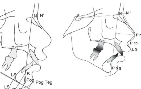
The following measurements were used Linear measurements with reference to the pogonium: (Figure 3)
LS’-Pog’ – measured from point LS’ to point Pog’, corresponding to the greatest anteroposterior dimension or distance of the lateral image of the mandibular symphysis (total thickness of the symphysis).
LS’-B’ – measured from point LS’ to point B’, corresponding to the greatest anteroposterior dimension or distance of the lateral image of the mandibular symphysis.
B’-Pog’ – measured from point B’ to point Pog’, corresponding to the greatest anteroposterior dimension or distance of the lateral image of the mandibular symphysis.
Nasal Breathing Mean (Moment 1 - 2) Deviation Std. Deviation P
Lower Intercanine 1 – Lower Intercanine 2 26.45 – 26.40 0.05 0.81 – 0.90 0.75
Upper Intercanine1 – Upper Intercanine 2 34.82 – 34.61 0.21 1.22 – 1.16 0.40
Lower Intermolar 1 – Lower Intermolar 2 45.49 – 46.07 -0.58 1.14 – 1.28 0.02*
Upper Intermolar 1 – Upper Intermolar 2 51.36 – 51.95 -0.59 0.77 – 0.98 0.00*
Mouth Breathing
Lower Intercanine 1 – Lower Intercanine 2 26.71 – 26.54 0.17 0.75 – 0.59 0.36
Upper Intercanine1 – Upper Intercanine 2 34.05 – 34.40 -0.35 0.62 – 0.75 0.03*
Lower Intermolar 1 – Lower Intermolar 2 45.89 – 46.04 -0.15 1.02 – 1.53 0.53
Upper Intermolar 1 – Upper Intermolar 2 50.99 – 51.05 -0.04 0.60 - .87 0.69
Note: * p<0.05 indicates statistically difference
Table 1- Comparison between intercanine and intermolar distances in nasal and mouth breathing according to the moments
Breathing Mode N Mean Std. Deviation P
Lower Intercanine Deviation Nasal 24 -0.21 3.10
Mouth 17 -0.64 2.82 0.33
Upper Intercanine Deviation Nasal 24 -0.68 3.62
Mouth 17 1.08 1.83 0.04*
Lower Intermolar Deviation Nasal 24 1.30 2.49
Mouth 17 0.34 2.29 0.11
Upper Intermolar Deviation Nasal 24 1.15 1.48
Mouth 17 0.13 1.14 0.01*
Note: * p<0.05 indicates statistically difference
Table 2- Deviation comparison according to breathing mode
Figure 3- Pogonium cephalometric tracing with linear measurements
Pog’-PogTeg’ – measured from point Pog’ to point Pog Teg’, corresponding to the greatest anteroposterior dimension or distance of the lateral image of the mandibular symphysis.
Line NB – union of points N and B.
Pog-NB – distance from point Pog to line NB. Indicates the bone prominence in the most anterior region of the mentum as from line NB. Positive values when located in front of the line and negative when situated beyond line NB.
Linear measurements with reference to the nose: (Figure 4)
N’-Prn – measured from the tip of the nose in relation to the nasium. Determined by nose
projection.
Prn-NPog – measured from the nasal depth in relation to the facial plane.
Angular measurements with reference to the nose:
SN-Prn-Sn – measured from the prominence of the nose in relation to the nasium - sella.
Prn-Sn-LS – nasium - labial angle – measured from the inclination of the columella in relation to the upper lip.
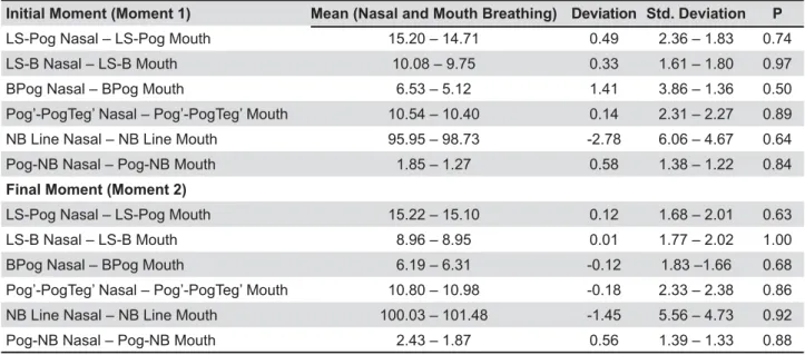
Initial Moment (Moment 1) Mean (Nasal and Mouth Breathing) Deviation Std. Deviation P
LS-Pog Nasal – LS-Pog Mouth 15.20 – 14.71 0.49 2.36 – 1.83 0.74
LS-B Nasal – LS-B Mouth 10.08 – 9.75 0.33 1.61 – 1.80 0.97
BPog Nasal – BPog Mouth 6.53 – 5.12 1.41 3.86 – 1.36 0.50
Pog’-PogTeg’ Nasal – Pog’-PogTeg’ Mouth 10.54 – 10.40 0.14 2.31 – 2.27 0.89
NB Line Nasal – NB Line Mouth 95.95 – 98.73 -2.78 6.06 – 4.67 0.64
Pog-NB Nasal – Pog-NB Mouth 1.85 – 1.27 0.58 1.38 – 1.22 0.84
Final Moment (Moment 2)
LS-Pog Nasal – LS-Pog Mouth 15.22 – 15.10 0.12 1.68 – 2.01 0.63
LS-B Nasal – LS-B Mouth 8.96 – 8.95 0.01 1.77 – 2.02 1.00
BPog Nasal – BPog Mouth 6.19 – 6.31 -0.12 1.83 –1.66 0.68
Pog’-PogTeg’ Nasal – Pog’-PogTeg’ Mouth 10.80 – 10.98 -0.18 2.33 – 2.38 0.86
NB Line Nasal – NB Line Mouth 100.03 – 101.48 -1.45 5.56 – 4.73 0.92
Pog-NB Nasal – Pog-NB Mouth 2.43 – 1.87 0.56 1.39 – 1.33 0.88
Note: p>0.05 indicates statistically difference
Table 4- Comparison between pogonium measurements in moments according to breathing mode
Nasal Breathing Mean (Moment 1 - 2) Deviation Standard Deviation P
LS-Pog 1 – LS-Pog 2 15.20 – 15.22 -0.02 2.36 – 1.68 1.42
LS’-B’ 1 – LS’-B’ 2 10.08 – 8.96 1.12 1.61 – 1.77 0.00*
BPog 1 – BPog 2 6.53 – 6.19 0.34 3.86 – 1.83 0.39
Pog’-PogTeg’ 1 – Pog’-PogTeg’ 2 10.54 – 10.80 -0.26 2.31 – 2.33 0.33
NB Line 1 – NB Line 2 95.95 – 100.03 -4.08 6.06 – 5.56 0.00*
Pog-NB 1 – Pog-NB 2 1.85 – 2.43 -0.58 1.38 – 1.39 0.01*
Mouth Breathing
LS-Pog 1 – LS-Pog 2 14.71 – 15.10 -0.39 1.83 – 2.01 0.35
LS’-B’ 1 – LS’-B’ 2 9.75 – 8.95 0.80 1.80 – 2.02 0.00*
BPog 1 – BPog 2 5.12 – 6.31 -1.19 1.36 –1.66 0.25
Pog’-PogTeg’ 1 – Pog’-PogTeg’ 2 10.40 – 10.98 -0.58 2.27 – 2.38 0.05
NB Line 1 – NB Line 2 98.73 – 101.48 -2.75 4.67 – 4.73 0.00*
Pog-NB 1 – Pog-NB 2 1.27 – 1.87 -0.50 1.22 – 1.33 0.01*
Note: * p<0.05 indicates statistically difference
RESULTS
Dental Measurements
The Kolmogorov-Smirnov statistical test indicated that all the variables presented homogeneity of variances between the respiratory modes (p<0.05). Therefore, moments 1 and 2 were compared to verify whether there were differences both for the nasal and mouth breathing modes, the Student’s-t test for paired samples was used. For the nose breathers, there was statistically $%%&# distance between moments 1 and 2 for the mandible and maxilla (Table 1). For the mouth breathers, $%%&# for the maxillary intercanine distance, there being an increase from moment 1 to moment 2 (Table 1). In the comparison between the variations from moment 1 to moment 2, it was observed that there to the respiratory mode, and for the analysis, the Student’s-t test for independent samples was used. The result of the test indicated that there was $%%&#
variable maxillary intercanine distance, with the group of mouth breathers presenting a positive and larger variation than the group of nose breathers, which presented negative variation. With regard to the variable maxillary intermolar distance, there $%%&# group of mouth breathers presenting a positive variation, smaller than that of the group of nose breathers (Table 2).
Facial Measurements Pogonium
Since the Kolmogorov-Smirnov statistical test indicated that all the variables, in all the moments presented normal distribution, with the exception of B’-Pog’ and Pog-NB, comparison of the mean values according to moments was performed by two-way ANOVA with repeated measures and Tukey HSD multiple comparison test.
The results of this test demonstrated that the variables LS’-B’, Pog’-PogTeg’, line NB, and *+=> $%%&# interference of the respiratory mode. The variables
Nasal Breathing Mean (Moment 1 - 2) Deviation Standard Deviation P
NPRN 1 – NPRN 2 48.46 – 51.17 -2,71 1.55 – 1.95 0.00*
PRNNPOG 1 – PRNNPOG 2 31.44 – 33.37 -1.93 3.05 – 1.66 0.06
NPRNSN 1 – NPRNSN 2 99.74 – 96.09 3.65 1.59 – 1.98 0.00*
PRNSNL 1 – PRNSNL 2 125.76 – 126.48 -0.72 3.90 – 4.08 0.04*
Mouth Breathing
NPRN 1 – NPRN 2 48.65 – 51.09 -2,44 2.55 – 2.85 0.00*
PRNNPOG 1 – PRNNPOG 2 31.16 – 33.54 -2.38 2.24 – 2.48 0.04*
NPRNSN 1 – NPRNSN 2 100.88 – 98.00 2.88 4.24 – 3.89 0.02*
PRNSNL 1 – PRNSNL 2 120.59 – 122.76 -2.17 4.60 – 4.75 0.14
Note: * p<0.05 indicates statistically difference
Table 5- Comparison between nose measurement in nasal and mouth breathing according to the moments
Note: * p<0.05 indicates statistically difference
Initial Moment (Moment 1) Mean (Nasal and Mouth Breathing) Deviation Std. Deviation P
NPRN Nasal – NPRN Mouth 48.46 – 48.65 -0,19 1.55 – 2.55 1.00
PRNNPOG Nasal – PRNNPOG Mouth 31.44 – 31.16 0.28 3.05 – 2.24 1.00
NPRNSN Nasal – NPRNSN Mouth 99.74 – 100.88 -1.14 1.59 – 4.24 0.96
PRNSNL Nasal – PRNSNL Mouth 125.76 – 120.59 5.17 3.90 – 4.60 0.43
Final Moment (Moment 2)
NPRN Nasal – NPRN Mouth 51.17 – 51.09 0.08 1.95 – 2.85 1.00
PRNNPOG Nasal – PRNNPOG Mouth 33.37– 33.54 -0.17 1.66 – 2.48 1.00
NPRNSN Nasal – NPRNSN Mouth 96.09 – 98.00 -1.91 1.98 – 3.89 0.85
PRNSNL Nasal – PRNSNL Mouth 126.48 – 122.76 3.72 4.08 – 4.75 0.37
>?+*? @J?+*Q? difference (p>0.05) (Table 3 and 4).
Nose
Since the Kolmogorov-Smirnov statistical test indicated that all the variables, with the exception of Prn-NPog, in all the moments presented normal distribution, comparison of the mean values according to moments was performed by two-way ANOVA with repeated measures and Tukey HSD multiple comparison test.
The results of this test showed that the variables presented statistically significant difference $%%&# without any interference of the breathing mode (Tables 5 and 6).
DISCUSSION
X development of orofacial structures has been a subject of debates in articles, texts and researches for over a century. Various studies have investigated the possible repercussions of the respiratory pattern on the functional, neuromuscular, skeletal and dental functions13,14,18,28.
Nasal respiration is primordial in order for correct growth and development of the craniofacial complex to occur10,20, but some authors that disagree that
facial morphology and the respiratory mode are intimately related14,18,28. According to Klein13 (1986),
there is no conclusive evidence that obstruction of nasal respiration alters facial growth and development.
Harvold, et al.12 (1981), in a study with animals,
observed that after 9 and 15 months of eminently mouth breathing, the maxillary intercanine distance was significantly smaller in the monkeys with obstructed nostrils in comparison with the control animals. The mandibular intercanine distance Y monkeys.
In this study, there was an increase in the dental distances assessed in nose and mouth breather individuals, except for the variable maxillary intercanine distance in nose breathers, in which there was narrowing, as demonstrated by Carter and McNamara Júnior5 (1998). On the other hand,
Trask and Shapiro26 (1987) found no differences
in the intercanine and intermolar widths between mouth and nose breathers.
The importance of maintaining dental arch shape and the intercanine and intermolar distances in the maxilla, and particularly, the intercanine distance in the mandible is described in the literature over the course of time, as it is a fact that teeth moved by orthodontic appliances tend to revert to their original positions3,23. For some authors, moderate
increases in these distances can be tolerated, while
for others, any change, even if minor, may cause instability.
The age range in this study was from 10 and 16 years, differing from Formby, et al.9 (1994), who
studied the range between 18 and 42 years and hard and soft tissues in both genders, especially, an increase in all the dimensions of the nose.
Tourne and Schweiger25 (1996) found differences
in the craniofacial development between mouth and nose breathers, with predominantly mouth breathers presenting greater inclination of the mandibular plane angle and a vertical growth pattern.
In the present research, there was alteration in the skeletal pogonium and tegumentary measurements according to the moments, in agreement with Koch16 (1979).
Increase in the measurement LS’-Pog’ was observed, as a result of differentiated bone remodeling with greater bone apposition in the lingual region of the pogonium, resulting in an increase in the tegumentary pogonium (Pog’-PogTeg’). These changes characterize skeletal growth, as proved by the histological studies of Enlow and Harris7 (1964) and the metal implants
of Bjork2 (1963).
In nose breathers, a decrease in the measurement >?+*? was an increase. Longitudinal studies by Bjork2
(1963)and Enlow and Harris7 (1964) proved that
the thickening of the symphysis is due to the greater bone apposition in the lingual region, while the region of the pogonium is more stable and grows very little or not at all. However, in this study, it was not possible to assess the decrease ratio in B’-Pog’. One supposes that the decrease in B’-Pog’ and LS’-B’ occurred because of the smaller displacement in the posterior direction of point B, as a result of downward and backward rotation of the mandible, not ignoring the possibility of less appositional growth in the region of the pogonium.
J *+ => the position of point B, due to the combination of displacement towards the posterior direction of point B and bone apposition in the pogonium, in addition to variations in the inclination of the mandibular plane angle.
With regard to analysis of the nose, there was [%%&# and depth, measured by means of the values of =?+**+=* mode.
present study. These data are in agreement with those of Brant, et al.4 (2006), who demonstrated
significant changes in N-Prn-Sn when they compared the groups with and without extraction of premolars.
The nasolabial angle (Prn-Sn-LS) revealed [%%&# moments, however, without presenting interaction with the respiratory mode. The data are in agreement with Salgado22 (2003), who observed
angle in individuals with Angle’s Class II and III malocclusions, the values in Class II being higher.
The interrelation between the tegumentary tissue of the nose and pogonium, as well as the dental alterations must be considered in the diagnosis, when establishing the plan and performing the treatment in individuals in the growth phase, particularly in patients with predominantly mouth breathing.
CONCLUSIONS
There was change in the nose and skeletal and tegumentary pogonium in the anteroposterior plane, during the course of growth, without interference of the breathing pattern. On the other hand, the maxillary and mandibular intercanine differences between the moments and breathing pattern.
REFERENCES
1- Aksu M, Kocadereli I. Arch width changes in extraction and nonextraction treatment in Class I patients. Angle Orthod. 2005;75(6):948-52.
2- Bjork A. Variations in the growth pattern of the human mandible: longitudinal radiographic study by the implant method. J Dent Res. 1963;42(1):400-11.
3- Boley JC, Mark JA, Sachdeva RC, Buschang PH. Long-term stability of Class I premolar extraction treatment. Am J Orthod Dentofacial Orthop. 2003;124(3):277-87.
4- Brant JCO, Siqueira VCV. Soft tissue changes evaluated in Class II, division 1 cases, after orthodontic treatment. Rev Dent Press Ortodon Ortopedi Facial. 2006;11(2):93-102.
5- Carter GA, McNamara JA Jr. Longitudinal dental arch changes in adults. Am J Orthod Dentofacial Orthop. 1998;114(1):88-99. 6- Chaconas SJ. A statistical evaluation of nasal growth. Am J Orthod. 1969;56(4):403-14.
7- Enlow DH, Harris DB. A study of the postnatal growth of the human mandible. Am J Orthod. 1964;50(1):25-50.
\+] ^]=XJ_`{]X patients treated with extraction and nonextraction of premolars. Am J Orthod Dentofacial Orthop. 2006;129(6):775-84
9- Formby WA, Nanda RS, Currier GF. Longitudinal changes ^ | } ~ } 1994;105(5):464-76.
10- Graber TM, Vanarsdall RL Jr. Orthodontics: current principles and techniques. 3rd ed. St. Louis: Mosby; 2000.
11- Hambleton RS. The soft tissue covering of the skeletal face as related to orthodontic problems. Am J Orthod. 1964;50(6):405-20. 12- Harvold EP, Tomer BS, Vargervik K, Chierici G. Primate experiments on oral respiration. Am J Orthod. 1981;79(4):359-72. 13- Klein JC. Nasal respiratory function and craniofacial growth. Arch Otolaryngol Head Neck Surg. 1986;112(8):843-9.
14- Kluemper GT, Vig PS, Vig KW. Nasorespiratory characteristics and craniofacial morphology. Eur J Orthod 1995;17(6):491-5. 15- Knott VB. Longitudinal study of dental arch widths at four stages of dentition. Angle Orthod. 1972;42(4):387-94.
16- Koch R, Gonzales A, Witt E. Profile and soft tissue changes during and after orthodontic treatment. Eur J Orthod. 1979;1(3):193-9.
17- Lee RT. Arch width and form: a review. Am J Orthod Dentofacial Orthop. 1999;115(3):305-13.
18- Nowak AJ, Warren JJ. Infant oral health and oral habits. Pediatr Clin North Am. 2000;47(5):1043-66.
19- Prahl-Andersen B, Ligthelm-Bakker AS, Wattel E, Nanda R. ^ ^ | } Dentofacial Orthop. 1995;107(5):476-83.
"%+*X!J~ 4th ed. St. Louis: Mosby; 2007.
21- Ricketts RM. Planning treatment on the basis of the facial pattern and an estimate of its growth. Angle Orthod. 1957;27(1):14-37.
22- Salgado JAP, Moraes LC, Castilho JCM, Moraes MEL. Nasolabial angle evaluation, in lateral cephalometric x-rays, divided in superior and inferior angle, by a parallel line to the Frankfort plane, in individuals bearers of Class II and Class III occlusion. Cienc Odontol Bras. 2003;6(3):40-9.
23- Sinclair PM, Little RM. Maturation of untreated normal occlusions. Am J Orthod. 1983;83(2):114-23.
24- Steiner CC. Cephalometrics in clinical practice. Angle Orthod. 1959;29(1):8-29.
25- Tourne LP, Schweiger J. Immediate postural responses to total nasal obstruction. Am J Orthod Dentofacial Orthop. 1996;110(6):606-11.
26- Trask GM, Shapiro GG, Shapiro PA. The effect of perennial allergic rhinitis on dental skeletal development: a comparison of sibling pairs. Am J Orthod Dentofacial Orthop. 1987;92(4):286-93. 27- Ward DE, Workman J, Brown R, Richmond S. Changes in arch width. A 20-year longitudinal study of orthodontic treatment. Angle Orthod. 2006;76(1):6-13.
28- Warren DW. Effect of airway obstruction upon facial growth. Otolaryngol Clin North Am. 1990;23(4):699-712.