Influence of intentional ankylosis of deciduous canines
to reinforce the anchorage for maxillary protraction
Luís Fernando Castaldi Tocci1, Omar Gabriel da Silva Filho2, Acácio Fuziy3, José Roberto Pereira Lauris4
How to cite this article: Tocci LFC, Silva Filho OG, Fuziy A, Lauris JRP. Inluence of intentional ankylosis of deciduous canines to reinforce the anchor-age for maxillary protraction. Dental Press J Orthod. 2013 Jan-Feb; 18(1):94-102.
Submitted: April 18, 2010 - Revised and accepted: June 30, 2011
» The author reports no commercial, proprietary or inancial interest in the prod-ucts or companies described in this article.
Contact address: Luís Fernando Castaldi Tocci
Rua Carneiro Lobo, 570 – Conj. 1003 – Batel - Curitiba/PR – Brazil CEP 80240.240 - E-mail: [email protected] 1 MSc in Orthodontics, UNIMAR.
2 MSc in Orthodontics, UNESP.
3 Post-Doc in Dentistry, FOB-USP.
4 Full Professor, USP. PhD in Human Communication Disturb, University
of São Paulo.
Introduction: This retrospective cephalometric study analyzed the inluence of intentional ankylosis of deciduous canines in pa-tients with Class III malocclusion and anterior crossbite, in the deciduous and early mixed dentition stages, treated by orthopedic maxillary expansion followed by maxillary protraction. Methods: Lateral cephalograms of 40 patients were used, divided in 2 groups paired for age and gender. The Ankylosis Group was composed of 20 patients (10 boys and 10 girls) treated with induced ankylosis and presenting initial and inal mean ages of 7 years 4 months and 8 years 3 months, respectively, with a mean period of maxillary protraction of 11 months. The Control Group comprised 20 patients (10 boys and 10 girls) treated without induced ankylosis, with initial and inal mean ages of 7 years 8 months and 8 years 7 months, respectively, with a mean period of maxil-lary protraction of 11 months. Two-way analysis of variance and covariance analysis were applied to compare the initial and inal cephalometric variables and the treatment changes between groups. Results: According to the results, the variables evidencing the signiicant treatment changes between groups conirmed that the intentional ankylosis enhanced the sagittal response of the apical bases (Pg-NPerp) and increased the facial convexity angles (NAP and ANB). Conclusions: The protocol involving intentional ankylosis of deciduous canines enhanced the sagittal response of the apical bases.
Keywords: Malocclusion. Angle Class III. Crossbite. Interceptive orthodontics.
Introdução: nesse estudo cefalométrico retrospectivo, analisou-se a inluência da anquilose intencional de caninos decíduos em pacientes com má oclusão de Classe III e mordida cruzada anterior, nos estágios de dentição decídua e mista precoce, tratados com expansão ortopédica da maxila, seguida de tração reversa. Métodos: foram utilizadas telerradiograias em norma lateral de 40 pacientes, divididos em 2 grupos pareados por idade e sexo. O Grupo Anquilose foi constituído de 20 pacientes (10 meninos e 10 meninas) tratados com anquilose induzida e que apresentavam as idades médias inicial e inal, respectivamente, de 7a 4m e 8a 3m, e o tempo médio de tração reversa de 11 meses. O Grupo Controle, composto de 20 pacientes (10 meninos e 10 meninas) tratados sem anquilose induzida e que apresentavam as idades médias inicial de 7a 8m e inal de 8a 7m, e tempo médio de tração reversa de 11 meses. Foram empregadas as análises de Variância a dois critérios e de Covariância para comparar as variáveis cefalométricas inicial e inal e as alterações de tratamento entre os grupos. Resultados: segundo os resultados, as variáveis que evidenciaram as mudanças de tratamento signiicativas entre os grupos conirmaram que o procedimento de anquilose intencional potencializou a resposta sagital das bases apicais (Pg-NPerp) e aumentou os ângulos de convexidade facial (NAP e ANB). Conclusão: o protocolo envolvendo a anquilose intencional de caninos decíduos potencializou a resposta sagital das bases apicais.
INTRODUCTION
Some questions still challenge the scientiic commu-nity concerning the Class III malocclusion: What will be the behavior of the face, and especially of the man-dible, during growth? Will the facial growth perpetuate or worsen the skeletal discrepancy? There are no estab-lished responses to these questions, raising controversies among orthodontists concerning the indication of early
treatment for the Class III malocclusion.1 The
treat-ment prognosis and posttreattreat-ment stability depend on
the skeletal pattern related with this malocclusion.16,18
Interceptive orthodontics (mechanics applied in the stages before the permanent dentition) has the general
objective to optimize the development of occlusion.18
The treatment with maxillary protraction is more efec-tive in patients with Class III malocclusion with maxillary
retrusion, which account for nearly 60% of the cases,16
and hypodivergent growth pattern,13,15 explaining the
increased professional interest for maxillary protraction. The orthopedic mechanics acts on the direction of
spon-taneous facial growth.16 If the treatment is initiated
dur-ing the period of eruption of maxillary central incisors, it
will contribute to stabilize the anterior relationship.9,13,15
Clinical results demonstrate that the utilization of maxillary protraction induces orthopedic and orth-odontic efects that provide an important improvement in the occlusion and face (Table 1). The immediate
favorable impact, though variable and individual,15 in
the deciduous and mixed dentition stages are related to
forward maxillary displacement,1,2,5,6,9-13,15,17,18,21,23
for-ward displacement of maxillary teeth,2,6,8,10,18 clockwise
mandibular rotation with corresponding signiicant
in-crease in the lower anterior facial height,5,10,18and
lin-gual tipping of mandibular incisors.2,3,5,9,16,18
Orthodontists are aware of the importance to op-timize the orthopedic efect of maxillary protraction,
rather than the orthodontic efects.6 Within this
con-text, the orthopedic maxillary expansion before maxil-lary protraction increases the efect of the face mask on
the maxilla1,2 due to the increase in the transverse width,
rupture of maxillary sutures and especially to the strong
anchorage provided by the expander16 (Table 1).
The intentional ankylosis of deciduous canines has also been used with the primary goal to potentiate the
orthopedic efects of maxillary protraction.18,19,20 This
led to the need to scientiically analyze its utilization, because though biocompatible and accepted by the
pa-tients,19 it is an invasive procedure.
This study evaluated the immediate efects of maxil-lary protraction applied in the deciduous and early mixed dentition stages to respond to the null hypotheses if there is diference between the immediate results produced by orthopedic maxillary protraction mechanics, using or not the induced ankylosis to reinforce the anchorage.
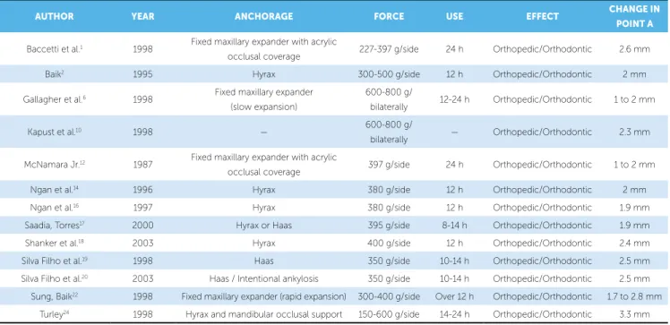
Table 1 - Literature on the utilization of maxillary protraction (with facial mask) applied immediately after orthopedic maxillary expansion.
AUTHOR YEAR ANCHORAGE FORCE USE EFFECT CHANGE IN
POINT A
Baccetti et al.1 1998 Fixed maxillary expander with acrylic
occlusal coverage 227-397 g/side 24 h Orthopedic/Orthodontic 2.6 mm
Baik2 1995 Hyrax 300-500 g/side 12 h Orthopedic/Orthodontic 2 mm
Gallagher et al.6 1998 Fixed maxillary expander
(slow expansion)
600-800 g/
bilaterally 12-24 h Orthopedic/Orthodontic 1 to 2 mm
Kapust et al.10 1998 — 600-800 g/
bilaterally — Orthopedic/Orthodontic 2.3 mm
McNamara Jr.12 1987 Fixed maxillary expander with acrylic
occlusal coverage 397 g/side 24 h Orthopedic/Orthodontic 1 to 2 mm
Ngan et al.14 1996 Hyrax 380 g/side 12 h Orthopedic/Orthodontic 2 mm
Ngan et al.16 1997 Hyrax 380 g/side 12 h Orthopedic/Orthodontic 1.9 mm
Saadia, Torres17 2000 Hyrax or Haas 395 g/side 8-14 h Orthopedic/Orthodontic 1.9 mm
Shanker et al.18 2003 Hyrax 400 g/side 12 h Orthopedic/Orthodontic 2.4 mm
Silva Filho et al.19 1998 Haas 350 g/side 10-14 h Orthopedic/Orthodontic 2.5 mm
Silva Filho et al.20 2003 Haas / Intentional ankylosis 350 g/side 10-14 h Orthopedic/Orthodontic 2.5 mm
Sung, Baik22 1998 Fixed maxillary expander (rapid expansion) 300-400 g/side Over 12 h Orthopedic/Orthodontic 1.7 to 2.8 mm
MATERIAL AND METHODS
The sample was composed of 40 Brazilian Cauca-sian children aged 5 years to 8 years 11 months, in the deciduous and early mixed dentition stages, retrospec-tively selected from the iles of the Postgraduate course in Preventive and Interceptive Orthodontics of the Hospital for Rehabilitation of Craniofacial Anomalies of University of São Paulo, at Bauru (HRAC-USP-Bauru), Brazil, among the records of 2,060 registered and treated patients.
The inclusion criteria were the following: 1– Bra-zilian Caucasian children in the deciduous and early mixed dentition stage; 2 – presenting Class III molar relationship; 3 – anterior crossbite; 4 – maxillary ret-rognathism with little or no mandibular involvement; and 5 – posterior crossbite in most cases. Patients with isolated mandibular impairment (prognathism) were not included.
The sample was divided into two groups of 20 pa-tients (10 boys and 10 girls) matched for age and gen-der. The Ankylosis Group was composed of patients
treated with induced ankylosis with initial (T1) and
inal (T2) mean ages of 7 years 4 months and 8 years
3 months, respectively, with mean period of maxil-lary protraction of 11 months. The Control Group was treated without induced ankylosis and presented initial mean age of 7 years 8 months and inal mean age 8 years 7 months, with a mean period of maxillary pro-traction of 11 months.
In the Ankylosis Group, among the 20 patients, 16 presented posterior crossbite, being 9 girls and 7 boys, and in the Control Group 17 out of the 20 patients pre-sented posterior crossbite, being 8 boys and 9 girls.
All 40 children were submitted to the same therapeu-tic protocol of rapid maxillary expansion with modiied ixed Haas expander, with activation of the screw until rupture of the midpalatal suture or up to correction of the posterior crossbite, when present. The therapeutic groups were distinguished by the accomplishment of intentional ankylosis of deciduous canines as anchorage reinforcement. Immediately ater completion of the ac-tive period of rapid maxillary expansion, the facial mask was placed using elastics delivering an approximate force of 500 g connected to hooks soldered at the ante-rior portion of the expander and bonded to the decidu-ous canines with resin. The patients were instructed to wear the facial mask for 16 hours/day.
The lateral cephalograms were obtained in centric occlusion, with the lips in relaxed and passive position. The anatomical tracings and identiication of the den-toskeletal points were manually performed by a single examiner and digitized on a UMAX 1220S scanner connected to a computer NB HP Pavillion dv6120BR (3400, HD 60 Gb, memory 512 Mb), with operational system Windows XP and sotware Radiocef Studio – Radiomemory, version 1 – release 16, Belo Horizonte,
MG, Brazil (Fig 1). Data were analyzed on this
sot-ware, which also corrected the magniication factors of radiographic images, which ranged from 6% to 9.8% according to the X-ray machine employed.
The cephalometric measurements selected are rep-resentative of the facial convexity (NAP, ANB), sagittal position of the apical bases (SNA, SNB, SND, SN.ANS, Co-A, A-NPerp, Pg-NPerp), mandibular and occlusal plane rotation (SN.GoGn, SN.Gn, SN.OP) and in-clination of maxillary and mandibular incisors (1.NA, 1-NA, 1.SN, 1.PP, 1.NB, 1-NB, IMPA) (Table 4).
Figure 1 - Cephalometric points used on the lateral tracing: 1) S: sella turcica; 2) N: nasion; 3) A: subspinale; 4) B: supramentale; 5) Pg: pogonion; 6) D: geo-metric center of the symphysis; 7) Gn: gnathion; 8) Me: menton; 9) Go: gonion; 10) Co: condylion; 11) ANS: anterior nasal spine; 12) PNS: posterior nasal spine; 13) MxIE: incisal edge of maxillary central incisor; 14) MxIA: apex of maxillary cen-tral incisor; 15) MdIE: incisal edge of mandibular cencen-tral incisor; 16) MdIA: apex of mandibular central incisor; 17) OCM1: mean occlusal contact between the maxillary and mandibular irst molars; 18) Po: porion; 19) Or: orbitale.
1
18 10
12 17
19 14
11
13 15
16 9
8 7 5 4
STATISTICAL ANALYSIS
Method error
For calculation of the intraexaminer error, at 20 days after the first tracing, 12 cephalograms of each group (30% of the sample) were randomly selected for achievement of new tracings, identification of points and achievement of linear and angular mea-surements. The intraexaminer systematic error was
evaluated by the paired t test. The casual error was
determined according to the Dahlberg formula
(er-ror = (∑d²/ 2n)1/2), in which d represents the
differ-ence between the first and second measurements and
n indicates the number of retraced cephalograms,
according to Houston.7 The significance level
ad-opted was 5% (p < 0.05).
The results of evaluations of systematic errors by the
paired t test and by the casual error measured by the
Dahlberg formula are presented in Table 4.
STATISTICAL METHODS
Descriptive statistics was performed for all data in
the sample: Initial and inal ages, treatment time (T2-T1)
and cephalometric variables analyzed in the study pe-riods. The normality of data distribution was assessed by the Kolmogorov-Smirnov, which revealed that all groups passed the normality criteria.
Two-way analysis of variance was applied for com-parison between groups and gender, initial age, treat-ment time, ANB and A-NPerp, to verify the similarity of data at treatment onset.
Since the groups has diferent severities (ANB and
A-NPerp) at T1 and the statistical analysis might be
in-luenced by this factor and not only by the diference between groups, comparison between groups was per-formed by the analysis of covariance (ANCOVA), using the measurements ANB and A-NPerp as co-variables, to take these initial diferences into account.
ns = non signiicant statistical diference. * = statistically signiicant diference (p < 0.05).
Table 2 - Mean, standard deviation of two measurements, paired t test and
Dahlberg formula to evaluate the systematic and casual errors.
measure-ment
first measurement
second
measurement t p Error
mean SD mean SD
NAP 3.77 6.54 3.65 6.41 0.621 0.538 ns 0.79
SNA 81.05 4.70 81.28 4.67 1.816 0.077 ns 0.58
SNB 79.24 4.62 79.41 4.59 1.705 0.097 ns 0.44
ANB 1.80 2.95 1.87 2.83 1.669 0.104 ns 0.53
SND 75.86 4.41 76.27 4.47 1.992 0.054 ns 0.94
SN.ANS 85.75 4.65 86.01 4.62 1.854 0.072 ns 0.63
Co-A 81.09 6.82 81.24 6.73 2.029 0.051 ns 0.33
A-NPerp 1.02 3.27 0.93 3.38 0.967 0.340 ns 0.41
Pg-NPerp -1.43 6.80 -1.53 6.99 0.620 0.539 ns 0.68
SN.GoGn 36.38 5.99 36.22 6.07 1.667 0.104 ns 0.42
SN.Gn 67.63 4.80 67.52 4.85 1.551 0.129 ns 0.33
SN.OP 22.33 10.41 22.09 10.48 1.603 0.117 ns 0.67
1.NA 24.65 7.85 25.02 7.52 1.966 0.057 ns 0.84
1-NA 2.27 3.33 2.41 3.37 1.907 0.064 ns 0.33
1.SN 105.70 8.12 106.02 8.16 1.514 0.138 ns 0.94
1.PP 111.12 8.01 111.09 8.20 0.158 0.875 ns 0.72
1.NB 23.45 5.60 23.84 5.85 1.557 0.128 ns 1.10
1-NB 3.15 3.00 3.29 3.08 2.489 0.017* 0.28
IMPA 85.39 6.76 85.65 6.77 1.083 0,286 ns 1.03
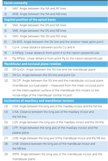
Table 3 - Dental and skeletal cephalometric variables.
Facial convexity
1) NAP: Angle between the NA and AP lines
2) ANB: Angle between the NA and NB lines
Sagittal position of the apical bases
3) SNA: Angle between the SN and NA lines
4) SNB: Angle between the SN and NB lines
5) SND: Angle between the SN and ND lines
6) SN.ANS: Angle between the SN line and the anterior nasal spine point
7) Co-A: Linear distance between points Co and A
8) A-NPerp: Linear distance from point A to the nasion-perpendicular
9) Pg-NPerp: Linear distance from point Pg to the nasion-perpendicular
Mandibular and occlusal plane rotation
10) SN.GoGn: Angle between the SN line and the mandibular plane
11) SN.Gn: Angle between the SN line and point Gn
12) SN.OP: Angle between the SN line and the mandibular occlusal plane
(mandibular occlusal plane – measured from the mean occlusal point on the intercuspation surface of the mandibular irst molars to the
incisal edge of the mandibular central incisor)
Inclination of maxillary and mandibular incisors
13) 1.NA: Angle between the long axis of the maxillary incisor and the NA line
14) 1-NA: Distance between the long axis of the maxillary incisor and the NA line
15) 1.SN: Angle between the long axis of the maxillary incisor and the SN line
16) 1.PP: Angle between the long axis of the maxillary incisor and the
palatal plane
17) 1.NB: Angle between the long axis of the mandibular incisor and the NB line
18) 1-NB: Distance between the long axis of the mandibular incisor and the NB line
19) IMPA: Angle between the long axis of the mandibular incisor and the
Therefore, comparison between groups was per-formed by two-way analysis of variance (Group – Con-trol and Ankylosis, and Gender – Female and Male), ixed model. If the analysis of variance indicated statisti-cally signiicant diference, the Tukey test for multiple comparisons was applied. A signiicance level of 5% (p < 0.05) was considered for all tests.
All statistical analyses were performed on the sot-ware Statistica version 5.1 StatSot Inc. (Tulsa, USA).
RESULTS
Table 2 presents the analysis of systematic and casual
errors, by analysis by the paired t test and the Dahlberg7
formula applied to all study variables. Only the variable (1-NB) presented statistically signiicant systematic er-ror, yet with a diference of only 0.14 mm. The casual error ranged from 0.28 mm (1-NB) to 1.10° (1.NB).
Table 5 displays the cephalometric measurements
analyzed during the treatment period (T1 and T2) for
Table 4 - Comparison of initial and inal ages, treatment time, ANB and A-NPerp between groups.
Group Gender Initial age Treatment time ANB A-NPerp
Mean SD Mean SD Mean SD Mean SD
Control M 91.90 9.76 11.60 8.14 2.90 2.86 1.71 4.32
F 93.00 10.13 9.40 4.45 1.86 1.66 2.44 3.45
Ankylosis M 87.25 16.02 11.10 4.38 -0.54 3.52 -1.13 3.15
F 85.38 13.50 10.50 3.21 -0.65 3.00 -0.66 2.79
Anova
p group 0.147 ns 0.861 ns 0.002 * 0.010 *
p gender 0.926 ns 0.416 ns 0.524 ns 0.587 ns
p interaction 0.721 ns 0.641 ns 0.607 ns 0.906 ns
Mean and standard deviation of measurements obtained for the sample groups
Control female
Control male Ankylosis female Ankylosis male Cephalometric measurement
T1 T2 T1 T2 T1 T2 T1 T2
NAP 3.49 ± 4.89 5.22 ± 4.63 5.61 ± 5.98 6.00 ± 5.13 -0.45 ± 7.67 7.07 ± 8.18 -0.54 ± 8.41 6.49 ± 7.44
SNA 80.35 ± 4.81 81.13 ± 5.91 83.2 ± 5.31 83.47 ± 5.44 81.25 ± 5.05 84.19 ± 5.35 80.19 ± 2.87 82.26 ± 3.27
SNB 78.49 ± 4.65 78.35 ± 5.18 80.4 ± 5.49 80.11 ± 4.82 82.13 ± 4.16 81.05 ± 4.04 80.71 ± 3.45 79.25 ± 2.61
ANB 1.86 ± 1.66 2.78 ± 1.80 2.80 ± 2.81 3.36 ± 2.47 -0.65 ± 3.00 3.23 ± 3.61 -0.52 ± 3.55 3.02 ± 3.73
SND 75.23 ± 4.77 75.18 ± 5.24 77.09 ± 5.41 77.18 ± 4.89 77.81 ± 3.31 77.20 ± 3.39 76.65 ± 3.45 75.7 ± 2.59
SN.ANS 85.25 ± 4.41 87.50 ± 6.54 87.30 ± 4.24 88.1 ± 4.82 86.25 ± 4.85 88.20 ± 5.41 84.6 ± 4.01 86.8 ± 4.03
Co-A 79.05 ± 7.80 81.44 ± 7.70 83.20 ± 4.88 86.71 ± 5.4 74.68 ± 3.57 79.15 ± 4.77 76.42 ± 2.26 80.43 ± 4.76
A-NPerp 1.71 ± 4.32 2.90 ± 4.82 2.44 ± 3.45 2.74 ± 3.07 -1.14 ± 3.15 1.40 ± 3.50 -0.66 ± 2.79 0.67 ± 3.12
Pg-NPerp 0.28 ± 8.66 1.12 ± 8.85 -0.39 ± 7.66 -0.12 ± 6.44 -2.16 ± 5.89 -3.46 ± 6.33 -1.28 ± 6.51 -4.13 ± 5.42
SN.GoGn 35.84 ± 5.39 36.44 ± 6.21 35.26 ± 6.81 35.86 ± 5.48 35.80 ± 5.88 37.71 ± 6.17 33.83 ± 5.55 35.5 ± 4.71
SN.Gn 67.76 ± 3.89 68.59 ± 4.23 67.02 ± 6.27 67.50 ± 5.36 65.71 ± 4.05 67.24 ± 3.85 65.00 ± 4.78 66.77 ± 3.85
SN.OP 21.43 ± 5.54 19.18 ± 4.97 23.02 ± 9.76 21.23 ± 9.1 31.63 ± 11.83 21.21 ± 11.77 24.92 ± 11.07 22.19 ± 8.35
1.NA 27.05 ± 6.30 25.97 ± 4.87 18.26 ± 7.51 22.29 ± 3.91 25.42 ± 10.47 27.51 ± 7.6 18.72 ± 9.50 19.83 ± 9.59
1-NA 1.69 ± 2.40 3.47 ± 1.63 0.30 ± 3.77 2.83 ± 1.86 0.04 ± 4.47 2.74 ± 3.61 -1.60 ± 2.26 0.84 ± 3.42
1.SN 107.4 ± 7.09 107.1 ± 4.63 101.46 ± 7.48 105.76 ± 7.61 106.67 ± 10.24 111.63 ± 6.33 98.99 ± 9.29 102.1 ± 8.45
1.PP 116.75 ± 5.66 112.9 ± 6.08 105.95 ± 7.24 109.45 ± 5.01 110.9 ± 7.43 115.2 ± 5.85 104.25 ± 8.93 107.25 ± 9.49
1.NB 28.03 ± 4.69 23.63 ± 7.44 27.00 ± 7.40 26.18 ± 5.38 21.72 ± 6.13 25.29 ± 6.09 19.56 ± 8.21 20.77 ± 5.66
1-NB 3.93 ± 2.04 3.7 ± 2.28 3.82 ± 2.84 3.84 ± 2.7 0.52 ± 3.35 3.78 ± 3.43 1.24 ± 2.83 1.97 ± 3.46
IMPA 91.25 ± 5.73 86.51 ± 8.96 89.11 ± 8.43 87.84 ± 6.33 81.05 ± 7.94 86.61 ± 10.56 82.31 ± 7.42 83.57 ± 5.11
each study group (female control, female ankylosis, male control, male ankylosis).
The statistical comparison between groups reveals that they were compatible considering the initial age and treatment time. However, there was signiicant dif-ference in the variables ANB and A-NPerp (Table 4), which led to the utilization of analysis of covariance for comparison between groups.
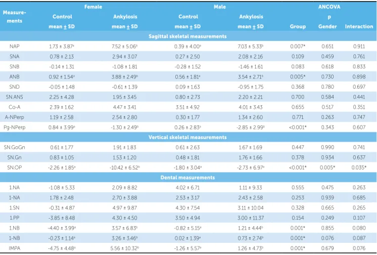
The statistical analysis revealed that the cephalomet-ric measurements were not inluenced by gender, except for the variable SN.OP. The changes in cephalometric measurements occurred in the same direction and mag-nitude for both genders (Table 6). There was statistically signiicant diference between groups in the variation of sagittal measurements NAP, ANB and Pg-NPerp, the vertical measurement SN.OP, and the dental measure-ments 1.NB, 1-NB and IMPA (Table 6).
DISCUSSION
The early treatment of anterior crossbite with Class III malocclusion has the orthopedic goal to promote forward displacement of the maxillary dental arch, with downward and forward advancement of the growth
di-rection of the maxilla.1,2,5,6,9-13,15-18,21,23 For that purpose,
the maxillary protraction ater orthopedic maxillary expansion has been used by most orthodontists, with favorable immediate results in 90% of patients treated
in the deciduous and mixed dentitions4 with a relatively
short treatment time of nearly 8 months.18 Regardless
of the inluence of facial growth on the long-term post-treatment stability, the immediate goal of orthopedic treatment for the Class III malocclusion is to potentiate the skeletal changes rather than the dental
compensa-tion, by the utilization of strong anchorage1This study
addresses the maxillary protraction, more speciically to
Measure-ments
Female Male ANCOVA
Control Ankylosis Control Ankylosis p
mean ± SD mean ± SD mean ± SD mean ± SD Group Gender Interaction
Sagittal skeletal measurements
NAP 1.73 ± 3.87a 7.52 ± 5.06b 0.39 ± 4.00a 7.03 ± 5.33b 0.007* 0.651 0.911
SNA 0.78 ± 2.13 2.94 ± 3.07 0.27 ± 2.50 2.08 ± 2.16 0.109 0.459 0.761
SNB -0.14 ± 1.31 -1.08 ± 1.81 -0.28 ± 1.52 -1.46 ± 1.61 0.083 0.618 0.833
ANB 0.92 ± 1.54a 3.88 ± 2.49b 0.56 ± 1.81a 3.54 ± 2.71b 0.005* 0.730 0.898
SND -0.05 ± 1.48 -0.61 ± 1.39 0.09 ± 1.63 -0.95 ± 1.75 0.368 0.780 0.697
SN.ANS 2.25 ± 4.28 1.95 ± 3.45 0.80 ± 2.73 2.20 ± 2.21 0.700 0.584 0.441
Co-A 2.39 ± 1.62 4.47 ± 3.41 3.51 ± 4.92 4.01 ± 3.43 0.655 0.517 0.351
A-NPerp 1.19 ± 2.58 2.54 ± 2.80 0.30 ± 1.77 1.34 ± 2.60 0.771 0.263 0.747
Pg-NPerp 0.84 ± 3.99a -1.30 ± 2.49b 0.26 ± 2.83a -2.85 ± 2.99b <0.001* 0.343 0.607
Vertical skeletal measurements
SN.GoGn 0.61 ± 1.77 1.91 ± 1.83 0.61 ± 2.63 1.67 ± 1.69 0.447 0.990 0.741
SN.Gn 0.83 ± 1.05 1.53 ± 1.20 0.48 ± 1.81 1.76 ± 1.66 0.378 0.934 0.637
SN.OP -2.26 ± 1.85a -10.42 ± 6.52b -1.80 ± 3.04a -2.73 ± 6.97a <0.001* 0.005* 0.035*
Dental measurements
1.NA -1.08 ± 5.33 2.09 ± 8.82 4.02 ± 6.71 1.11 ± 9.33 0.555 0.475 0.263
1-NA 1.78 ± 2.48 2.70 ± 3.88 2.53 ± 3.17 2.43 ± 2.58 0.253 0.939 0.685
1.SN -0.31 ± 4.87 4.97 ± 9.87 4.30 ± 7.54 3.11 ± 10.04 0.328 0.665 0.265
1.PP -3.85 ± 8.48 4.30 ± 4.50 3.50 ± 4.94 3.00 ± 11.37 0.154 0.249 0.107
1.NB -4.40 ± 3.99a 3.57 ± 6.83b -0.82 ± 5.15a 1.21 ± 4.44b 0.001* 0.855 0.080
1-NB -0.23 ± 1.14a 3.26 ± 3.46b 0.02 ± 1.39a 0.73 ± 2.74b 0.001* 0.076 0.087
IMPA -4.75 ± 4.48a 5.56 ± 10.32b -1.26 ± 5.57a 1.26 ± 4.73b 0.001* 0.679 0.076
* – statistically signiicant diference (p < 0.05). Groups with similar letters do not have statistically signiicant diference to each other.
analyze the inluence of intentional ankylosis of decidu-ous canines to reinforce the anchorage, a protocol estab-lished at the Hospital for Rehabilitation of Craniofacial
Anomalies of University of São Paulo.19,20 In this
treat-ment protocol, the deciduous canines are ankylosed be-fore orthopedic expansion and maxillary protraction, in
the deciduous or early mixed dentition stages.19
This therapeutic possibility was developed to opti-mize the forward displacement of point A and involves diferent specialties in addition to Orthodontics, such as Surgery and Endodontics, thus not being promptly
accepted by patients and caretakers.19 For this reason,
the cost-beneit relationship of intentional ankylosis should be individually considered, being indicated for cases of anterior crossbite and greater severity of maxil-lary deiciency, especially when dental anchorage in the
maxillary arch is not reliable and satisfactory.19,21 The
ankylosis should be contraindicated in cases of Class III malocclusion assigned only to mandibular prognathism, because of the unpredictable mandibular growth ater treatment and the normal maxillary positioning on the
face of these patients.19,21
The results revealed that the convexity angles, rep-resented by the angular measurements NAP and ANB had a signiicant impact by the orthopedic mechanics of maxillary protraction (Table 6), which is also observed in studies analyzing the efects of maxillary protraction
in the treatment of Class III malocclusion.3,10,18,20 In
addition to the alterations in facial convexity, the sot tissue proile was improved. The intentional ankylo-sis of deciduous canines increased the facial convexity (Table 6), as an immediate efect of maxillary protrac-tion, which does not depend on facial growth.
The improved facial convexity is assigned to the sag-ittal change in the maxillary and mandibular apical bas-es. The literature demonstrated that the maxilla tends to present forward displacement with maxillary
protrac-tion.3,5,6,8,9,10,12,13,17,18,21,22 However, this displacement was
not statistically signiicant for the alveolar portion, rep-resented by the SNA angle and the basal portion, char-acterized by the SN.ANS angle (Table 6). Even though no statistically signiicant diference was observed be-tween groups in the SNA angle, this variable presented a greater mean alteration in the Ankylosis Group (2.94 in females and 2.07 in males) than the Control Group (0.78 in males and 0.27 in males), demonstrating that the forward displacement of point A was greater in the
Ankylosis Group (Table 5),explaining the signiicant increase in facial convexity evaluated by the variables NAP and ANB in the Ankylosis Group compared to the Control Group (Table 6).
The SNB angle presented more posterior position-ing, especially in the group with intentional ankylosis of deciduous canines (Table 5). The sagittal improvement in point B is related to the mandibular rotation during
maxillary protraction.13 Indirectly, the increased angle
of facial convexity is also inluenced by the mandibular rotation. These changes promoted by maxillary
pro-traction have been reported in the literature.2,3,16,18,20,22,24
In the present study groups, the angles SN.GoGn and NS.Gn demonstrated that the mandible presented
clockwise rotation, as mentioned in the literature,3,4,5,8
yet without statistical signiicance. This behavior was similar in the two groups, indicating that ankylosis did not inluence the mandibular rotation (Table 6).
The occlusal plane in the present study did not fol-low the mandibular rotation. This occurred in clock-wise direction, while the occlusal plane presented coun-terclockwise rotation (Table 5). Due to the dental age of the sample, in the deciduous and early mixed dentition stages, the references taken to identify the mandibular occlusal plane, (mean occlusal point on the intercuspa-tion surface of maxillary and mandibular irst molars to the incisal edge of the permanent mandibular central in-cisors), were still in the period of eruption, with impor-tant variation in vertical direction. The vertical instabil-ity of the reference teeth at this period of occlusal de-velopment may explain the divergent behavior between the mandibular and occlusal planes. There is concern to maintain the inclination of the occlusal plane dur-ing maxillary protraction, by applydur-ing the elastic at the region of deciduous canines, directly on the ixed Haas
expander.15 The maxillary protraction from the
poste-rior region is contraindicated in most patients because it
lowers the posterior portion of the maxilla,8 while
pro-traction from the canines region controls this rotation
efect during maxillary protraction.21
increase in maxillary length (ANS-PNS) compared to
an untreated control group.21However, the forward
dis-placement of point A was not inluenced by the inten-tional ankylosis of deciduous canines, even though the maxillary displacement was greater in the group with ankylosis (Tables 5 and 6). Conversely, the point Pog exhibited more posterior positioning in relation to the line NPerp in the group with ankylosis (Table 6). The importance of the behavior of points A and Pog refers to their inluence on facial convexity.
The interpretations related to the dental changes should consider that the permanent incisors were still in the period of eruption, since the patients were in the de-ciduous and early mixed dentition stages. Some changes in the tipping of these teeth occur during occlusion de-velopment and thus should be carefully analyzed.
The intentional ankylosis of deciduous canines did not influence the maxillary incisors. The litera-ture unanimously reports the dental effect induced by orthopedic mechanics, with buccal tipping of
max-illary incisors,5,6 regardless of the accomplishment
of intentional ankylosis of deciduous canines.20 No
exclusively orthopedic effect may be produced by
tooth-supported appliances.19 The question is if the
intentional ankylosis of deciduous canines would re-duce the orthodontic effect. Comparison between intentional ankylosis and the control group did not reveal difference between groups, indicating that
an-kylosis did not influence the dental compensation in the maxillary arch (Table 6). This result clearly dem-onstrates that the intentional ankylosis of deciduous canines may potentiate the orthopedic effect induced by maxillary protraction, yet does not avoid dental compensation in the maxilla, represented by the buc-cal tipping of maxillary incisors, except for the sample group female control (Table 5).
The mandibular incisors exhibited an unexpected change, with an increase in buccal tipping during ortho-pedic treatment in the group with intentional ankylosis. In general, reduced tipping of these teeth is expected in the orthopedic treatment for the Class III
malocclu-sion, as part of the compensatory mechanism.13 This
re-sult may be related to the treatment period, in the early mixed dentition stage, when the permanent incisors erupt. Another probable explanation for the behavior of mandibular incisors is the increased overjet provided by the maxillary advancement in the group with inten-tional ankylosis, providing addiinten-tional space for buccal tipping of mandibular incisors.
CONCLUSIONS
1. Baccetti T, McGill JS, Franchi L, McNamara JA Jr, Tollaro I. Skeletal efects of early treatment of Class III malocclusion with maxillary expansion and face-mask therapy. Am J Orthod Dentofacial Orthop. 1998;113(3):333-43. 2. Baik HS. Clinical results of the maxillary protraction in Korean children.
Am J Orthod Dentofacial Orthop. 1995;108(6):583-92.
3. Chong YH, Ive JC, Artun J. Changes following the use of protraction
headgear for early correction of Class III malocclusion. Angle Orthod. 1996;66(5):351-62.
4. Delaire J. Maxillary development revisited: relevance to the orthopedic treatment of Class III malocclusions. Eur J Orthod. 1997;19(3):289-311.
5. Dahlberg G. Statistical methods for medical and biological students. New
York: Interscience; 1940.
6. Gallagher RW, Miranda F, Buschang PH. Maxillary protraction:
treatment and postreatment efects. Am J Orthod Dentofacial Orthop. 1998;113(6):612-9.
7. Göyenç Y, Ersoy S. The efect of a modiied reverse headgear force
applied with a facebow on the dentofacial structures. Eur J Orthod. 2004;26(1):51-7.
8. Houston WJ. The analysis of errors in orthodontic measurements. Am J
Orthod. 1983;83(5):382-90.
9. Ishii H, Morita S, Takeuchi Y, Nakamura S. Treatment efect of combined
maxillary protraction and chincap appliance in severe skeletal Class III cases. Am J Orthod Dentofacial Orthop. 1987;92(4):304-12. 10. Kapust AJ, Sinclair PM, Turley PK. Cephalometric efects of face mask/
expansion therapy in Class III children: a comparison of three ages groups. Am J Orthod Dentofacial Orthop. 1998;113(2):204-12. 11. Kiliçoglu H, Kirliç Y. Proile changes in patients with Class III
malocclusions after Delaire mask therapy. Am J Orthod Dentofacial Orthop. 1998;113(4):453-62.
12. McNamara JA Jr. An orthopedic approach to the treatment of Class III malocclusion in young patients. J Clin Orthod. 1987;21(9):598-608. 13. Mermigos J, Full CA, Andreasen G. Protraction of the maxillofacial
complex. Am J Orthod Dentofacial Orthop. 1990;98(1):47-55.
REFERENCES
14. Ngan P, Hägg U, Yiu C, Merwin D, Wei SH. Soft tissue and dentoskeletal proile changes associated with maxillary expansion and protraction headgear treatment. Am J Orthod Dentofacial Orthop. 1996;109(1):38-49. 15. Ngan P, Hägg U, Yiu C, Merwin D, Wei SH. Treatment response to
maxillary expansion and protraction. Eur J Orthod. 1996;18(2):151-68. 16. Ngan PW, Hagg U, Yiu C, Wei SH. Treatment response and long-term dentofacial adaptations to maxillary expansion and protraction. Semin Orthod. 1997;3(4):255-64.
17. Saadia M, Torres E. Sagittal changes after maxillary protraction with expansion in Class III patients in the primary, mixed and late mixed dentitions: a longitudinal retrospective study. Am J Orthod Dentofacial Orthop. 2000; 117(6):669-80.
18. Shanker S, Ngan P, Wade D, Beck M, Yiu C, Hägg U, Wei SH. Cephalometric A point changes during and after maxillary protraction and expansion. Am J Orthod Dentofacial Orthop. 1996;110(4):423-30. 19. Silva Filho OG, Magro AC, Capelozza Filho L. Early treatment of the Class
III malocclusion with rapid maxillary expansion and maxillary protraction. Am J Orthod Dentofacial Orthop. 1998;113(2):196-203.
20. Silva Filho OG, Ozawa TO, Okada CH, Okada HY, Carvalho RM. Intentional ankylosis of deciduous canines to reinforce maxillary protraction. J Clin Orthod. 2003;37(6):315-20.
21. Silva Filho OG, Ozawa TO, Okada CH, Okada HY, Dahmen L. Anquilose intencional dos caninos decíduos como reforço de ancoragem para a tração reversa da maxila. Estudo cefalométrico prospectivo. Rev Dental Press Ortod Ortop Facial. 2006;11(6):35-44.
22. Sung SJ, Baik HS. Assessment of skeletal and dental changes by maxillary protraction. Am J Orthod Dentofacial Orthop. 1998;114(5):492-502. 23. Takada K, Petdachai S, Sakuda M. Changes in dentofacial morphology
in skeletal Class III children treated by a modiied maxillary protraction headgear and a chin cup: a longitudinal cephalometric appraisal. Eur J Orthod. 1993;15(3):211-21.