Study performed at the General Pathology Disciplines from Federal University of Triangulo Mineiro, Uberaba MG, Brazil (UFTM) and from Tropical Pathology and Public Health Institute from the Federal University of Goais, Goiânia GO, Brazil (IPTSP/UFG):
1Pathology Doctor, Professor at the General Pathology Discipline from IPTSP/UFG; 2Pathology Master, Professor at Cellular Biology
Discipline from UFTM; 3Tropical Medicine Master, doctorate student from the Tropical Medicine Post-Graduation Programme from
IPTSP/UFG; 4Pathology Doctor and Professor at the General Pathology Discipline from UFTM. Financial support: Conselho Nacional
de Desenvolvimento Científico e Tecnológico (CNPq), Fundação de Amparo à Pesquisa do Estado de Minas Gerais (FAPEMIG), Fundação de Ensino e Pesquisa de Uberaba (FUNEPU).
Received 20 July 2006, received in final form 3 October 2006. Accepted 26 October 2006.
Dr. Ruy de Souza Lino Junior - Universidade Federal de Goiás / Instituto de Patologia Tropical e Saúde Pública / Disciplina de Patologia Geral - Rua 235 esquina com 1ª Avenida S/N / Setor Universitário - 74605-050 Goiânia GO - Brasil. E-mail: [email protected]
ANATOMOPATHOLOGICAL ASPECTS OF
NEUROCYSTICERCOSIS IN AUTOPSIED PATIENTS
Ruy de Souza Lino-Junior
1, Ana Carolina Guimarães Faleiros
2,
Marina Clare Vinaud
3, Flávia Aparecida de Oliveira
1,
Janaína Valadares Guimarães
1, Marlene Antônia dos Reis
4,
Vicente de Paula Antunes Teixeira
4ABSTRACT - The aim of this paper was to describe the occurrence and morphology of neurocysticercosis (NCC) in autopsies. We revised 2218 autopsies performed at the School Hospital from Federal Unversity of Triangulo Mineiro, 1970-2003. Data referring to age, gender and color of patients were reported and NCC was microscopically and macroscopically analyzed. We found 53 (2.4%) NCC cases. The mean age was 50 years old, 34 (64.1%) individuals were male and 36 (67.9%) white. Macroscopically, 17 cysticerci were ana-lyzed. The most frequent location was meningocortical in 12 (70.6%) cases. Microscopically, the cysticerci presented an ovoid shape, containing the larvae preserved in 4 (23.5%) cases or in destruction degrees in 13 (76.5%) cases. Therefore, in NCC was found several general pathologic processes (necrosis, interstitial deposits, fibrosis, gliosis, inflammation) amongst which are highlighted beta-fibrillose in 13 (76.5%) cases associated to inflammatory process in 16 (94.1%) cases caused by the parasite, not yet related to NCC, and calcification present in viable and destruction parasites.
KEY WORDS: autopsy neurocysticercosis, general pathologic processes, cysticercosis, central nervous system.
Aspectos anatomopatológicos da neurocisticercose em pacientes autopsiados
RESUMO - O objetivo desse trabalho foi descrever ocorrência e morfologia da neurocisticercose (NCC) au-tópsias. Revisou-se 2218 autópsias realizadas no Hospital Escola da Universidade Federal do Triângulo Mi-neiro (UFTM), 1970-2003. Registrou-se idade, gênero e cor dos pacientes, analisou-se macroscopia e micros-copia da NCC. Encontrou-se 53 (2,4%) casos de NCC. A média das idades foi 50 anos, sendo 34 (64,1%) do sexo masculino e 36 (67,9%) brancos, não havendo diferença significante na comparação da idade, gênero e cor dos pacientes. Analisou-se macroscopicamente 17 cisticercos. A localização mais comum foi a meningo-cortical em 12 (70,6%) casos. Microscopicamente, os cisticercos apresentaram forma oval contendo a larva íntegra em 4 (23,5%) casos ou em grau de destruição em 13 (76,5%) casos. Portanto, na NCC foram veri-ficados vários processos patológicos gerais (necrose, depósitos intersticiais, fibrose, gliose, inflamação) desta-cando-se: beta-fibrilose em 13 (76,5%) casos associada ao processo inflamatório em 16(94,1%) casos cau-sado pelo parasito, ainda não relatada na NCC, e calcificação presente no parasito viável e em destruição.
PALAVRAS-CHAVE: autópsia, neurocisticercose, processos patológicos gerais, cisticercose, sistema nervoso central.
Human cysticercosis occurs due to ingestion, with contaminated food, of eggs containing oncospheres. In the intestinal portion the oncospheres hatch, pen-etrate in the intestinal wall and disseminate through blood and lymph vessels until it gets to several tis-sues, however with great tropism to the central nerv-ous system (CNS)1,2. Neurocysticercosis (NCC) is
char-acterized by presenting several clinic manifestations3.
The polymorphism of its clinic manifestations depends not only on its localization but as well as on the num-ber of parasites, on the developmental stages of cys-ticercus (viable or in destruction) and on the organ-ic characteristorgan-ic of the patient4.
The frequency of NCC in autopsies in Brazil varies from 0.12 to 19%, in clinic manifestations varies from 0.03 to 7.5% and in serum-epidemiologic studies from 0.68 to 5.2%. The areas with high frequency reports of this disease are: São Paulo, Rio de Janeiro, Parana, Minas Gerais, Espirito Santo, Bahia, Rio Grande do Norte, Paraiba and Distrito Federal. The age brack-et prevails bbrack-etween 21 to 40 years old. The male gen-der is the most common and the rural area prece-dence prevails. The predominant clinic manifestation is epilepsy followed by intracranial hypertension5-13.
Cysticerci may be found in an active form causing arachnoiditis, hydrocephaly by meningeal inflamma-tion, parenchymatous cysts and cerebral infarct; or in an inactive form that can be constituted by pa-renchymatous calcifications. The most common signs and symptoms of NCC are epilepsy, cephalalgia, papilledema, vomit and pyramidal signs14-19.
Ac-cordingly to Takayanagui and Leite20, in a review
arti-cle, the frequency of these manifestations are: epilep-tic crisis (62%), intracranial hypertension syndrome (38%), cysticercotic meningitis (35%), psychic distur-bance (11%), apoplectic or endarteritic form (2.8%) and spinal cord syndrome (0.5%). The lacunar cere-bral infarct, which is one of the NCC cerebrovascular complications, results from the arterial occlusion sec-ondary to intense inflammatory reaction. The arach-noiditis due to NCC is in many cases associated to hydrocephaly14,15.
The aims of this study were to describe the fre-quency and the anatomopathological aspects of NCC in autopsied individuals.
METHOD
Autopsy protocols performed at the School Hospital from Federal University of Triangulo Mineiro (UFTM), Ube-raba, Minas Gerais, were revised from 1970 to 2003. From individuals with or without cysticercosis were reported: age, color, gender and localization of the cysticerci in the encephala. After approval of the Ethics in Research Com-mittee from UFTM, macroscopic and microscopic analysis were performed of the lesions associated to cysticerci in the encephala recovered from anatomic organs archive at the General Pathology Discpiline, UFTM. Macroscopically, were evaluated the number of cysticerci in each organ, the implantation site of the cysticercus and the general patho-logic processes (GPP). To the microscopic analysis, the ence-phalic fragments with cysticercosis were submitted to rou-tine processing followed by cutting of 6 µm width sections. These sections were stained by hematoxylin-eosin (HE) tech-nique, or histochemical techniques when necessary such as Sirius red, to fibrosis identification, periodic acid-Schiff (PAS), to glicidic radicals deposits identification, von Kossa, to calcium salts deposits identification, Congo red, to beta-fibrilosis and Giemsa accordingly to established techniques21.
The microscopic analysis was performed aiming the iden-tification of GPP that could occur in three distinguished sites: at the parasite, at the host-parasite interface and at the host tissue.
To the statistical analysis an electronic sheet was elab-orated. Afterwards the variable were tested to verify the normal distribution and variance through Kolmogorov-Smirnov test. When the distribution was normal, paramet-ric tests were used: in the comparison of two groups the “t” Student’s test and in the comparison of three or more groups the variance analysis for multiple groups. When the distribution was not normal, non parametric tests were used: in two groups comparison the Mann-Whitney test and in the three groups comparison the Kruskal-Wallis test. The proportions were compared by the χ2test followed by
the exact Fisher test. Statistically significant differences were considered when p<0.05
RESULTS
2218 autopsies protocols were revised and 71 (3.2%) cysticercosis reports were found. From these cases, 53 (74.6%) presented NCC representing 2.4% from the total number of autopsies. The mean of age was 50 years old, varying from 21 to 70 years old in NCC individuals: from 21 to 30 years old, 1 NCC case; from 31 to 40 years old, no NCC case; from 41 to 50 years old, 4 NCC cases; from 51 to 60 years old, 3 NCC cases and from 61 to 70 years old, 2 NCC cases. The mean of age of individuals without NCC was 48 years old varying from 15 to 99 years old. From the NCC individuals 34 (64.1%) were male and 36 (67.9%) were white. There were no statistically significant differences in the age, gender and color comparisons between patients (p>0.05).
From the 53 patients with NCC it was possible to recover 10 encephala and the greater number of cys-ticerci found in one encephala was five. The macro-scopic analysis was made in 17 cysticerci. The most common localization was meningocortical with 12 (70.6%) cases, followed by interventricular with 2 (11.7%) cases, hypothalamic with 2 (11.7%) cases and white matter with 1 (6%) case. In the host tissue it was most frequently found the focal leptomeningi-tis because the cysticerci were causing compression and subsequent deformation of the nearby tissue.
so it was possible to identify eosinophils and masto-cytes. It was also found hyperemia, specially on host tissue vessels, in 23.5% of cases; thickening of vessel walls exclusively on host tissue in 35.5% of cases; ede-ma in host tissue in 100% of cases; hypotrophy in host tissue in 5.9% of cases; cellular growth and dif-ferentiation alterations in the host-parasite interface in 5.9% of cases presented as increase of cellularity of glia cells, discrete cellular pleomorphism with lit-tle variation on the distribution and quantity of chro-matin accompanied by mild vascular proliferation. There was no statistically significant difference bet-ween the GPP and the localization of the cysticerci in the encephala.
DISCUSSION
In this study an occurrence of 2.4% of NCC was found in autopsies, this is in accordance with values described also in autopsy material that varies from
0.12% to 9%22. The patients mean age in this study
was 50 years old which represents a higher age brack-et when compared to the one described in a review
Table. Distribution of general pathologic processes found in 17 cysticerci from autopsied individuals at the School Hospital of the Federal University of Triangulo Mineiro, Uberaba (MG), in the period from 1970 to 2003, accordingly to committed site.
Sites
Pathologic processes
Parasite Host-parasite interface n (%)
Host n (%) Viable n(%) In destruction n(%)
Cell death – 1 (5.9) 1 (5.9) 5 (29.4)
Glicidid radical deposits 2 (11.8) 9 (52.9) 11 (64.7) –
Beta-fibrillose – 2 (11.8) 13 (76.5) 1 (5.9)
Gliosis 2 (11.8) 4 (23.5) 16 (94.1) 16 (94.1)
Calcification 1 (5.9) 5 (29.4) 3 (17.6) 1 (5.9)
Hemosiderosis – 4 (23.5) 3 (17.6) –
Inflammation 1 (5.9) 4 (23.5) 16 (94.1) 14 (82.4)
Vascular proliferation – – 15 (88.2) 13 (76.5)
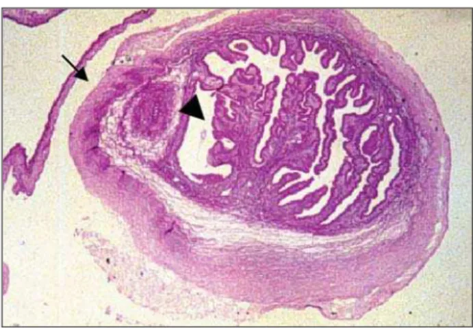
Observation: The sum is higher than 100% because the pathologic processes were found in more than one localization. Fig 1. Viable cysticercus in meningocortical localization. Glicidic
radicals deposits are observed, as well as PAS positive, in the duct area (arrow head) and membranes (arrow) of the para-site (periodic acid-Schiff, 50X).
Fig 2. Cysticercus in destruction in meningocortical localiza-tion. Fibrous connective tissue (arrow) neoformation is observed in the parasite, in the host-parasite interface and in the host tissue (Picro red, 50X).
article varying from 21 to 40 years old . Maybe this difference can be explained because this review arti-cle gathered clinic, serum-epidemiologic and autop-sies data while we only considered autopautop-sies data. Overall the white NCC individuals prevailed in com-parison to the non-white ones, which is compatible to other authors’ data12,23. Furthermore, the male
gender was more frequent than the female one accordingly to the reported literature24.
The organism reaction surrounding the cysticer-cus may occur accordingly to its localization in sev-eral organs as well as in the same organ the lesions can vary accordingly to its implantation site. The meningocortical localization of encephala cysticerci is highlighted because it causes more severe lesions while inside the parenchyma more discrete lesions were observed. Differently from the data described by Chimelli et al.25reporting the parenchymatous
localization as the most frequent in NCC.
The inflammatory infiltrate of mononuclear cells in the cystic wall, the gliosis, the tissue edema and the thickening of small vessels near to the cysticerci were discrete in viable cysticerci. While the host’s inflammatory response became more intense the par-asite began to suffer a destruction process that result-ed in its death. With the parasite’s disintegration the inflammatory reaction tended to decrease although the persistence of giant cells surrounded by the fi-brous capsule, gliosis and edema. According to the literature this fact indicates a continuum of the host’s reaction against the parasite’s debris without, how-ever, an association to the type or intensity of the inflammatory response22.
The necrosis of neurons near the cysticercus im-plantation site is probably due to the compression caused by the parasite or by the secretion of media-tors from the cysticercus or by the inflammatory response in the nearby tissue which is in accordance to other studies26.
Glicidic radicals’ deposits were observed in the parasite and in the host-parasite interface. Accordin-gly to Thomas et al.27these deposits inside
intrapar-asitary vacuoles are connected to the parasite’s metabolism. Therefore there is a relation to the via-bility of the cysticercus such as the greater number of intraparasitary vacuoles the more viable is the par-asite. Besides these gathering indicates a probable crossed reactivity with the embryonic tissue that com-poses the larvae28.
In this study, the beta-fibrillose most frequent site
was the host-parasite interface which corresponds to the main inflammatory site. However, in the litera-ture there are no reports found related to beta-fib-rillose associated to cysticercosis. Accordingly to some authors this type of deposit in other diseases occurs due to complications of a chronic inflammatory proc-ess or of a procproc-ess that destroys the tissue and, there-fore, being called secondary beta-fibrillose29. The
association of beta-fibrillose to the inflammatory process may be due to the presence of precursor of the serum A amyloidal substance (SAA) which have several immunomodulatories attributions such as chemotaxis induction and the expression of adhe-sion molecules besides the cytokine like proprieties. The SAA acts through some mechanisms in the lesion sites aiming the repair of the tissue. The increase and consequent deposit of these substance products are the process we call beta-fibrillose30.
The calcification was identified more frequently in cysticerci in destruction stage, however we also observed calcification in viable cysticerci and with discrete intensity. Accordingly to other studies that describe the calcification as an important process in NCC31, specially in more advanced evolutive stages
and representing the most important finding in com-puterized tomography diagnosis26. Accordingly to
Vargas-Parada et al.31the calcification found in viable
cysticerci may be explained by the calcareous corpus-cles produced by the parasite and deposited in the lumen of protonephridial ducts.
The lesions in the growth and cellular diferenti-ation were identified in the host-parasite interface in one NCC case and is in accordance to the literatu-re32which indicates a significant association between
NCC and gliomas.
In conclusion, in NCC were found several patho-logic processes that differ in localization, cysticercus viability, aggression mechanism and immunologic response, besides its intensity is varied accordingly to three distinguished sites: the parasite, the host-parasite interface and the host’s tissue. Beta-fibril-lose associated to the inflammatory process caused by the parasite is highlighted and have not yet been described related to cysticercosis. The calcification is seen differently from the description of other authors revealing the possibility of a pattern that may result in a greater NCC diagnosis difficulty.
Aknowledgments – The authors thank Lorena
REFERENCES
1. Del Brutto OH, Sotelo J. Neurocysticercosis: an update. Rev Infect Dis 1988;10:1075-1087.
2. Flisser A. Taeniasis-cysticercosis: an introdution. Southeast Asian J Trop Med Public Health 1991;22:233-235.
3. Pedretti L, Bedaque EA, Sotelo J, Del Brutto OH. Cisticercose. In Veronesi R, Focaccia R (Eds). Tratado de infectologia, 2.reimpressão. São Paulo: Editora Atheneu, 1999:1332-1347.
4. Nascimento E. Teníase e cisticercose. In Neves DP (Ed). Parasitologia humana. 9.Ed. São Paulo: Editora Atheneu, 1998:244-256.
5. Gobbi H, Adad SJ, Neves RR, Almeida HO. Ocorrência de cisticercose (Cysticercus cellulosae) em pacientes necropsiados em Uberaba, MG. Rev Patol Trop 1980;9:51-59.
6. Peregrino AJP, Porto SO. Neurocisticercose no sudeste da Bahia. Arq Neuropsiquiatr 1985;43:55-60.
7. Costa-Cruz JM, Rocha A, Silva AM, et al. Ocorrência de cisticercose em necropsias realizadas em Uberlândia, Minas Gerais, Brasil. Arq Neuro-psiquiatr 1995;53:227-232.
8. Albuquerque ES, Galhardo I. Neurocisticercose no Estado do Rio Grande do Norte. Arq Neuropsiquiatr 1995;53:464-470.
9. Takayanagui OM, Castro E, Silva AAMC, et al. Notificação compul-sória da cisticercose em Ribeirão Preto - SP. Arq Neuropsiquiatr 1996; 54:557-564.
10. Gonçalves-Coelho TD, Coelho MDG. Cerebral cysticercosis in Campina Grande, Paraíba - northern Brazil. Arq Neuropsiquiatr 1996;54:94-97. 11. Gomes I, Veiga M, Embiruçu EK, et al. Taeniasis and cysticercosis
preva-lence in a small village from northeastern Brazil. Arq Neuropsiquiatr 2002;60:219-223.
12. Agapejev S. Aspectos clínicos-epidemiológicos da neurocisticercose no Brasil: análise crítica. Arq Neuropsiquiatr 2003;61:822-828.
13. Lino-Junior RS, Reis MA, Teixeira VPA. Ocorrência de cisticercose (Cysticercus cellulosae) encefálica e cardíaca em necropsias. Rev Saúde Publ 1999;33:60-63.
14. Queiroz AC, Martinez AMB. Envolvimento do sistema nervoso cen-tral na cisticercose. Arq Neuropsiquiatr 1979;37:34-41.
15. Rubio-Donnadieu F. Aspectos clínicos de la neurocisticercose. Gac Med Mex 1988;124:194-197.
16. Silva AV, Martins HH, Marques CM, et al. Neurocysticercosis and
micro-scopic hippocampal dysplasia in a patient with refractory mesial tem-poral lobe epilepsy. Arq Neuropsiquiatr 2006;64:309-313.
17. Trentin AP, Teive HAG. Achados tomográficos em 1000 pacientes con-secutivos com antecedentes de crises epilépticas. Arq Neuropsiquiatr 2002;60:416-419.
18. Rocha MSG, Brucki SMD, Ferraz AC, Piccolo AC. Doença cerebrovas-cular e neurocisticercose. Arq Neuropsiquiatr 2001;59:778-783. 19. Takayanagui OM. Neurocisticercose: II. Avaliação da terapêutica com
praziquantel. Arq Neuropsiquiatr 1990;48:11-15.
20. Takayanagui OM, Leite JP. Neurocisticercose. Rev Soc Bras Med Trop 2001;34:283-290.
21. Prophet EB, Mills B, Arrington JB, Sobin LH. Laboratory methods in histotechnology. Amer Registry Pathol, Washington 1994:278. 22. Agapejev S. Epidemiology of neurocysticercosis in Brazil. Rev Inst Med
Trop São Paulo 1996;38:207-216.
23. Spina-França A, Livramento JA, Machado LR. Cysticercosis of the cen-tral nervous system and cerebrospinal fluid. Arq Neuropsiquiatr 1993; 51:16-20.
24. Montemor MR Netto, Gaspareto EL, Faoro LN, et al. Neurocisticercose. Arq Neuropsiquiatr 2000;58:883-889.
25. Chimelli L, Lovalho AF, Takayanagui OM. Neurocisticercose: con-tribuição da necropsia na consolidação da notificação compulsória em Ribeirão Preto-SP. Arq Neuropsiquiatr 1998;56:577-584.
26. Pittella JEH. Neurocysticercosis. Brain Pathol 1997;4:681-693. 27. Thomas JA, Kothare SN, Baptist SJ. Cysticercus cellulosae. J Trop Med
Hyg 1973;76:106-110.
28. Thomas JA, Knoth R, Schwechheimer K, et al. Disseminated human neurocysticercosis. Acta Neuropathol 1989;78:594-604.
29. Cotran RS, Kumar V, Robbins SL, et al. Doenças da imunidade. In Cotran RS, Kumar V, Robbins SL, et al (Eds). Patologia estrutural e fun-cional. 6.Ed. Rio de Janeiro: Editora Guanabara Koogan, 2000:152-212. 30. Cunnane G, Whitehead, AS. Amyloide precursors and amyloidosis in rhelmatoid arthritis. Baillieres Best Pract Res Clin Rheumatol 1999; 13:615-28.
31. Vargas-Parada L, Merchant MT, Willms K, et al. Formation of calcare-ous corpuscles in the lumen of excretory canals of Taenia solium cys-ticerci. Parasitol Res 1999;85:88-92.