AR
TIGO ORIGINAL / ORIGINAL AR
TICLE
INTRODUCTION
The treatment of locally advanced extra-peritoneal rectal adenocarcinoma used for the last two decades, has been the association of preoperative chemora-diotherapy followed by resection surgery with total mesorectal excision(6). The neoadjuvant therapy leads to a decrease of the lesion, facilitating the surgical resection and increasing the rates of sphincter preser-vation and maintenance of urinary and sexual func-tions(11). This multimodal approach is more effective in the control of local recurrence, but some studies also suggest an increase of survival rates.
The response of neoadjuvant therapy leads to re-duction of tumor staging until the complete regression,
NEOADJUVANT THERAPY AND SURGERY
FOR RECTAL CANCER.
Comparative study between partial and
complete pathological response
Vitor Augusto de ANDRADE, Claudio Saddy Rodrigues COY, Raquel Franco LEAL,
João José FAGUNDES, Carlos Augusto Real MARTINEZ and
Maria de Lourdes Setsuko AYRIZONO
Received 13/1/2016 Accepted 28/3/2016
ABSTRACT - Background - The approach of locally advanced extra-peritoneal rectal adenocarcinoma implies a treatment with neoadjuvant chemoradiotherapy associated with total mesorectal excision surgery. However, the tumors respond variably to this neoadjuvant therapy, and the mechanisms for response are not completely understood. Objective - Evaluate the variables related to the complete tumor response and the outcomes of patients who underwent surgery, comparing those with partial tumor regression and those with total remission of rectal lesion, at the pathological examination. Methods - Retrospective analysis of medical records of 212 patients operated between 2000 and 2010, in which 182 (85.9%) obtained partial remission at neoadjuvant therapy (Group 1) and 30 (14.1%), total remission (Group 2). Results - No difference was found between the groups in relation to gender, ethnicity, age, tumor distance from the anal verge, occurrence of metastases and synchronous lesions on preoperative staging, dose of radiotherapy and performed surgery. In Group 2, was veriied high rate of complete remission when the time to surgery after neoadjuvant therapy was equal or less than 8 weeks (P=0.027), and a tendency of lower levels of pretreatment carcinoembryonic antigen (P=0.067). In pathological analysis, the Group 1 presented in relation to Group 2, more affected lymph nodes (average 1.9 and 0.5 respectively; P=0.003), more
angiolymphatic (19.2% and 3.3%; P=0.032) andperineural involvement (15.4% and 0%; P=0.017) and greater number of lymph nodes examined (16.3 and 13.6; P=0.023). In the late follow-up, Group 1 also had lower overall survival than Group 2 (94.1 months and 136.4 months respectively; P=0.02) and disease-free survival (85.5 months and 134.6 months; P=0.004). There was no statistical difference between Group 2 and Group 1 in local recurrence (15% and 3.4%, respectively) and distant metastasis (28% and 13.8%, respectively). Conclusion - In this study, the only factor associated with complete remission of rectal adenocarcinoma was the time between neoadjuvant therapy and surgery. This group of patients had less affected lymph nodes, less angiolymphatic and perineural involvement, a longer overall and disease-free survival, but no signiicant statistical difference was observed in local recurrence and distant metastasis. Although the complete pathologic remission was associated with better prognosis, this not implied in the cure of the disease for all patients.
HEADINGS - Neoadjuvant therapy. Colorectal surgery. Rectal neoplasms.
Declared conflict of interest of all authors: none Disclosure of funding: no funding received
Universidade Estadual de Campinas (UNICAMP), Campinas, SP, Brasil.
Correspondence: Vitor Augusto de Andrade. Universidade Estadual de Campinas (UNICAMP). Cidade Universitária – CEP: 13081-970 – Campinas, SP, Brasil. E-mail: [email protected]
deined as absence of cancer cells at the surgical speci-men. Rates of 10% to 30% of complete tumor remis-sion after neoadjuvant treatment are reported. For the metastatic lymph nodes, the neoadjuvant therapy also has effect at the regression, however, there might be maintenance of tumor cells even in patients with total regression of the tumor in the rectal wall, resulting in a reserved prognosis(3,11).
data and the follow-up of patients who showed partial regression and complete remission of rectum lesion, at the surgical specimen.
METHODS
Patients with extra-peritoneal rectal adenocarcinoma, submitted to neoadjuvant radiotherapy and chemotherapy (5-luorouracil and leucovorin) followed by total mesorectal excision surgery, were evaluated. The medical records of 212 patients operated from 2000 to 2010, by Colorectal Surgery Unit of the School of Medical Sciences, University of Campi-nas (UNICAMP) were analyzed. The study was approved by ethics committee of FCM-UNICAMP (Nº 727/2010).
Complete remission was deined as absence of tumor cells in the rectum, at the pathological study. The analysis of surgical specimen showed that there was presence of residual disease in 182 patients – 85.8% (Group 1) and total tumor remission in the rectum in 30 patients – 14.2% (Group 2). A protocol was applied containing the following information: general characteristics (gender, ethnicity, age); preoperative data (level of carcinoembryonic antigen – CEA, distance between the tumor and the anal verge, presence of synchronous lesions in colonoscopy, staging exams, time between neoadjuvant therapy and surgery); surgical data (intraoperative indings and performed surgery); pathologi-cal analysis of the surgipathologi-cal specimen (histologipathologi-cal grading of tumor, angiolymphatic and perineural involvement, number of examined and affected lymph nodes) and postoperative data (local recurrence, metastasis, deaths, disease-free sur-vival and overall sursur-vival).
The comparison between the two groups was performed through the Mann-Whitney test for the numerical variables and through the Chi-square or Fisher exact test, for the categorical variables. The association between variables was done through binary logistic regression. At the multiple analysis, the selection criteria of variables, was the stepwise. The Cox proportional risks model was used to compare the overall survival and disease-free survival between groups. For all statistical tests, a P value less that 0.05 was considered statistically signiicant.
RESULTS
The general characteristics and preoperative data are found in Table 1. In Group 1, 108 (59.3%) patients had lesions located below 5 cm from anal verge, 67 (36.8%) between 5 and 8 cm and 7 (3.8%) above 8 cm. In Group 2, 16 (53.3%) patients showed rectal lesion below 5 cm from anal verge, 14 (46.7%) between 5 and 8 cm and none above 8 cm (P=0.374).
Results of preoperative colonoscopy were not obtained in 7 (3.3%) patients, all of them from Group 1. In this group, 10 (5.7%) patients had stenosing lesion, not being possible to evaluate the proximal colon and in 43 (26.1%) the presence of synchronous lesions was observed. In the other group, 2 (6.7%) patients had stenosing lesion and 6 (21.4%) synchro-nous lesions (P= 0.60).
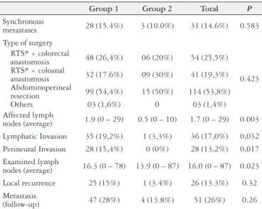
The CEA (carcinoembryonic antigen) level was not found in ive patients, all of them also from Group 1 and in 21 (9.9%) the interval between neoadjuvant therapy and surgery were not obtained (18 from in Group 1 and 3 from Group 2). Six (2.8%) patients were submitted to derivative colostomy because of intestinal obstruction before neoadjuvant therapy, four from the Group 1 and 2 from the Group 2. The surgical indings, pathological and postoperative follow-up data are shown in the Table 2.
Out of 182 patients with residual lesion, the histological grading of tumor was undifferentiated or poorly differenti-ated in 18 (9.9%) patients, moderately differentidifferenti-ated in 152 (83.5%) and well differentiated in other 12 (6.6%).
Regarding disease-free survival, there was signiicant difference between the Groups 1 and 2, with 85.5 months (ranging from 0 to 152) and 134.6 months (ranging from 1 to 166), respectively (P=0.004) (Figure 1). The overall survival was 94.1 months (ranging from 0 to 152) for the Group 1 and 136.4 months (ranging from 1 to 166) for the Group 2 (P=0.02) (Figure 2).
TABLE 1. General characteristics and preoperative data
Group 1 Group 2 Total P
Gender
(Male / Female) (58.2 / 41.7%)106 / 76 (60.0 / 40.0%)18 / 12 (58.5 / 41.5%)124 / 88 0.856
Ethnicity
(White / No white)
152 / 30 (83.5 / 16.5%)
27 / 3 (90.0 / 10.0%)
179 / 33
(84.4 / 15.6%) 0.586
Mean age
(years) (29 – 88)60.3 (33 – 79)57.6 (29 – 88)59.9 0.338
Metastases
Lung 7 (3.8%) 0 (0%) 7 (3.3%) 0.597
Liver 23 (12.6%) 1 (3.3%) 24 (11.3%) 0.212
CEA*
≤ 5 ng/mL 86 (48.6%) 20 (66.7%) 106 (51.2%) 0.067 > 5 ng/mL 91 (51.4%) 10 (33.3%) 101 (48.8%)
Interval between neoadjuvant therapy and surgery
≤ 8 weeks 84 (51.2%) 20 (71.1%) 104 (54.4%) 0.027 > 8 weeks 80 (48.8%) 7 (25.9%) 87 (45.5%)
Radiotherapy dose
≤ 4000 cGy 5 (3.11%) 1 (4.0%) 6 (3.23%)
0.499 4000 -
5000 cGy 151 (93.79%) 23 (92.0%) 174 (93.55%)
> 5000 cGy 5 (3.11%) 1 (4.0%) 6 (3.23%)
DISCUSSION
The treatment for extra-peritoneal rectal cancer has been preferably carried out by a multimodal approach that includes chemoradiotherapy followed by total mesorectal excision surgery. Studies comparing neoadjuvant therapy with radiotherapy and chemotherapy, and radiotherapy alone before surgery, or even, with radio and chemotherapy postoperative, showed that these are worse outcomes than the neoadjuvant therapy, and the adjuvant therapy is reserved only for patients after surgical treatment that showed disease in lymph nodes in the surgical specimen(21).
The introduction of neoadjuvant therapy was considered a hallmark in the management of rectal cancer as it resulted in considerable tumor remission rates, with reduced local recurrence and improved the patient’s survival. In the litera-ture, pathologic complete response varies from 0% to 42%, with the best results found when radiotherapy is associated with two chemotherapy drugs(10,24). The tumor regression observed in the rectal wall is not often accompanied by tumor remission in the lymph nodes. In our study, 10% of patients had metastatic lymph nodes in the group with total tumor remission, the same percentage found by Shwaartz et al.(25).
In the present study, complete remission was considered the absence of viable tumor cells in the rectal wall in the surgi-cal specimen, and were observed in 30 patients, representing 14% of the sample, as found by other authors Nyasavajjala et al.(20) (10%), Jeong et al.(15) (17%) and Garland et al.(9) (11.4 %). However, this data was slightly lower than the best results, perhaps suggesting differences in the neoadjuvant treatment and especially in the interval between neoadjuvant therapy and surgery.
This study aimed to assess the differences between groups of patients with complete regression of the rectal tumor with those who maintained residual lesion. The analysis of preoperative data revealed no differences between the two groups with respect to age, gender, ethnicity, distance between from the tumor and the anal verge, dose of radiotherapy, occurrence of synchronous lesions or metastases and type of surgery performed.
The level of CEA, in the comparison between the two groups, tended to a signiicant difference (P= 0.067), show-ing that in patients who responded with complete remission of the primary tumor, the level was less than or equal to 5.0 ng/mL in the preoperative in a larger number of patients. Park et al.(22) also found an association between higher CEA levels than 5.0 ng/mL and a worse response to neoadjuvant treatment and Yoon et al.(28) found relationship between a positive response and CEA levels less than 5.0 ng/mL. Zeng et al.(29), in a study conducted with 323 patients, the value of pretreatment CEA less than or equal to 5 ng/mL, was considered an independent clinical predictor for pathological complete response.
The beneit of preoperative chemoradiotherapy is well established in the literature, but the best interval between neo-adjuvant therapy and surgical treatment is not fully deined. The key point is that the necrosis induced by radiotherapy TABLE 2. Surgical, pathologic and follow-up data
Group 1 Group 2 Total P
Synchronous
metastases 28 (15.4%) 3 (10.0%) 31 (14.6%) 0.583 Type of surgery
RTS* + colorectal
anastomosis 48 (26,4%) 06 (20%) 54 (25,5%)
0.423 RTS* + coloanal
anastomosis 32 (17.6%) 09 (30%) 41 (19,3%) Abdominoperineal
resection 99 (54,4%) 15 (50%) 114 (53,8%) Others 03 (1,6%) 0 03 (1,4%) Affected lymph
nodes (average) 1.9 (0 – 29) 0.5 (0 – 10) 1.7 (0 – 29) 0.003 Lymphatic Invasion 35 (19,2%) 1 (3,3%) 36 (17,0%) 0,032 Perineural Invasion 28 (15,4%) 0 (0%) 28 (13,2%) 0,017 Examined lymph
nodes (average) 16.3 (0 – 78) 13.9 (0 – 87) 16.0 (0 – 87) 0.023 Local recurrence 25 (15%) 1 (3.4%) 26 (13.3%) 0.32 Metastasis
(follow-up) 47 (28%) 4 (13.8%) 51 (26%) 0.26
* RTS: rectosigmoidectomy
FIGURE 1. Disease-free survival of patients with and without total tumor
regression after neoadjuvant therapy (P= 0.004)
1.0
Time until metastasis or recorrence (months)
Estimat
ed sur
viv
al
0 50 100 150
0.0 0.2 0.4 0.6 0.8
Partial regression Complete regression
FIGURE 2. Overall survival of patients with and without total tumor
regression after neoadjuvant therapy (P= 0.02)
1.0
Time until death (months)
Estimat
ed sur
viv
al
0 50 100 150
0.0 0.2 0.4 0.6 0.8
appears to be time dependent, so a controlled postpone surgery would allow a potentiation of the effect, maximizing the beneits of neoadjuvant therapy(12,15).
At irst, in 1999, François et al.(8) through Trial Lyon R90-01, found a complete pathological response in 10.3% of the patients operated shortly time (2 weeks) after neoadju-vant therapy, and 26% with a longer interval (6 to 8 weeks). Based on these results, since then, most units adopted this interval of 6-8 weeks between the end of neoadjuvant therapy and surgery.
Saglam et al.(23) did not observe differences in pathological response rate, local recurrence of the disease, metastasis and overall survival between patients operated between 4 and 8 weeks, and after 8 weeks of neoadjuvant therapy. Similarly, Jeong et al.(15) found no differences in survival, local recur-rence, distant metastasis and tumor response rate, however they showed a better complete pathological response rate in the lymph nodes in the patients operated after 8 weeks of the completion of neoadjuvant therapy (66.7% vs 46.7%;
P=0.024).
Habr-Gama et al.(12), analyzing 250 patients, also observed a better pathological response in the lymph nodes when they were operated with a long time after the neoadjuvant therapy. They compared the group of patients operated with 12 or fewer weeks (48%) and after 12 weeks (52%) and found a lower signiicantly risk of lymph nodes involvement in the group operated after 12 weeks (P=0.015). There was no statistical difference between the two groups with respect to overall survival rate (86% and 81.6%) and disease-free survival (56.5% and 58.8%).
Still regarding the optimal time to perform surgery after the neoadjuvant therapy, Foster et al.(7), in a meta-analysis including 15 studies, concluded the evidence is insuficient to settle this time though there appear to be beneits after 6-8 weeks.
In our study, in patients with residual disease, there was no difference between the patients operated before and after 8 weeks from the end of neoadjuvant therapy, but the Group with total remission, 71.1%, were operated with intervals of less than 8 weeks, and this difference had statistical signii-cance (P=0.027). Therefore, we found that total remission occurred more frequently in patients operated with shorter intervals or equal to 8 weeks, contrary to the indings of other studies. We are conducting new surveys in our unit to explain these results, since the current trend is to wait more time between neoadjuvant therapy and surgery. Perhaps with a larger sample size and studying different intervals, and not just more or less than 8 weeks, we will have other conclusions.
Recurrence of the disease, locally or at distance, and quality of life of patients are important factors in the man-agement of rectal cancer. Poor histological differentiation, angiolymphatic and perineural invasion, advanced stage and elevation of CEA in the preoperative are factors associ-ated with worse prognosis(27). However, despite advances in detection and tumor staging, the characterization of lymph node involvement remains unclear. Considering patients with complete tumor response in the rectum, 7% to 17% will have
tumor involvement of lymph nodes(19,21); and in our study, this percentage was 10%. The preoperative analysis in order to select patients for a non-surgical treatment therefore, can jeopardize a percentage of patients with lymph node involve-ment, regardless of tumor regression in the rectum(1).
The pathologic evaluation of the lymph nodes of sur-gical specimens from patients with rectal cancer can have substantial impact on time to relapse and survival. The ideal number of lymph nodes evaluated in the surgical specimen has been controversial, but an inadequate number can lead to understaging, and then require adjuvant therapy(13). Cur-rently, it is considered a minimum 12 lymph nodes examined for proper staging(16,18,27).
Our series showed an average 16.3 lymph nodes in Group 1 and 13.9 in Group 2 (P=0.023) and average of affected lymph nodes 1.9 and 0.5, respectively (P=0.003). These results may suggest that neoadjuvant therapy was more effec-tive in the Group with complete pathological response also in relation to the lymph nodes, justifying a lower number of total and metastatic lymph nodes. Habr-Gama et al.(13) also observed that patients with more than 14 lymph nodes examined, and classiied as N0, presented signiicantly higher absence rates of residual disease and longer survival com-pared with lower number of lymph nodes. In this study, in the group with total remission of the rectal lesion, there was only a patient with angiolymphatic involvement and there was no perineural involvement.
Published data show that 50% to 60% of patients with rectal cancer will develop distant metastases, especially liver, lung, bone and brain(18). In our series, the global index of metastatic disease with median follow-up of 28.3 months was 26%, being 28% in patients with residual tumor and 13.8% in the Group with complete remission (P=0.26).
Local recurrence is another factor directly related to sur-vival, with rates ranging from 3% to 30%, with a signiicant reduction from the advent of the total mesorectal excision and neoadjuvant therapy. Relapse, usually occurs within the irst year, over 90 % occurring until third year of post-operative. Several factors may be related to these rates, as the presence of lymph nodes metastases, scanty or positive margins and penetration in the rectal wall(10).
In the overall analysis of our series, 13.3% of patients had pelvic recurrence, with no differences between the Groups (15% in Group 1 and 3.4% in Group 2; P=0.32). A meta-analysis conducted by Martin et al.(17), which included 16 studies and 3,363 patients, found in patients with complete tumor remission, a local recurrence of 0.7% and the distant metastases of 8.7%. Our percentage of local relapse of 3.4% in patients with complete remission refers to only one patient, so if it were a larger sample, this number would probably approach the rates found in this meta-analysis.
Our study has limitations, mainly because it is retrospec-tive and the information is collected from medical records with possible loss of some data. During this time also there was a great evolution especially in relation to chemotherapy and perhaps new studies will show a higher rate of com-plete pathological response and with the improvement in the accuracy of staging exams after neoadjuvant therapy, non-operative treatment might become the most appropriate option in the future.
However, up to the present date, chemotherapy combined with radiotherapy followed by total mesorectal excision sur-gery is still the pattern in the treatment of rectal cancer(2,21). In this study, patients with complete tumor remission in the
Andrade VA, Coy CSR, Leal RF, Fagundes JJ, Martinez CAR, Ayrizono MLS. Terapia neoadjuvante e cirurgia para câncer do reto. Estudo comparativo entre resposta patológica parcial e completa. Arq Gastroenterol. 2016,53(3):163-8.
RESUMO - Contexto - A abordagem do câncer retal extra-peritoneal localmente avançado implica em um tratamento com quimio e radioterapia neoadjuvante associada com a cirurgia de excisão total do mesorreto. Entretanto, os tumores respondem de maneiras variadas a esta terapia neoadjuvante, não se conhecendo completamente os mecanismos envolvidos nesta resposta. Objetivo - Avaliar os fatores relacionados à resposta tumoral completa e o seguimento de pacientes operados, comparando o grupo com regressão parcial com aqueles em que se evidenciou remissão total da lesão no reto, pelo estudo anatomopatológico. Métodos - Análise retrospectiva de prontuários médicos de 212 pacientes operados entre 2000 e 2010, sendo que 182 (85,9%) apresentaram remissão parcial a neoadjuvância (Grupo 1) e 30 (14,1%), remissão total (Grupo 2).
Resultados - Não foi encontrada diferença entre os grupos em relação a gênero, etnia, idade, distância do tumor a margem anal, ocorrência de metástases e lesões sincrônicas no estadiamento pré-operatório, dose de radioterapia e tipo de cirurgia realizada. No Grupo 2, foi veriicada alta taxa de remissão completa quando o paciente foi operado com intervalo menor ou igual a 8 semanas após a terapia neoadjuvante (P=0,027), e uma tendência a menor valor de antígeno carcinoembrionário pré-tratamento (P=0,067). Na análise patológica, o Grupo 1 apresentou em relação ao Grupo 2, mais linfonodos acometidos (média de 1,9 e 0,5 respectivamente; P=0,003), mais invasão angiolinfática (19,2% e 3,3%; P=0,032) e perineural (15,4% e 0%; P=0,017), e maior número de linfonodos examinados (16,3 e 13,6; P=0,023). No seguimento tardio, o Grupo 1 também apresentou menor sobrevida global do que o Grupo 2 (94,1 e 136,4 meses, respectivamente; P=0,02) e sobrevida livre de doença (85,5 e 134,6 meses; P=0,004). Não houve diferença estatística entre os Grupo 1 e Grupo 2 na ocorrência de recidiva local (3,4% e 15%, respectivamente; P=0,32) e metástases à distância (13,8 e 28%; P=0,26). Conclusão - Neste estudo, o único fator que foi associado à remissão completa do adenocarcimona retal, foi o tempo entre neoadjuvância e a cirurgia. Este grupo de pacientes apresentou menos linfonodos acometidos, menor invasão angiolinfática e perineural, maior sobrevida global e livre de doença, porém não apresentou diferença estatística signiicativa com relação à recorrência local e metástases à distância. Embora a remissão completa fosse associada com melhor prognóstico, não implicou na cura da doença em todos os pacientes.
DESCRITORES - Terapia neoadjuvante. Cirurgia colorretal. Neoplasias retais.
rectum wall had a lower number of metastatic lymph nodes, less angiolymphatic and perineural involvement, in addition to greater overall and disease-free survival. Moreover, they also showed a tendency of lower value in the preoperative CEA and better response when they were operated within 8 weeks or less after neoadjuvant therapy. Although the complete pathologic remission was associated with better prognosis, this not implied in the cure for all patients.
Authors’ contributions
REFERENCES
1. Andrade VA, Leal RF, Fagundes JJ, Coy CSR, Ayrizono MLS. Neoadjuvant therapy and surgery in rectal adenocarcinoma: Analysis of patients with complete tumor remission. J Coloproctol. 2013;33:222-7.
2. Artioukh DY. Controversial aspects of rectal cancer surgery. Colorectal Dis. 2010;12:25-9.
3. Chang GJ, Rodriguez-Bigas MA, Eng C, Skibber JM. Lymph Node Status After Neoadjuvant Radiotherapy for Rectal Cancer Is a Biologic Predictor of Outcome. Cancer. 2009;115:5432-40.
4. Chok KS, Law WL. Prognostic factors affecting survival and recurrence of patients with pT1 and pT2 colorectal cancer. World J Surg. 2007;31:1485-90.
5. Compton CC, Fielding LP, Burgart LJ, Conley B, Cooper HS, Hamilton SR, et al. Prognostic factors in colorectal cancer. Arch Pathol Lab Med. 2000;124:979-94. 6. Eich HT, Stepien A, Zimmermann C, Hellmich M, Metzger R, Hölscher A,
Müller RP. Neoadjuvant Radiochemotherapy and Surgery for Advanced Rec-tal Cancer Prognostic Signiicance of Tumor Regression. Strahlenther Onkol. 2011;187:225-30.
7. Foster JD, Jones EL, Falk S, Cooper EJ, Francis NK. Timing of surgery after long-course neoadjuvant chemoradiotherapy for rectal cancer: a systematic review of the literature. Dis Colon Rectum. 2013;56:921-30.
8. Francois Y, Nemoz CJ, Baulieux J, Vignal J, Grandjean JP, Partensky C, et al. Inluence of the interval between preoperative radiation therapy and surgery on downstaging and on the rate of sphincter-sparing surgery for rectal cancer: the Lyon R90-01 randomized trial. J Clin Oncol. 1999;17:2396.
9. Garland ML, Vather R, Bunkley N, Pearse M, Bissett IP. Clinical tumour size and nodal status predict pathologic complete response following neoadjuvant chemoradiotherapy for rectal cancer. Int J Colorectal Dis. 2014;29:301-7. 10. Habr-Gama A, Perez RO, Nadalin W, Sabbaga J, Ribeiro U Jr, Silva e Sousa AH
Jr, et al. Operative versus nonoperative treatment for stage 0 distal rectal cancer following chemoradiation therapy: long-term results. Ann Surg. 2004;240:711-7. 11. Habr-Gama A, Perez RO, Proscurshim I, Campos FG, Nadalin W, Kiss D,
Gama-Rodrigues J. Patterns of failure and survival for nonoperative treatment of stage c0 distal rectal cancer following neoadjuvant chemoradiation therapy. J Gastrointest Surg. 2006;10:1319-28.
12. Habr-Gama A, Perez RO, Proscurshim I, Nunes RMS, Kiss D, Gama-Rodrigues J, Cecconello I. Interval between surgery and neoadjuvant chemoradiation therapy for distal rectal cancer: does delayed surgery have an impact on outcome? Int J Radiat Oncol Biol Phys. 2008;71:1181-8.
13. Habr-Gama A, Perez RO, Proscurshim I, Rawet V, Pereira DD, Sousa AH, Kiss D, Cecconello I. Absence of lymph nodes in the resected specimen after surgery for distal rectal cancer and neoadjuvant chemoradiation therapy: what does it mean? Dis Colon Rectum.2008;51:277-83.
14. Hetnal M, Malecki K, Korzeniowski S, Zemelka T. Postoperative chemoradio-therapy in patients with rectal cancer, prognostic factors for disease control and survival. J Clin Oncol. 2006;24:13575.
15. Jeong DH, Lee HB, Hur H, Min BS, Baik SH, Kim NK. Optimal timing of surgery after neoadjuvante chemoradiation therapy in locally advanced rectal cancer. J Korean Surg Soc. 2013;84:338-45.
16. Lykke J, Jess P, Roikjaer O. A minimum yield of twelve lymph nodes in rectal cancer remains valid in the era of neo-adjuvant treatment. Int J Colorectal Dis. 2015;30:347-51.
17. Martin ST, Heneghan HM, Winter DC. Systematic review and meta-analysis of outcomes following pathological complete response to neoadjuvante chemoradiotherapy for rectal cancer. Br J Surg. 2012;99:918-28.
18. Meredith KL, Hoffe SE, Shibata D. The multidisciplinary management of rectal cancer. Surg Clin North Am.2009;89:177-215.
19. Nan KJ, Qin HX, Yang G. Prognostic factors in 165 elderly colorectal cancer patients. World J Gastroenterol. 2003;9:2207-10.
20. Nyasavajjala SM, Shaw AG, Khan AQ, Brown SR, Lund JN. Neoadjuvant chemo-radiotherapy and rectal cancer: can the UK watch and wait with Brazil? Colorectal Dis. 2009;12:33-6.
21. O’Neill BD, Brown G, Heald RJ, Cunningham D, Tait DM. Non-operative treatment after neoadjuvant hemoradiotherapy for rectal cancer. Lancet Oncol. 2007;8:625-33.
22. Park YA, Sohn SK, Seong J, Baik SH, Lee KY, Kim NK, Cho CW. Serum CEA as a predictor for the response to preoperative chemoradiation in rectal cancer. J Surg Oncol. 2006;93:145-50.
23. Saglam S, Bugra D, Saglam EK, Asoglu O, Balik E, Yamaner S, et al. Fourth versus eighth week surgery after neoadjuvant radiochemotherapy in T3-4/ N0+ rectal cancer: Istanbul R-01 study. J Gastrointest Oncol. 2014;5:9-17. 24. Sanghera P, Wong DW, McConkey CC, et al. Chemoradiotherapy for rectal
cancer: An updated analysis of factors affecting pathological response. Clin Oncol R Coll Radiol. 2008;20:176-83.
25. Shwaartz C, Haim N, Rosin D, Lawrence Y, Gutman M, Zmora O. Region-al lymph node status after neoadjuvant chemoradiation of rectRegion-al cancer producing complete or near complete rectal wall response. Colorectal Dis. 2015;17:595-9.
26. Stipa F, Chessin DB, Shia J. A pathologic complete response of rectal cancer to preoperative combined-modality therapy results in improved oncological outcome with those who achieve no downstaging on the basis of preoperative endorectal ultrasonography. Ann Oncol. 2006;13:1047-53.
27. Tepper JE, O’Connell MJ, Niedzwiecki D, Hollis D, Campton C, Benson III AB, et al. Impact of number of nodes retrieved on outcome in patients with rectal cancer. J Clin Oncol. 2001;19:157-63.