Radiol Bras. 2014 Mai/Jun;47(3):159–164 159
Current situation of high-dose-rate brachytherapy for cervical
cancer in Brazil
*
A atual situação da braquiterapia de alta taxa de dose em colo do útero realizada no Brasil
Silva RMV, Pinezi JCD, Macedo LEA, Souza DN. Current situation of high-dose-rate brachytherapy for cervical cancer in Brazil. Radiol Bras. 2014 Mai/ Jun;47(3):159–164.
Abstract
R e s u m o
Objective: To assess the current situation of high-dose-rate (HDR) brachytherapy for cancer of the cervix in Brazil, regarding apparatuses, planning methods, prescription, fractionation schedule and evaluation of dose in organs at risk.
Materials and Methods: In the period between March/2012 and May/2013, a multiple choice questionnaire was developed and sent to 89 Brazilian hospitals which perform HDR brachytherapy.
Results: Sixty-one services answered the questionnaire. All regions of the country experienced a sharp increase in the number of HDR brachytherapy services in the period from 2001 to 2013. As regards planning, although a three-dimensional planning software was available in 91% of the centers, conventional radiography was mentioned by 92% of the respondents as their routine imaging method for such a purpose. Approximately 35% of respondents said that brachytherapy sessions are performed after teletherapy. The scheme of four 7 Gy intracavitary insertions was mentioned as the most frequently practiced.
Conclusion: The authors observed that professionals have difficulty accessing adjuvant three-dimensional planning tools such as computed tomography and magnetic resonance imaging.
Keywords: Brachytherapy; Cancer; Cervix; Uterus; Research; Brazil.
Objetivo: Avaliar a situação atual da braquiterapia de alta taxa dose (BATD) realizada no Brasil para neoplasias do colo uterino, no que diz respeito aos aparelhos, métodos de planejamento, prescrições, fracionamentos e avaliações de dose nos órgãos de risco. Materiais e Métodos: Foi elaborado um questionário contendo questões de múltipla escolha, o qual, entre os meses de março de 2012 e maio de 2013, foi enviado a 89 instituições hospitalares de todo o Brasil que possuem equipamento de BATD.
Resultados: Sessenta e um serviços responderam o questionário. Todas as regiões do País experimentaram aumento acentuado no número de serviços que oferecem BATD entre os anos de 2001 e 2013. Quanto aos planejamentos, apesar de 91% dos centros afirmarem que possuem softwares capazes de suportar planejamento tridimensional, o aparelho de raios X convencional foi apontado
por 92% dos entrevistados como ferramenta de aquisição de imagens na rotina. Aproximadamente 35% dos entrevistados afirmaram que as sessões de braquiterapia são realizadas após a teleterapia. O esquema de quatro inserções de 7 Gy foi apontado como o mais praticado.
Conclusão: Neste estudo percebeu-se que há dificuldade dos profissionais ao acesso às ferramentas de aquisição de imagens adju-vantes aos planejamentos tridimensionais, como tomografia computadorizada e ressonância magnética.
Unitermos: Braquiterapia; Câncer; Colo; Útero; Pesquisa; Brasil.
* Study developed at Department of Physics, Universidade Federal de Sergipe (UFS), São Cristóvão, SE, Brazil.
1. Master, Fellow PhD degree in Physics, Universidade Federal de Sergipe (UFS), São Cristóvão, SE, Brazil.
2. Master, Professor, Pontifícia Universidade Católica de Goiás (PUC Goiás), Goiânia, GO, Brazil.
3. Specialist in Medical Physics, Physicist at Hospital Chama, Arapiraca, AL, Brazil. 4. PhD, Professor, Universidade Federal de Sergipe (UFS), São Cristóvão, SE, Brazil. Mailing Address: Rogério Matias Vidal da Silva. Departamento de Física – Univer-sidade Federal de Sergipe. Avenida Marechal Rondon, s/nº, Jardim Rosa Elze. São Cristóvão, SE, Brazil, 49100-000. E-mail: rmv.fisica@gmail.com.
Received July 9, 2013. Accepted after revision January 22, 2014.
the decade of 1960, but it was introduced in Brazil in the 1990s(1,2). This type of therapy has undergone significant
evolution in terms of treatment methods and planning sys-tems(3–6). Currently, the utilization of loading devices
com-patible with imaging techniques such as computed tomog-raphy (CT) and magnetic resonance imaging (MRI) allow for the acquisition of three-dimensional images of the entire pelvis with the loaders duly positioned for treatment. Such images associated with modern planning softwares allow for outlining the target volume and evaluating the radiation dose behavior on healthy structures with a high degree of fidel-ity, thus allowing for the optimization of the points and dwell time of the source(6). Studies developed by Wang et al.(6)
and Zwahlen et al.(7) have related the plannings developed
with basis on CT and MRI, respectively, with the
two-di-Rogério Matias Vidal da Silva1, Juliana Castro Dourado Pinezi2, Luiz Eduardo Andrade Macedo3, Divanízia
do Nascimento Souza4
INTRODUCTION
mensional plannings based on conventional radiographic images, demonstrating better tumor coverage and lower tox-icity on healthy tissues as such modern imaging techniques are utilized for the planning.
Considering such developments, the present study pre-sents the results of a research developed with hospitals and clinics in Brazil in order to obtain data on the current situa-tion of uterine cervix brachytherapy in the country. Data on the number of apparatuses and their distribution through-out the country were obtained. The present study also de-scribes the methods utilized by the Brazilian radiotherapists to prescribe and evaluate brachytherapy doses in their clini-cal routines. Preliminarily, post-treatment complications that may be related to overdoses to the bladder were also inves-tigated.
MATERIALS AND METHODS
In March 2012, 220 hospitals which operate radiotherapy services were identified at the website of Comissão Nacional de Energia Nuclear (National Commission of Nuclear En-ergy). Such institutions were contacted by phone in order to confirm the existence of active HDR brachytherapy equip-ment in their premises. Of the contacted institutions, 89 hospitals and clinics confirmed the existence of HDR brachytherapy facilities. Presentation letters with a question-naire (Exhibit 1) addressed to the radiotherapists of the in-stitution were mailed.
In the first part of the questionnaire, the respondents an-swered questions on the planning routine and on the charac-teristics of imaging and therapy apparatuses available at their institutions. On the second part, the questionnaire was fo-cused on data about the moment of treatment when the brachytherapy sessions are performed and on whether there are differences in the prescription in cases where the lesion persists after teletherapy, in addition to adopted fractionation schedule types. The third part of the questionnaire ap-proached questions on the methods to evaluate the dose to risk organs. In this part, a figure similar to that included in the ICRU-38 was presented, representing a lateral view of the whole pelvis at a session of intracavitary brachytherapy for cervical cancer(4). Several checkboxes were inserted into
such a figure, some of them in locations defined by the ICRU-38 and others at random spots, in order for the respondents to identify at which points they routinely evaluate the radia-tion dose delivered by HDR brachytherapy.
Considering that investigating the occurrence of all pos-sible side effects resulting from this type of treatment would considerably increase the number of propositions, the fourth and last part of the questionnaire was restricted to the impli-cations of HDR brachytherapy effects on the urinary tract, more specifically, on the bladder. Such an organ was also selected on account of its reference point dose apparently having little relevance in the estimation of the actual dose absorbed by this organ during this type of brachytherapy(8– 10). In this questionnaire part, a blank space was left for the
professionals to freely express their suggestions, criticism and opinions on aspects related to the questionnaire.
RESULTS
Out of the 89 radiotherapy services which confirmed the existence of HDR brachytherapy equipment in operation, 61 sent back filled-out questionnaires, corresponding to a total of 68% of the brachytherapy services.
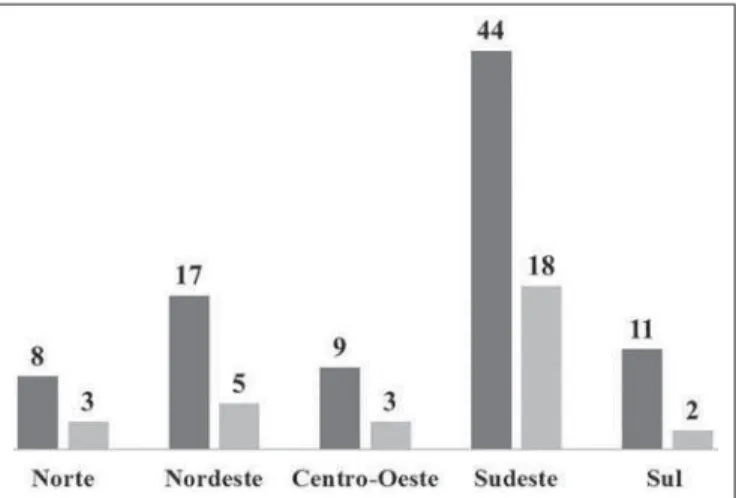
Figure 1 presents a comparative chart of the number of institutions equipped with HDR brachytherapy in the five geographic regions of the country in the years 2001 and 2013, with basis on the comparison of data obtained by the present investigation and those reported by Esteves et al.(3) in 2001.
On Figure 2 one can observe the ratio between estimates of new cases of uterine cervix cancer and the number of avail-able HDR brachytherapy apparatuses per region in 2013, considering the national estimates provided by Instituto Nacional de Câncer(11).
By comparing the number of apparatuses installed in 2001 (31) and in 2013 (89), a global increase of approxi-mately 300% is observed. All the regions presented a sig-nificant increase in the number of services; however the Southern region had the highest increase with 550%. The Southeastern region has the highest number of apparatuses,
Figure 1. Number of HDR brachytherapy apparatuses by region in 2013 and 2001. Number of apparatuses in 2013 (dark gray columns) and number of ap-paratuses in 2001 (light gray columns).
Exhibit 1
QUESTIONNAIRE
1. Routine brachytherapy planning X-ray apparatus
Computed tomography apparatus Magnetic resonance imaging apparatus Software supporting 3D planning 3D planning is routinely performed 2. When brachytherapy sessions are performed
Before teletherapy Alternated with teletherapy After teletherapy
3. Regarding doses prescription and fractionation
Are there differences in dose prescription (fractionation) if reccurrence is observed after teletherapy?: 4. Fractionation for stages IB-IIA – Early stage disease
No. of HDR fractions
06
07
08
05
06
Others:...
5. Fractionation for stages IIB-IVA – Advanced stage disease No. of HDR fractions
05
06
04
05
06
Others:...
6. Definition of dose evaluation points (routinely) Utilization of points defined at ICRU 38 Utilization of ESTRO recommendations Utilization of alternative methods
The tolerance limit is respected (dose in the bladder) < 80%
7. On figure below, mark the dose evaluation points utilized in the routine (in case the definition by means of points is utilized)
8. Patient’s complaints regarding abdominal pain or dysuria which may be associated with overdose in the bladder during and after brachytherapy
There were no reports Frequent reports Scarce reports
9. In the case of reports about discomfort, which are the most common ones? Acute, predominantly abdominal pain (< 6 months)
Sub-acute (> 6 months) Late (cistytis)
10. Comments (optional)
Dose/Fraction
7,5
6,5
6,0
6,0
5,3
Others:...
Dose/Fraction
6,5
5,8
7,0
6,0
5,3
Others:...
Obs.:...
Obs.:...
Yes
No
Obs.:...
Obs.:...
Obs.:...
Obs.:...
Obs.:...
Obs.:...
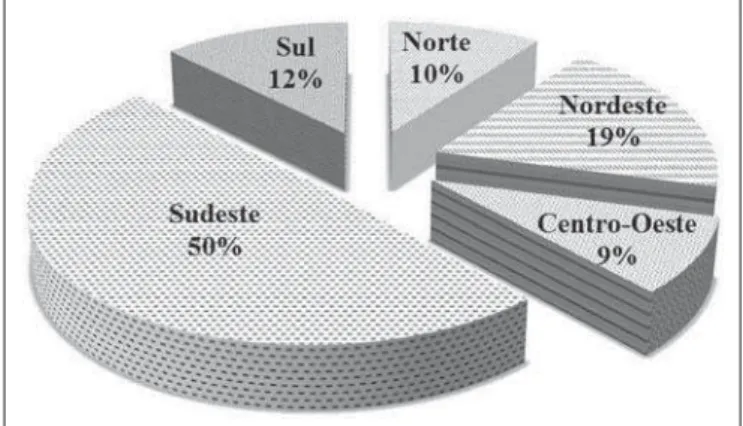
while it also presents with the highest estimates of new uter-ine cervix cancer cases. The percent distribution of opera-tional apparatuses in the Brazilian regions in 2013 is shown on Figure 3.
As the respondents were asked about which points or methods are utilized to prescribe and evaluate the planning in the routine, 75% of them indicated the utilization of only the points defined by the ICRU-38, while 22% indicated the utilization of alternative methods such as other points on the vesical probe surface to estimate the dose delivered to the bladder.
The professionals were asked to mark the points which they use to evaluate the dose during planning on a figure of a sagittal section of the pelvis identical to the one included in the ICRU-38. The utilization of such an image after the question related to the methods habitually utilized for moni-toring was intentional, with the purpose of identifying false-positives in the answer to the previous question. Even when informing that they utilized the points recommended by the ICRU-38, 6% of the respondents marked points different from those recommended by such a document. The highest number of mistakes in the identification of such points were related to the point of the rectum. The dose evaluation point in the sigmoid colon, also proposed by the ICRU-38, was not represented on the figure; but 65% of the respondents pointed it out graphically.
When asked whether they hear reports of abdominal pain which might be associated with overdoses to the bladder, 32% of the respondents answered that patient complains regard-ing some type of discomfort are frequent, while 61% re-sponded that they rarely hear some type of report and 7%, that they never heard any reports. As regards the most fre-quent complaints, 86% of the respondents informed that those were related to acute side effects of the therapy, which are predominantly abdominal pain during the first six months after the treatment. Other respondents (14%) pointed out late effects, such as occurrence of cystitis.
DISCUSSION
Equipment distribution
Without considering the differences in the assistance at public and private services, and the utilization of brachy-therapy equipment for treating other types of cancer, the Southeastern region generally presents the lowest number of new cases per installed apparatus (150 new cases per HDR brachytherapy apparatus). On the other hand, the Northeast-ern region, in spite of having experienced an increase of 340% in the number of apparatuses, presents the highest number of new cases per installed HDR brachytherapy apparatus (297). Currently, in the Northern region, the States of Roraima and Amapá do not have any HDR brachytherapy service, although those states present estimated 120 new cases of cervical cancer for 2013.
Characteristics of the planning routine in Brazil
In the literature, MRI has been described as one of the most appropriate techniques for staging patients with cervi-cal cancer(12). Such a method is also described as being the
best tool for the acquisition of images during the routine
Figure 3. Percentage distribution of HDR apparatuses in operation by Brazilian regions in 2013.
The Southeastern region concentrates one half of all HDR brachytherapy apparatuses in the country, followed by the Northeastern region with 19%. The Midwestern region with has the lower number of apparatuses, with only 9% of the total number of apparatuses in the country.
The authors observed that 91% of the centers are equipped with three-dimensional planning softwares; how-ever, conventional radiography was mentioned by 92% of the respondents as the main imaging tool after insertion of the loading device in their routines, while 23% informed that CT was sporadically utilized for the planning, and only 8% informed that exclusively CT was utilized. None of the re-spondents reported the utilization of MRI in the HDR brachytherapy planning routine.
The present investigation showed that in approximately 35% of the institutions brachytherapy is performed only after teletherapy is concluded. Most institutions opt for four sessions, with doses ranging between 6.5 and 7.5 Gy per fraction. Table 1 presents the percentages of institutions according to frac-tionation schedule and doses per session in the routine.
Table 1—Fractionation and doses per session: percentage of institutions.
Fractionation for stages IB-IIA – Early stage disease
Number of sessions
4 4 5 Other
Dose per session (Gy)
7.0 6.5 6.0 Other
Percentage of institutions
73% 19% 6% 2%
Fractionation for stages IIB-IVA – Advanced stage disease
Number of sessions
4 4 5 Other
Dose per session (Gy)
7.0 7.5 6.5 or 6.0
Other
Percentage of institutions
three-dimensional planning, overcoming conventional radi-ography and the images obtained by means of CT(12–15). On
the other hand, a recent study developed by Viswanathan et al.(16), involving institutions in Asia, North America, Europe
and Oceania, demonstrated that obtaining CT images after the insertion of the loading device is the most common ap-proach. An important characteristic in this type of planning that also must be observed is that the systematic description of the disease extent, i.e., the follow-up of changes in the gross tumor volume as the treatment progresses, particularly after teletherapy, requires time for planning and costs asso-ciated with images acquisition. The fact that a high number of institutions have softwares for three-dimensional plannings while only a small part of them actually utilize such softwares can be justified by the inappropriateness of the values paid by Sistema Único de Saúde. Specifically, one observes that the cost associated with CT and MRI required for such pro-cedures are not covered by the regulations from the Health Ministry about procedures and values associated with radio-therapy(17,18).
Several studies demonstrate that extending the total treatment time may negatively influence the local manage-ment and the patient survival(19–21). In Brazil, most
inter-viewed institutions alternate HDR brachytherapy sessions with teletherapy sessions. However, as reported by some ra-diotherapists, a high number of radiotherapy services in the country are not equipped with brachytherapy apparatuses, a fact that leads to many patients being referred to other insti-tutions in order to complete their treatment, which in some situations increases the treatment time.
In spite of the lack of consensus about fractionation sched-ules and doses per session that should be utilized for a given clinical staging, generally the relative doses prescribed for teletherapy and brachytherapy must depend upon the initial volume of the disease, the geometric variations of the blad-der and rectum, the degree of tumor regression during pel-vic irradiation and also upon the therapeutic protocol fol-lowed by the institution(16,22–24).
As regards fractionation and doses per session practiced by the investigated institutions, few differences were identi-fied among them. A small number of institutions reported that it was possible to change the number of sessions and doses per session in particular situations, according to the disease extent and response to radiation. In the Brazilian context, such a small variation regarding the number of HDR brachytherapy sessions performed by the institutions may be directly associated with the guidelines imposed by Sistema Único de Saúde, which only supports the cost of four ses-sions for the hospital institution, regardless of the clinical staging of the patient(17,18).
Dose monitoring points
In HDR brachytherapy, special attention must be placed on the definition of dose monitoring points in the bladder and in the rectum, as proposed by ICRU-38. The originally
defined dose evaluation point in the bladder has demonstrated to be of little representativeness(8–10). With the purpose of
improving the estimation of the dose received by this organ, another point located 1.5 cm above the current one has re-cently been suggested as being most appropriate for the monitoring(23). As regards the point in the rectum, the
lit-erature also reports moderate representativeness for evalu-ating the dose received by this organ during a HDR brachytherapy session. In the present study, 98% (59) of the respondents utilize the points recommended by ICRU-38, while only 3% (2) informed that they utilize the doses in volume recommended by the GEC-ESTRO guidelines for cervical cancer brachytherapy(25).
Observation of side effects possibly related to the Brazilian clinical practice
During the treatment of uterine cervical cancer, the re-gion of the lower abdomen is weakened due to factors such as the previous extent of the disease, the injury caused by radiation itself, besides the trauma caused by the insertion of loading devices and probes for brachytherapy. Consider-ing the wide range of factors leadConsider-ing to temporary fragility in this region of the body, relating the causes with injuries caused by inappropriate insertions and unsuccessful plannings associated with the clinical practice would be a complex task. However, relying on the clinical experience of the respon-dents, the authors have preliminarily sought to investigate the efficiency of the performed brachytherapy. In spite of some studies pointing towards the lack of representativeness of the dose monitoring point in the bladder proposed by ICRU-38, in the present study most of the respondents in-formed that patients reports about abdominal pain which might be associated with overdose to this organ were scarce or non-existent.
CONCLUSIONS
The present study confirms that over the past decade there was a significant increase in the number and distribu-tion of services offering HDR brachytherapy for uterine cer-vical cancer in Brazil. The Southeastern region is still ahead of the other regions as regards number of services in opera-tion.
Despite some studies reporting lack of representative-ness of the dose monitoring point in the bladder as proposed by ICRU-38, most of the respondents informed that patients reports about abdominal pain which might be associated with overdose to this organ were scarce or non-existent.
The performance of HDR brachytherapy only after tele-therapy is completed and the practice of referring the pa-tients to undergo the procedure in other institutions, as re-ported by some of the respondents, may induce an increase in uterine cervical cancer recurrence and decrease in patients’ survival.
Acknowledgements
To Medical Physicist Homero Lavieri Martins, for all suggestions and guidance for the present study, and to Coordenação de Aperfeiçoamento de Pessoal de Nível Su-perior (Capes) and Fundação de Apoio à Pesquisa e Inovação Tecnológica do Estado de Sergipe (Fapitec-SE), for the fi-nancial support.
REFERENCES
1. Fu KK, Philips TL. High-dose-rate versus low-dose-rate intracavi-tary brachytherapy for carcinoma of the cervix. Int J Radiat Oncol Biol Phys. 1990;19:791–6.
2. Ferrigno R, Nishimoto IN, Novaes PE, et al. Comparison of low and high dose rate brachytherapy in the treatment of uterine cervix cancer. Retrospective analysis of two sequential series. Int J Radiat Oncol Biol Phys. 2005;62:1108–16.
3. Esteves SCB, Oliveira ACZ, Feijó LFA. Braquiterapia de alta taxa de dose no Brasil. Radiol Bras. 2004;37:337–41.
4. International Commission on Radiation Units and Measurements. Dose and volume specification for reporting intracavitary therapy in gynecology (Report 38). Bethesda, MD: ICRU; 1985. 5. Fellner C, Pötter R, Knocke TH, et al. Comparison of
radiogra-phy- and computed tomograradiogra-phy-based treatment planning in cer-vix cancer in brachytherapy with specific attention to some quality assurance aspects. Radiother Oncol. 2001;58:53–62.
6. Wang B, Kwon A, Zhu Y, et al. Image-guided intracavitary high-dose-rate brachytherapy for cervix cancer: a single institutional ex-perience with three-dimensional CT-based planning. Brachytherapy. 2009;8:240–7.
7. Zwahlen D, Jezioranski J, Chan P, et al. Magnetic resonance imag-ing-guided intracavitary brachytherapy for cancer of the cervix. Int J Radiat Oncol Biol Phys. 2009;74:1157–64.
8. Pelloski CE, Palmer M, Chronowski GM, et al. Comparison be-tween CT-based volumetric calculations and ICRU reference-point estimates of radiation doses delivered to bladder and rectum during intracavitary radiotherapy for cervical cancer. Int J Radiat Oncol Biol Phys. 2005;62:131–7.
9. Wilkinson JM, Harris MA, Davidson SE, et al. A retrospective study of bladder morbidity in patients receiving intracavitary brachytherapy as all or part of their treatment for cervix cancer. Br J Radiol. 2003; 76:897–903.
10. Yoshimura R, Hayashi K, Ayukawa F, et al. Radiotherapy doses at
special reference points correlate with the outcome of cervical can-cer therapy. Brachytherapy. 2008;7:260–6.
11. Brasil. Ministério da Saúde. Secretaria de Atenção à Saúde. Instituto Nacional de Câncer. Incidência do câncer no Brasil: estimativa 2012. Rio de Janeiro, RJ: INCA; 2012.
12. Camisão CC, Brenna SMF, Lombardelli KVP, et al. Ressonância magnética no estadiamento dos tumores de colo uterino. Radiol Bras. 2007;40:207–15.
13. Justino PB, Carvalho HA, Baroni RH, et al. Valor da ressonância magnética no planejamento radioterápico dos tumores de colo de útero: resultados preliminares. Radiol Bras. 2005;38:399–402. 14. Haie-Meder C, Pötter R, Van Limbergen E, et al.
Recommenda-tions from Gynaecological (GYN) GEC-ESTRO Working Group* (I): concepts and terms in 3D image based 3D treatment planning in cervix cancer brachytherapy with emphasis on MRI assessment of GTV and CTV. Radiother Oncol. 2005;74:235–45.
15. Hellebust TP, Tanderup K, Lervag C, et al. Dosimetric impact of interobserver variability in MRI-based delineation for cervical can-cer brachytherapy. Radiother Oncol. 2013;107:13–9.
16. Viswanathan AN, Creutzberg CL, Craighead P, et al. International brachytherapy practice patterns: a survey of the Gynecologic Cancer Intergroup (GCIG). Int J Radiat Oncol Biol Phys. 2012;82:250–5. 17. Brasil. Ministério da Saúde. Portaria Nº 346, de 23 de junho de 2008. [acessado em 20 de maio de 2013]. Disponível em: http:// dtr2001.saude.gov.br/sas/PORTARIAS/Port2008/PT-346.htm. 18. Brasil. Ministério da Saúde. Portaria Nº 1.095/GM, de 5 de julho
de 2005. [acessado em 20 de maio de 2013]. Disponível em: http:// dtr2001.saude.gov.br/sas/PORTARIAS/Port2005/GM/GM-1095.htm.
19. Girinsky T, Rey A, Roche B, et al. Overall treatment time in ad-vanced cervical carcinomas: a critical parameter in treatment out-come. Int J Radiat Oncol Biol Phys. 1993;27:1051–6.
20. Perez CA, Grigsby PW, Castro-Vita H, et al. Carcinoma of the uterine cervix. I. Impact of prolongation of overall treatment time and timing of brachytherapy on outcome of radiation therapy. Int J Radiat Oncol Biol Phys. 1995;32:1275–88.
21. Petereit DG, Sarkaria JN, Chappell R, et al. Adverse effect of treat-ment prolongation in cervical carcinoma. Int J Radiat Oncol Biol Phys. 1995;32:1301–7.
22. Petereit DG, Pearcey R. Literature analysis of high dose rate brachytherapy fractionation schedules in the treatment of cervical cancer: is there an optimal fractionation schedule? Int J Radiat Oncol Biol Phys. 1999;43:359–66.
23. Viswanathan AN, Thomadsen B; American Brachytherapy Society Cervical Cancer Recommendations Committee. American Brachy-therapy Society consensus guidelines for locally advanced carcinoma of the cervix. Part I: general principles. Brachytherapy. 2012;11:33– 46.
24. Quinn MA, Benedet JL, Odicino F, et al. Carcinoma of the cervix uteri. FIGO 26th Annual Report on the Results of Treatment in Gynecological Cancer. Int J Gynaecol Obstet. 2006;95 Suppl 1: S43–103.