IX
Radiol Bras. 2011 Jan/Fev;44(1):IX–XI
Editorial
Arterial spin labeling (ASL) is a recently developed magnetic resonance (MR) technique that assesses cere-bral blood flow (CBF)(1,2). This method has been mainly
used for investigative purposes with very few centers in the world performing it on a routine clinical basis. ASL has already been validated and proven to be use-ful in the assessment of a growing number of diseases and conditions(3). As with any recently established
technique, ASL has some limitations that need to be overcome to become more widely used and to be part of the daily routine of the neuroimaging specialist.
Off line post processing is probably the single most important factor that has limited ASL accessibility. The low signal-to-noise ratio (SNR) is another inherent problem in ASL imaging. The lack of transit time infor-mation (using current methods), susceptibility artifacts derived from blood products, metal deposition, calcifi-cation and surgical material can potentially worsen this situation, as well as areas of interfaces between brain and air containing cavities (paranasal sinuses and mastoid air cells).
There are multiple ways of studying CBF includ-ing positron emission tomography, computed tomog-raphy (CT) perfusion, MR perfusion (susceptibility-weighted imaging or bolus tracking), single photon
0100-3984 © Colégio Brasileiro de Radiologia e Diagnóstico por Imagem
Novelties in radiology – Arterial spin labeling,
the gadolinium-free MR perfusion
Novidades em radiologia – Arterial spin labeling, a perfusão por RM sem contraste
Fabrício Guimarães Gonçalves1
, Joseph A. Maldjian2
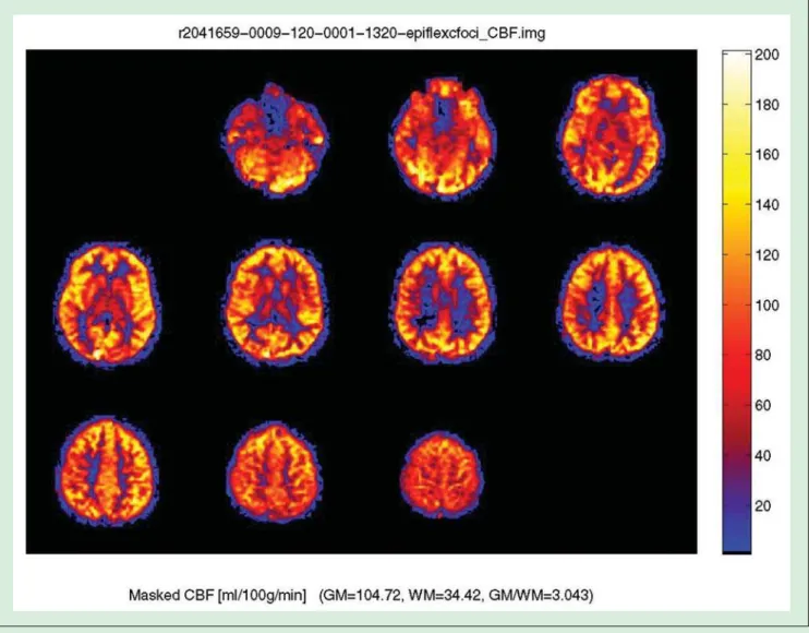
emission and xenon CT. The main advantage of ASL is the lack of the need for an injection of intravenous con-trast material, which makes it a gadolinium-free method and injection device independent. Furthermore, ASL provides a measure of absolute blood flow (as op-posed to relative cerebral blood volume from gado-linium based methods) (please refer to the Figure 1) and can also be repeated many times without the fear of nephrogenic systemic fibrosis, especially in patients with significant renal disease(4).
Currently, four major current ASL techniques are available: pulsed ASL (PASL), continuous ASL (CASL), pseudo-continuous ASL (PCASL) and velocity-selective ASL (VSASL)(5).
In ASL imaging, the spins (protons) of the flowing blood supplying the brain are labeled (or tagged) be-fore entering the region to be imaged. The extent of pa-renchyma to be studied and the location where the spins are labeled may vary with the different methods. Images are acquired in two phases: baseline (control) and post-labeling phases, with the time gap following the labeling tag known as “post-labeling delay”. The signal is obtained by subtracting the control from the post-labeling images. The resulting difference in signal is small and dependent on the cardiac output, travel time of the labeled spins to arrive in the area to be imaged and on the velocity of flow. To compensate for the low SNR, multiple sets of pre- and post-labeling images are typically acquired to ensure enough perfusion signal(5).
In PASL, the radiofrequency (RF) pulses used to la-bel the spins are single and short (2- to 5 milliseconds) with a wider labeling area, located proximal to the imaging plane. PASL has the advantage of having higher tagging efficiency, less power deposition and improved 1. MD, Visiting Fellow Advanced Neuroscience Imaging Research
Core – Wake Forest University School of Medicine, Winston-Sa-lem, NC, USA, Neuroradiology Clinical Fellow – McGill Univer-sity Health Center, Montreal, Que., Canada.
X Radiol Bras. 2011 Jan/Fev;44(1):IX–XI transit time for the labeled spins to travel from the
la-beling area to the imaging plane. The main difficulties in PASL are low SNR and increased transit delay.
In CASL, the RF pulses are longer (1-2 seconds) and the labeling area narrower, located just below the im-aging plane. In this technique, the spins are labeled con-tinuously as they pass through the labeling area. This results in more SNR than in PASL, but more RF power deposition. Lower labeling efficiency and the necessity of continuous RF transmitting hardware are the main downsides of CASL.
PCASL was developed to have high labeling effi-ciency as with CASL, but with less RF power deposi-tion. In comparison with CASL, this technique has a good trade-off between power deposition and SNR. PCASL has a higher SNR than PASL, higher labeling
power than CASL, but presently has the disadvantage of being less clinically available.
VSASL has the ability to measure low flow. In com-parison with PASL, CASL and PCASL, which label the spins at a specific location, this technique labels all the inflowing spins that are moving faster than a specific threshold. Disadvantages are lower SNR and difficul-ties in determining the specific encoding velocity(5).
Many pathological processes have been studied using ASL. Two major patterns are seen: hypo- and hyperperfusion, which can be focal or diffuse. Processes that demonstrate focally decreased perfusion include the ischemic core and penumbra, chronic cerebrovas-cular occlusive disease, chronic microvascerebrovas-cular is-chemia, encephalomalacia, hematoma, infection, arte-riovenous malformation steal phenomena, posterior
XI
Radiol Bras. 2011 Jan/Fev;44(1):IX–XI
reversible encephalopathy syndrome (in the late phase) and post-ictal states. Diffusely decreased perfusion can be seen in brain death, poor cardiac output, aging with cerebral atrophy, vasculitis, cerebral vasospasm, as well as exogenous agents (caffeine)(6).
Focal hyperperfusion can be seen in conditions such as: luxury perfusion, posterior reversible encephalopa-thy syndrome (in the acute phase), reperfusion in is-chemic areas, seizure activity, and in some cases of vas-cular malformations (including developmental venous anomalies). Diffuse increased perfusional states can be seen in normal individuals, global cerebral hyperper-fusion complicating embolic stroke, carotid endarter-ectomy with the post-CEA hyperperfusion syndrome, traumatic head injury, and in the setting of hypercap-nia. Brain tumors can demonstrate patterns of hypo-or hyperperfusion(7).
In conclusion, ASL is a promising and evolving tech-nique used to measure CBF. This gadolinium-free MR perfusion method has several advantages including the lack of a contrast injection and the ability to provide absolute blood flow measurement. The absence of the
risk of nephrogenic systemic fibrosis, non-invasiveness and the possibility of repeating the study several times if necessary, certainly will play a role in the popular-ization of this technique.
REFERENCES
1. Chalela JA, Alsop DC, Gonzalez-Atavales JB, et al. Magnetic resonance perfusion imaging in acute ischemic stroke using con-tinuous arterial spin labeling. Stroke. 2000;31:680–7.
2. Brown GG, Clark C, Liu TT. Measurement of cerebral perfusion with arterial spin labeling: Part 2. Applications. J Int Neuropsychol Soc. 2007;13:526–38.
3. Wintermark M, Sesay M, Barbier E, et al. Comparative over-view of brain perfusion imaging techniques. J Neuroradiol. 2005; 32:294–314.
4. Deibler AR, Pollock JM, Kraft RA, et al. Arterial spin-labeling in routine clinical practice, part 1: technique and artifacts. AJNR Am J Neuroradiol. 2008;29:1228–34.
5. Pollock JM, Tan H, Kraft RA, et al. Arterial spin-labeled MR per-fusion imaging: clinical applications. Magn Reson Imaging Clin N Am. 2009;17:315–38.
6. Deibler AR, Pollock JM, Kraft RA, et al. Arterial spin-labeling in routine clinical practice, part 2: hypoperfusion patterns. AJNR Am J Neuroradiol. 2008;29:1235–41.