7 Radiol Bras. 2011 Jan/Fev;44(1):7–12
Correlation between frontal cortical thickness
and executive functions performance in patients
with human immunodeficiency virus infection
*
Correlação entre espessura cortical frontal e desempenho de funções executivas em pacientes com infecção pelo vírus da imunodeficiência humana
Tania Maria Netto1, Denise Vieira Greca2, Rafael Ferracini3, Denis B. Pereira3, Bernardo Bizzo4, Thomas Doring5, Tadeu Kubo6, Paulo R. V. Bahia7, Rochele Paz Fonseca8, Emerson L. Gasparetto9
Objective: To investigate the correlation between frontal regions cortical thickness measured by magnetic resonance imaging of HIV-positive patients and their performance on instruments for assessing executive functions. Materials and Methods: The present study included 22 HIV-positive patients in the age range from 45 to 65, schooling ranging between three and 20 years, with executive functions deficit and undergoing antiretroviral therapy. Magnetic resonance imaging was performed with conventional T1-weighted, 3D sequences and the images were processed with the Freesurfer software to measure cortical thickness. The following instruments were utilized to evaluate the patients’ executive functions: Trail Making, Wisconsin, Hayling, working memory (WAIS-III), verbal fluency and Stroop tests. The Pearson’s correlation coefficient was utilized in the data statistical analysis. Results: Significant correlations were found between: Wisconsin scores and the thickness of the right pre-central, lateral and left pre-central orbitofrontal regions; Trail Making scores and thickness of right pre-central and left anterior caudal cingulate areas; and Hayling Test scores and thickness of the left lateral orbitofrontal area. Conclusion: Correlations between cortical thickness measurements by magnetic resonance imaging and cognitive performance suggest that the executive function deficit in HIV-positive patients are related to a reduction in the thickness of the frontal cortex.
Keywords: Magnetic resonance imaging; Cortical thickness; Frontal lobe; Neuropsychological; Executive functions; HIV.
Objetivo: Investigar a relação entre a espessura cortical medida pela ressonância magnética em regiões frontais e o desempenho em instrumentos que avaliam funções executivas em pacientes com HIV positivo. Materiais e Méto-dos: Participaram deste estudo 22 pacientes HIV-positivos, com déficits em funções executivas, sob terapia antirretro-viral, idades entre 45 e 65 anos e escolaridade entre 3 e 20 anos. Foi realizada ressonância magnética com sequências convencionais, T1 3D, processado pelo Freesurfer para verificar espessura cortical. Instrumentos de avaliação das funções executivas: Teste de Trilhas, Wisconsin, Hayling, Dígitos (WAIS-III), fluência verbal ortográfica e Stroop. Para análise da relação espessura versus cognição, utilizou-se coeficiente de correlação de Pearson. Resultados: Correlações signi-ficativas foram encontradas entre escores de: Wisconsin e espessura das regiões pré-central e orbitofrontal lateral à direita e pré-central esquerda; Teste de Trilhas e espessura da área pré-central direita e cíngulo anterior caudal es-querdo; e Teste Hayling e espessura da área lateral orbitofrontal esquerda. Conclusão: As correlações existentes entre medidas de espessura cortical pela ressonância magnética e desempenho cognitivo sugerem que os déficits executi-vos em pacientes HIV-positiexecuti-vos relacionam-se a uma redução da espessura cortical das regiões frontais.
Unitermos: Imagem por ressonância magnética; Espessura cortical; Lobo frontal; Avaliação neuropsicológica; Fun-ções executivas; HIV.
Abstract
Resumo
* Study developed at the Unit of Radiodiagnosis of Hospital Universitário Clementino Fraga Filho da Universidade Federal do Rio de Janeiro (HUCFF-UFRJ), Rio de Janeiro, RJ, Brazil.
1. PhD, Post-doctorate fellow, Program of Post-graduation in Medicine (Radiology), Universidade Federal do Rio de Janeiro (UFRJ), Rio de Janeiro, RJ, Brazil.
2. Psychologist, Fellow Master degree, Program of Post-gradu-ation in Medicine (Radiology), Universidade Federal do Rio de Janeiro (UFRJ), Rio de Janeiro, RJ, Brazil.
3. MDs, Radiologists, Fellow Master degree, Program of Post-graduation in Medicine (Radiology), Universidade Federal do Rio de Janeiro (UFRJ), Rio de Janeiro, RJ, Brazil.
Netto TM, Greca DV, Ferracini R, Pereira DB, Bizzo B, Doring T, Kubo T, Bahia PRV, Fonseca RP, Gasparetto EL. Correlation between frontal cortical thickness and executive functions performance in patients with human immunodeficiency virus infection. Radiol Bras. 2011 Jan/Fev;44(1):7–12.
4. Student, Graduation Course of Medicine, Universidade Federal do Rio de Janeiro (UFRJ), Rio de Janeiro, RJ, Brazil.
5. Master, Fellow PhD degree, Program of Post-graduation in Medicine (Radiology), Universidade Federal do Rio de Janeiro (UFRJ), Medical Physicist at Clínica de Diagnóstico Por Imagem (CDPI), Rio de Janeiro, RJ, Brazil.
6. Medical Physicist at Clínica de Diagnóstico Por Imagem (CDPI), Rio de Janeiro, RJ, Brazil.
7. Post-doctorate, Associate Professor, Department of Radi-ology, Universidade Federal do Rio de Janeiro (UFRJ), Head of the Unit of Radiodiagnosis, Hospital Universitário Clementino Fraga Filho da Universidade Federal do Rio de Janeiro
(HUCFF-UFRJ), MD, Radiologist at Clínica Menezes da Costa, Rio de Janeiro, RJ, Brazil.
8. Post-doctorate, Associate Professor, School of Psychology, Coordinator for the Group of Clinical and Experimental Neuro-psychology, Pontifícia Universidade Católica do Rio de Janeiro (PUC-Rio), Rio de Janeiro, RJ, Brazil, Post-doctorate fellow at the Neuroimaging Center of Université de Montréal, Quebec, Ca-nada.
INTRODUCTION
The human immunodeficiency virus (HIV) is a retrovirus that penetrates the ner-vous system cells producing lesions(1) and impairing cognitive, motor and behavioral functions, many times resulting in demen-tia(2). The progress in the development of the highly active antiretroviral therapy (HAART) has allowed a significant reduc-tion in the mortality of patients infected with HIV and, consequently, has aided them to achieve the aging phase of the lifecy-cle(3,4). According to the World Health Or-ganization(5,6), the prevalence of cases of HIV/AIDS still remains quite high, affect-ing about 730 thousand individuals in 2008, just in Brazil. Such data indicate the greater longevity of these patients, but also demon-strate an increase in the incidence of neuro-cognitive complications frequently observed in the elderly populations. Such complica-tions, in association with aging, become a significant problem for HIV-positive pa-tients(3,7–9). Thus, the current reality en-hances the need for a better clinical and imaging evaluation of patients with HIV/ AIDS with possible cognitive deficit(10).
Advanced techniques of magnetic reso-nance imaging (MRI), including images acquisition and processing, have been uti-lized in the assessment of patients at ad-vanced stages of cognitive deficit, particu-larly those who have progressed to a de-mential state(11). Such techniques seek to find out morphological or functional alter-ations in patients with no significant find-ing at conventional MRI. Studies approach-ing such imagapproach-ing techniques have demon-strated a good correlation with neuropsy-chological tests (NPT)(11,12), clinically vali-dating the eventual findings. Investigations approaching imaging methods have already been developed with some samples of HIV/ AIDS patients(12,13). Thompson et al.(12) have found significant cortical atrophy in the gray matter of AIDS patients, involving the following areas: primary sensorimotor, pre-motor, frontopolar, language-related
fron-tal and temporal lobes, pre-fronfron-tal and pa-rietal areas, with significant correlation be-tween these two latter areas and perfor-mance in neuropsychological tests. Comple-mentarily, Chiang et al.(13) have observed severe atrophy in both, the primary and sensorimotor association areas of both hemi-spheres. Such alterations, particularly those in the white matter, observed on images, also present significant correlation with neuropsychological tests results. In such study, additionally to the mentioned find-ings, the authors have observed volumet-ric decrease in the frontal, medial and basal regions, in the middle segment of the cin-gulate gyrus and corpus callosum genu(13). Also, the frontostriatal areas may be in-jured by the human immunodeficiency vi-rus at early stages of the disease. Generally, lesions in such areas are associated with ex-ecutive functions deficits(14,15). Moreover, the frontal cortex is closely related to the processing of executive functions, particu-larly the dorsolateral prefrontal system as well as the basal ganglia and the posterior parietal cortex(16). The literature has sug-gested that executive functions deficits has been one of the core neurocognitive impair-ments in HIV-positive patients(17). Such functions comprehend several cognitive processes which, as a whole, control and monitor other cerebral functions to achieve goal-oriented behaviors. These functions facilitate the planning and adaptation to new situations(18,19) and support appropri-ate functioning of other relappropri-ated cognitive domains, such as, memory.
Despite the possible damages caused by HIV to the executive functions, few stud-ies are found in the literature investigating the correlation between cortical thickness in such regions and performance in neurop-sychological tests that evaluate cognitive components of executive functions in HIV/ AIDS patients(20,21).
Considering the relevance of this type of investigation, the present study was aimed to correlate the frontal cortical thick-ness with performance in neuropsychologi-cal tests that evaluate executive functions in HIV-positive patients. As a hypothesis, it was expected that HIV patients presented correlations between cortical thickness of frontal areas and their performances, par-ticularly in tests evaluating inhibition.
MATERIALS AND METHODS
Study sample
The present study evaluated 22 HIV-positive patients (5 women and 17 men; mean age 52.91 years, standard deviation (SD) = 5.879; mean education level = 11.95 years, SD = 4.541). The patients were undergoing clinical follow-up at Hospital Universitário Clementino Fraga Filho (HUCFF) da Universidade Federal do Rio de Janeiro (UFRJ), Rio de Janeiro, Brazil. Inclusion criteria were the following: age range between 45 and 65 years, diagnosis of HIV infection for at least five years, absence of previous history of any type of neurological disease, currently undergoing treatment with HAART, absence of uncor-rected auditory and/or visual alterations, mini-mental state examination score ≥ 17, for patients with schooling ≤ 4 years, or ≥ 24 for patients with schooling ≥ 5 years. In compliance with ethical principles, all the individuals in this study participated on a voluntary basis, received no remunera-tion, and signed a term of free and informed consent. This study was approved by the Committee for Ethics in Research of HUCFF-UFRJ (CEP/UFRJ No. 151/08).
Data collection
The evaluation of these patients was conducted in two parts: MRI and neurop-sychological assessment. The MRI studies were performed at HUCFF in a 1.5 tesla Avanto system (Siemens Medical Systems, Erlangen, Germany) with an eight-channel head coil. The following sequences were acquired: axial FLAIR [repetition time (TR): 9000 ms; echo time (TE): 83 ms; inversion time (TI): 2500 ms; flip angle: 180°; matrix: 256 × 256; field-of-view (FOV): 230 mm), 3D sagittal T1-weighted (TR: 2530 ms; TE: 3.39 ms; TI: 1100 ms; flip angle: 7°; voxel: 1.33 mm3), coronal T2-weighted (TR: 3500
ms; TE: 99 ms; flip angle: 136°; matrix: 256 × 256; FOV: 210 mm),diffusion ten-sor imaging (TR: 1900 ms; TE: 81 ms; matrix: 256 × 256; FOV: 230 mm; B = 0 s/mm2 and B = 1000 s/mm2, six gradient
directions). All the conventional magnetic resonance images were evaluated by two experienced radiologists and were consid-ered as normal, except for the presence of different degrees of cortical atrophy.
Janeiro (HUCFF-UFRJ), MD, Neurologist, Clínica de Diagnóstico Por Imagem (CDPI), Rio de Janeiro, RJ, Brazil.
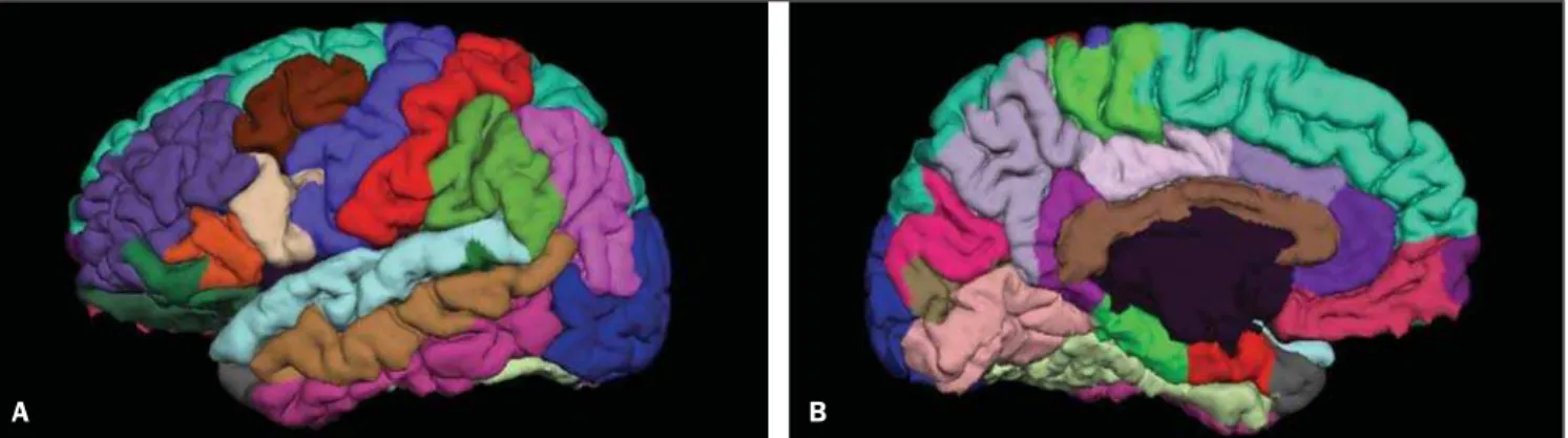
The MRI T1-weighted images for the study of cortical thickness were processed by means of the FreeSurfer v4.0.5 (Marti-nos Center, Boston, USA). Technical post-processing details were described in previ-ous publications(22,23). In summary, the images processing included the following steps: motion correction, removal of non-cerebral tissue, automated Talairach trans-formation, intensity normalization, sub-cortical white matter and deep grey matter and deep grey matter segmentation, corti-cal reconstruction and subdivision, brain insufflation and cortical thickness map-ping. Specifically for the present study the mean cortical thicknesses of the following regions in both hemispheres were evalu-ated: pre-frontal, lateral orbitofrontal, up-per frontal and caudal level of anterior cin-gulum (Figure 1).
The neuropsychological battery was ad-ministered by experienced and trained neu-ropsychologists. The evaluation was accom-plished in a single session of approximately 90 minutes. Furthermore, the standardized instruments selected to measure cognitive components of the executive functions, were the ones most frequently found in the literature on HIV-positive patients(24), such as, processing speed, inhibition, verbal flu-ency (initiation and verbal planning), cen-tral executive component of working memory, cognitive flexibility, organization, selection and strategies maintenance, among others. The neuropsychological in-struments administration sequence was planned in a way to minimize the effects of interference among them. Instruments with predominantly verbal tasks were alternated with non-verbal tasks, as follows:
1. Trail Making Test(25) – comprises two parts as follow: part (A) analyzes visual-motor coordination, processing speed and concentrated attention; part (B) besides the cognitive functions of part A, it also inves-tigates alternate attention, cognitive flex-ibility and inhibition.
2. Wisconsin Card Sorting Test(26) – Evaluates organization, planning, catego-rization, inhibition, cognitive flexibility and rules learning.
3. Hayling test(27) – Evaluates verbal ini-tiation (part A), verbal inhibition and pro-cessing speed (part B).
4. Digits subtest of the Wechsler Adult Intelligence Scale (WAIS-III)(28) – Analy-ses concentrated attention and short-term memory (direct order recall), besides the central executive component of working memory (indirect order recall).
5. Subtest of orthographic verbal flu-ency of Montreal Communication Assess-ment Battery (MAC Battery)(29) – investi-gates verbal initiation, inhibition, lexical memory and language.
6. Stroop Color and Word Test(30) – Veri-fies concentrated attention, inhibition, pro-cessing speed and cognitive flexibility.
7. Mini-Mental State Exam(31) – is a test
for cognitive screening of suggestive signs of dementia, employed for characterizing the sample.
Statistical analysis
The data obtained by neuroimaging and neuropsychological assessments were cor-related by the Pearson’s correlation coef-ficient, p ≤ 0.05. The Statistical Package for the Social Sciences (SPSS) 16.0 was used for this analysis.
RESULTS
Mean values and standard deviation of the cortical thickness in the different re-gions evaluated are shown on Table 1.
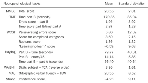
Table 2 shows mean scores and standard deviations of the neuropsychological per-formance observed in the executive func-tions tests.
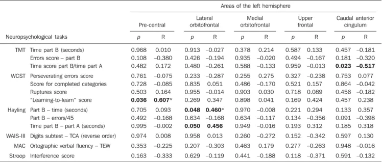
Results regarding the correlation be-tween cortical thickness and performance on instruments for assessing executive functions are shown on Tables 3 (right hemisphere) and 4 (left hemisphere).
Tables 3 and 4 demonstrate moderately to strongly significant positive correlation between Wisconsin test scores and cortical thickness in the pre-central and lateral orbitofrontal regions at right, and left pre-central region. Also, a significant negative correlation was observed between the Trail Making Test scores and the cortical thick-ness in the right pre-central region and left anterior caudal cingulum. Finally, moder-ately significant correlation was observed between Hayling Test scores and cortical thickness in the left lateral orbitofrontal region.
DISCUSSION
The present study investigated the asso-ciation between measures of cortical thick-ness of frontal and pre-frontal regions and performance scores on cognitive tasks that assessed executive functions of HIV-posi-tive patients. The hypothesis of an associa-tion between morphological brain mea-surements at MRI and neuropsychological measurements was confirmed, and
signifi-Figure 1. Lateral (a) and medial (b) views of left cerebral hemisphere, with segmentation of the different cerebral gyri with the aid of the FreeSurfer software.
Table 3 Results of correlational analysis of frontal cortical thickness with neuropsychological executive performance – right hemisphere. Areas of the right hemisphere
TMT WCST Hayling WAIS-III MAC Stroop Neuropsychological tasks
Time part B (seconds) Errors score – part B Time score part B/time part A
Perseverating errors score Score for completed categories Ruptures score
“Learning-to-learn” score
Part B – time (seconds) Part B – errors/45
Time part B – part A (seconds)
Digits subtest – TCA (reverse order)
Ortographic verbal fluency – TEW
Interference score Pre-central Lateral orbitofrontal Middle orbitofrontal Caudal anterior cingulum Upper frontal p 0.312 0.013 0.970 0.397 0.996 0.898 0.005 0.966 0.233 0.930 0.648 0.464 0.095 R –0.245 –0.556* –0.009 –0.206 0.001 0.032 0.755** –0.010 –0.287 –0.022 –0.112 –0.179 –0.394 p 0.723 0.672 0.806 0.287 0.702 0.707 0.039 0.929 0.072 0.842 0.104 0.270 0.696 R 0.087 –0.104 0.060 –0.257 –0.094 0.092 0.601* –0.022 –0.421 –0.049 –0.385 –0.266 –0.096 p 0.935 0.608 0.191 0.469 0.492 0.501 0.831 0.514 0.283 0.488 0.996 0.846 0.506 R –0.020 –0.126 –0.314 0.177 –0.168 0.165 0.069 0.160 –0.260 0.169 0.001 0.048 –0.163 p 0.761 0.353 0.828 0.070 0.272 0.585 0.208 0.090 0.061 0.077 0.192 0.778 0.124 R –0.075 –0.226 0.054 –0.425 0.266 0.134 0.391 0.400 –0.437 0.415 –0.313 –0.069 –0.366 p 0.307 0.593 0.068 0.630 0.756 0.991 0.459 0.135 0.253 0.089 0.114 0.951 0.908 R –0.247 –0.131 –0.427 –0.118 0.076 0.003 –0.237 0.356 –0.276 0.400 0.374 0.015 0.029
TMT, Trail Making Test; WCST, Wisconsin Card Sorting Test; WAIS-III, Wechsler Adult Intelligence Scale; TCA, Total of correct answers; MAC, Montreal Communication Assessment Battery; TEW, Total of evoked words. Note: * p = 0.05; ** p = 0.01.
Table 2 Mean and standard deviation of scores for performance in cognitive tasks. Neuropsychological tasks MMSE TMT WCST Hayling WAIS-III MAC Stroop Total score
Time part B (seconds) Errors score – part B Time score part B/time part A
Perseverating errors score Score for completed categories Ruptures score
“Learning-to-learn” score
Part B – time (seconds) Part B – errors/45
Time part B – part A (seconds)
Digits subtest – TCA (reverse order)
Ortographic verbal fluency – TEW
Interference score Mean 26.55 170.35 1.95 2.87 5.86 3.50 1.36 –0.59 79.77 14.14 56.40 3.95 20.55 –4.25 Standard deviation 2.01 85.04 3.92 1.28 12.62 2.15 1.32 9.63 40.61 5.85 40.64 1.61 8.52 9.11
MMSE, Mini-Mental State Exam; TMT, Trail Making Test; WCST, Wisconsin Card Sorting Test; WAIS-III, Wechsler Adult Intelligence Scale; TCA, Total of correct answers; MAC, Montreal Communication Assessment Battery; TEW, Total of evoked words.
Table 1 Mean and standard deviation of cortical thickness measurements by cerebral hemisphere and frontal areas. Area Right hemisphere Left hemisphere Pre-central Lateral orbitofrontal Middle orbitofrontal Upper frontal Caudal anterior cingulum
Pre-central Lateral orbitofrontal Middle orbitofrontal
Upper frontal Caudal anterior cingulum
Mean (mm) 2.24 2.36 2.12 2.43 2.23 2.26 2.39 2.18 2.47 2.35 Standard deviation 0.16 0.18 0.19 0.13 0.24 0.16 0.15 0.19 0.13 0.31
cant (both positive and negative) correla-tions were found.
With regard to correlations found in the Trail Making Test, the score that was nega-tively associated with cortical thickness of the left caudal anterior cingulate,was the time required to complete Part B of this task. Such index represents the measure-ment of the processing speed, connected with inhibition, cognitive flexibility and alternate attention. Thus, the longer the time required for HIV-positive patients to complete the part B of this test, the thinner was the cortical thickness of this region, suggesting that slowing down in the pro-cessing speed of these patients, may be related to the decrease in the thickness of the anterior third of the cingulate gyrus. Such region has been related to the execu-tive functions, particularly the inhibi-tion(32). As a complement, the score corre-sponding to the number of errors in the most complex part of the Trail Making Test was negatively correlated with the cortical thickness of the right pre-central region, indicating that the greater the amount of errors, the thinner is the cortical thickness of this area in the evaluated patients. Also, such region has frequently been associated with executive components, particularly in patients with personality disorders(33).
Negative correlations observed between performance in the Trail Making test part B and pre-frontal areas of the right hemi-sphere and caudal anterior cingulum, in the left hemisphere, are similar to some find-ings reported in the literature on the corre-lation between decrease in the cortical thickness and worsening of the processing speed(21,21). As regards the Stroop Color and Word test evaluating concentrated atten-tion, inhibiatten-tion, processing speed and cog-nitive flexibility, the present study did not observed any correlation between cortical thickness, particularly in the frontal cortex, and executive functions. However, this type of correlation was observed in a study with HIV-positive patients, but without using antiretroviral therapy(36).
Overall, the present study may be con-sidered as relatively pioneering at national level, with tangential investigations in the international literature. However, the present findings have a preliminary, explor-atory character. Thus, one must consider the limitations represented by the small size of the sample and the correlational delin-eation that did not include a control group of healthy individuals or a comparative group of HIV-positive patients with no antiretroviral treatment.
Another implication is the cross-sec-tional design of the study, which restricts comparisons of the cognitive deficits
pro-gression over time and also restricts a wealthy source of information of compar-ing the progression of HIV related cogni-tive deficits with the patient’s baseline.
Longitudinal investigations are required to evaluate how antiretroviral therapies affect white substance lesions in HIV-posi-tive patients(37), and also can more clearly demonstrate the association between fron-tal cortex atrophy and the executive func-tioning. Finally, another limitation involved the correlation between many cognitive and neuroanatomical variables which, for the purpose of the present preliminary study, demonstrate promising relationships that should be more deeply investigated and specified in future studies. Consider-ing that executive functions are critical in the daily life, further studies in this area may guide practitioners not only in their clinical practice, but also in the research of effective therapies to reduce cognitive defi-cits in HIV-positive individuals. Such interventional actions with both, medica-tions and at cognitive level, may contrib-ute to prevent or minimize executive func-tions impairments before the progression to dementia associated with HIV infection.
Finally, based on the results of the present study with 22 HIV-positive pa-tients, a correlation can be observed be-tween frontal cortical thickness and perfor-mance in neuropsychological executive Table 4 Results of correlational analysis of frontal cortical thickness with neuropsychological executive performance – left hemisphere.
Areas of the left hemisphere
TMT
WCST
Hayling
WAIS-III
MAC
Stroop
Neuropsychological tasks
Time part B (seconds) Errors score – part B Time score part B/time part A
Perseverating errors score Score for completed categories Ruptures score
“Learning-to-learn” score
Part B – time (seconds) Part B – errors/45
Time part B – part A (seconds)
Digits subtest – TCA (reverse order)
Ortographic verbal fluency – TEW
Interference score
Pre-central
Lateral orbitofrontal
Medial orbitofrontal
Caudal anterior cingulum Upper
frontal
p
0.968 0.108 0.482
0.761 0.728 0.503 0.036 0.705 0.492 0.995
0.974
0.353
0.163 R
0.010 –0.380
0.172
–0.075 –0.085 0.164 0.607*
0.093 –0.168 –0.002
0.008
–0.225
–0.333
p
0.913 0.426 0.480
0.233 0.835 0.955 0.269
0.048 0.634 0.050 0.958
0.207
0.629 R
–0.027 –0.194 –0.261
–0.287 0.051 –0.014
0.347
0.460* –0.168 0.456 0.013
–0.303
–0.119
p
0.378 0.935 0.588
0.255 0.486 0.903 0.898
0.970 0.634 0.949
0.260
0.463
0.441 R
0.214 –0.020 –0.133
0.275 –0.170
0.030 0.041
–0.008 –0.117 –0.016
–0.272
0.179
–0.188
p
0.587 0.494 0.959
0.327 0.521 0.718 0.169
0.221 0.134 0.193
0.152
0.277
0.118 R
0.133 –0.167 –0.013
–0.238 0.157 0.089 0.424
0.294 –0.356 0.312
–0.342
–0.263
–0.371
p
0.457 0.181 0.023 0.753 0.864 0.456 0.457
0.133 0.091 0.185
0.597
0.948
0.591 R
–0.181 –0.320 –0.517 0.077 –0.042 –0.182 0.238
0.357 –0.398
0.318
0.130
–0.016
–0.132
TMT, Trail Making Test; WCST, Wisconsin Card Sorting Test; WAIS-III, Wechsler Adult Intelligence Scale; TCA, Total of correct answers; MAC, Montreal Communication Assessment Battery; TEW, Total of evoked words. Note: * p = 0.05; ** p = 0.01.
of the Trail Making Test, that the learning strategies scores were positively correlated with the cortical thickness of the pre-central region (bilateral). This may suggest that the HIV-positive patients’ difficulty in learning with the previously employed strategy, as well as, their difficulty in solving problems (learning-to-learn score)(34) must be related to the decrease in the cortical thickness of this region. Additionally to this correlation, a positive association was observed be-tween this score and the right lateral orbitofrontal thickness, a region that is re-lated to executive components measured by the learning-to-learn score(35).
functions tests. The correlations demon-strated in the present study suggest that executive deficits in HIV-positive patients are related to a decrease in the cortical thickness of several frontal regions, most significantly in the left hemisphere. Posi-tive correlations indicating increase in the thickness at some regions may be related to the neuronal plasticity in HIV pa-tients(38). Further studies are required to corroborate such results, demonstrating more clearly evidences of the interface between neuroradiology and neuropsychol-ogy in the setting of neurodegenerative disorders related to HIV infection.
Acknowledgements
To the following research funding insti-tutions: Coordenação de Aperfeiçoamento de Pessoal de Nível Superior (Capes), Con-selho Nacional de Desenvolvimento Cien-tífico e Tecnológico (CNPq) and Fundação de Amparo à Pesquisa do Estado do Rio de Janeiro (Faperj).
REFERENCES
1. Christo PP. Alterações cognitivas na infecção pelo HIV e AIDS. Rev Assoc Med Bras. 2010;56:242– 7.
2. Bottiggi KA, Chang JJ, Schmitt FA, et al. The HIV dementia scale: predictive power in mild demen-tia and HAART. J Neurol Sci. 2007;260:11–5. 3. Cysique LA, Brew BJ. Neuropsychological
func-tioning and antiretroviral treatment in HIV/AIDS: a review. Neuropsychol Rev. 2009;19:169–85. 4. Woods SP, Moore DJ, Weber E, et al. Cognitive
neuropsychology of HIV-associated cognitive dis-orders. Neuropsychol Rev. 2009;19:152–68. 5. World Health Organization. Global summary of
the HIV/AIDS epidemic, December 2008. [aces-sado em 26 de outubro de 2010]. Disponível em: http://www.who.int/hiv/data/2009_global_ summary.gif
6. World Health Organization. Epidemological fact sheet on HIV and AIDS: core data on epidemiol-ogy and response – Brazil, December 2008. [acessado em 26 de outubro de 2010]. Disponível em: http://www.who.int/countries/bra/en/ 7. Tozzi V, Balestra P, Bellagamba R, et al.
Persis-tence of neuropsychologic deficits despite long-term highly active antiretroviral therapy in pa-tients with HIV-related neurocognitive impair-ment: prevalence and risk factors. J Acquir Im-mune Defic Syndr. 2007;45:174–82.
8. Deeks SG. HIV infection, inflammation, immuno-senescence, and aging. Annu Rev Med. 2011;62: 141–55.
9. Brew BJ, Crowe SM, Landay A, et al. Neuro-degeneration and ageing in the HAART era. J Neuroimmune Pharmacol. 2009;4:163–74. 10. Kochunov P, Robin DA, Royall DR, et al. Can
structural MRI indices of cerebral integrity track cognitive trends in executive control function during normal maturation and adulthood? Hum Brain Mapp. 2009;30:2581–94.
11. Sjöbeck M, Elfgren C, Larsson EM, et al. Alzheimer’s disease (AD) and executive dysfunc-tion. A case-control study on the significance of frontal white matter changes detected by diffusion tensor imaging (DTI). Arch Gerontol Geriatr. 2010;50:260–6.
12. Thompson PM, Dutton RA, Hayashi KM, et al. Thinning of the cerebral cortex visualized in HIV/ AIDS reflects CD4+ T lymphocyte decline. Proc Natl Acad Sci U S A. 2005;102:15647–52. 13. Chiang MC, Dutton RA, Hayashi KM, et al. 3D
pattern of brain atrophy in HIV/AIDS visualized using tensor-based morphometry. Neuroimage. 2007;34:44–60.
14. Gunning-Dixon FM, Murphy CF, Alexopoulos GS, et al. Executive dysfunction in elderly bipo-lar manic patients. Am J Geriatr Psychiatry. 2008; 16:506–12.
15. Melrose RJ, Tinaz S, Castelo JMB, et al. Com-promised fronto-striatal functioning in HIV: an fMRI investigation of semantic event sequencing. Behav Brain Res. 2008;188:337–47. 16. Stuss DT, Levine B. Adult clinical
neuropsychol-ogy: lessons from studies of the frontal lobes. Annu Rev Psychol. 2002;53:401–33. 17. Dawes S, Suarez P, Casey CY, et al. Variable
pat-terns of neuropsychological performance in HIV-1 infection. J Clin Exp Neuropsychol. 2008;30: 613–26.
18. Collette F, Van der Linden M, Laureys S, et al. Exploring the unity and diversity of the neural substrates of executive functioning. Hum Brain Mapp. 2005;25:409–23.
19. Collette F, Hogge M, Salmon E, et al. Exploration of the neural substrates of executive functioning by functional neuroimaging. Neuroscience. 2006;139:209–21.
20. Harrison MJG, Newman SP, Hall-Craggs MA, et al. Evidence of CNS impairment in HIV infection: clinical, neuropsychological, EEG, and MRI/ MRS study. J Neurol Neurosurg Psychiatry. 1998;65:301–7.
21. Poutiainen E, Elovaara I, Raininko R, et al. Cog-nitive performance in HIV-1 infection: relation-ship to severity of disease brain atrophy. Acta Neurol Scand. 1993;87:88–94.
22. Fischl B, Salat DH, Busa E, et al. Whole brain segmentation: automated labeling of neuroana-tomical structures in the human brain. Neuron. 2002;33:341–55.
23. Desikan RS, Ségonne F, Fischl B, et al. An auto-mated labeling system for subdividing the human cerebral cortex on MRI scans into gyral based re-gions of interest. Neuroimage. 2006;31:968–80. 24. Rippeth JD, Heaton RK, Carey CL, et al.
Meth-amphetamine dependence increases risk of neupsychological impairment in HIV infected persons. J Int Neuropsychol Soc. 2004;10:1–14. 25. Fonseca RP, Grassi-Oliveira R, Oliveira CR, et al. Instruments of executive functions assessment: preliminary normative data and sociodemo-graphic studies. Dement Neuropsychol. In Press. 26. Cunha JA, Trentini CM, Argimon IL, et al. Teste Wisconsin de classificação de cartas – adaptação e padronização brasileira. São Paulo, SP: Casa do Psicólogo; 2005.
27. Fonseca RP, Oliveira CR, Gindri G, et al. Teste Hayling: um instrumento de avaliação de compo-nentes das funções executivas. In: Hutz CS, or-ganizador. Avanços em avaliação psicológica e neuropsicológica de crianças e adolescentes. 1ª ed. São Paulo, SP: Casa do Psicólogo; 2010. p. 337–64.
28. Nascimento E. Adaptação, validação e normati-zação do WAIS-III para uma amostra brasileira. In: Wechsler D. WAIS-III: manual para adminis-tração e avaliação. São Paulo, SP: Casa do Psi-cólogo; 2004. p. 161–92.
29. Fonseca RP, Parente MAMP, Cote H, et al. Bate-ria Montreal de avaliação da comunicação – Ba-teria MAC. Barueri, SP: Pró-Fono; 2008. 30. Strauss E, Sherman EMS, Spreen O. Executive
functions. In: Strauss E, Sherman EMS, Spreen O, editors. A compendium of neuropsychological tests: administration, norms, and commentary. 3rd ed. New York, NY: Oxford University Press; 2006. p. 401–545.
31. Almeida OP. Mini exame do estado mental e o diagnóstico de demência no Brasil. Arq Neu-ropsiquiatr. 1998;56:605–12.
32. Hoerst M, Weber-Fahr W, Tunc-Skarka N, et al. Correlation of glutamate levels in the anterior cin-gulate cortex with self-reported impulsivity in patients with borderline personality disorder and healthy controls. Arch Gen Psychiatry. 2010;67: 946–54.
33. Swick D, Jovanovic J. Anterior cingulate cortex and the Stroop task: neuropsychological evidence for topographic specificity. Neuropsychologia. 2002;40:1240–53.
34. Szbot CM, Eizirik M, Cunha RD, et al. Neuro-imagem no transtorno de déficit de atenção/hipe-ratividade. Rev Bras Psiquiatr. 2001;23(Supl I): 32–5.
35. Keller M, Werlang BSG. Flexibilidade na resolu-ção de problemas em tentadores de suicídio. J Bras Psiquiatr. 2005;54:128–36.
36. Chang L, Ernst T, Witt MD, et al. Relationships among brain metabolites, cognitive function, and viral loads in antiretroviral-naïve HIV patients. Neuroimage. 2002;17:1638–48.
37. Li X, Lu ZL, D’Argembeau A, et al. The Iowa Gambling Task in fMRI images. Human Brain Mapp. 2010;31:410–23.