13
Radiol Bras. 2011 Jan/Fev;44(1):13–19
Computed tomography findings of pulmonary tuberculosis
in adult AIDS patients
*
Aspectos tomográficos da tuberculose pulmonar em pacientes adultos com AIDS
Lanamar Aparecida de Almeida1, Mario Flores Barba2, Fernando Alves Moreira3, Sidney Bombarda4, Sebastião André de Felice5, Edenilson Eduardo Calore6
Objective: The present study is aimed at describing computed tomography findings pulmonary tuberculosis in adult AIDS patients assisted at a radiology unit of a reference infectious diseases hospital, in an attempt to establish the association between such findings and CD4 count. Materials and Methods: Forty-five patients were evaluated by chest computed tomography over a four-year period. Results: Mediastinal and/or hilar lymph node enlargement was found in 31 (68.8%) cases, pleural effusion in 29 (64.4%), centrilobular nodules with segmental distribution in 26 (57.7%), consolidation in 24 (53.3%), confluent micronodules in 17 (37.7%), poorly defined nodules with centrilobular distribution in 16 (35.5%), tree-in-bud pattern in 13 (28.9%), bronchial wall thickening in 12 (26.6%), thick-walled cavity in 10 (22.2%), miliary nodules in 9 (20%), and cylindrical bronchiectasis in 6 (13.3%). Among the 45 patients, 35 (77.8%) presented CD4 count < 200 cel/mm3 and 10 (22.2%) presented CD4 count ≥ 200 cel/mm3. Conclusion: Differently from reports in the literature, the authors conclude that mediastinal and/or hilar lymph node enlargement and consolidation were significantly most frequent in patients with CD4 count ≥ 200 cel/mm3. However, lymph nodes with hypodense center were most often observed in severely immunosuppressed patients with CD4 count < 200 cel/mm3.
Keywords: Tuberculosis; Tomography; Chest; HIV; AIDS; SIDA.
Objetivo: Este trabalho tem como finalidade descrever os achados tomográficos da tuberculose pulmonar em pacien-tes adultos com AIDS atendidos no serviço de radiologia de um hospital de referência em doenças infecciosas, procu-rar associações desses achados e a contagem de CD4. Materiais e Métodos: Foram estudados 45 pacientes por meio de tomografia computadorizada de tórax durante quatro anos. Resultados: Foram encontrados linfonodomega-lia mediastinal e/ou hilar em 31 (68,8%) dos casos, derrame pleural em 29 (64,4%), nódulos centrolobulares de dis-tribuição segmentar em 26 (57,7%), consolidação em 24 (53,3%), confluência de micronódulos em 17 (37,7%), nódulos mal definidos com distribuição centrolobular em 16 (35,5%), padrão de “árvore em brotamento” em 13 (28,9%), espessamento de parede brônquica em 12 (26,6%), cavidade de parede espessa em 10 (22,2%), nódulos miliares em 9 (20%) e bronquiectasias cilíndricas em 6 (13,3%). Dos 45 pacientes, 35 (77,8%) apresentaram CD4 < 200 cel/mm3
e 10 (22,2%) apresentaram CD4 ≥ 200 cel/mm3
. Conclusão: Concluímos que neste estudo, diversamente do descrito na literatura, linfonodomegalia mediastinal e/ou hilar e consolidação foram significativamente mais fre-quentes em pacientes com CD4 ≥ 200 cel/mm3
. No entanto, linfonodos com centro hipodenso foram mais frequen-temente observados em pacientes com severa imunodepressão, ou seja, CD4 < 200 cel/mm3
.
Unitermos: Tuberculose; Tomografia; Tórax; HIV; AIDS; SIDA.
Abstract
Resumo
* Study developed at Instituto de Infectologia “Emílio Ribas” (IIER), São Paulo, SP, Brazil.
1. Master, Director, Department of Diagnostic and Therapeu-tic Support of Instituto de Infectologia “Emílio Ribas” (IIER), São Paulo, SP, Brazil.
2. PhD, MD, Director, Unit of Imaging Diagnosis and Graphic Methods of Instituto de Infectologia “Emílio Ribas” (IIER), São Paulo, SP, Brazil.
3. PhD, MD, Radiologist responsible for the Unit of Imaging Diagnosis at Hospital Paulistano, São Paulo, SP, Brazil.
4. PhD, MD, Physician Assistant at Division of Pneumology, Hospital das Clínicas da Faculdade de Medicina da Universidade de São Paulo (HC-FMUSP), São Paulo, SP, Brazil.
5. Private Docent, Technical Director, Instituto de Infectologia “Emílio Ribas” (IIER), São Paulo, SP, Brazil.
6. Private Docent, Head of Sector at the Service of Pathologi-cal Anatomy, Instituto de Infectologia “Emílio Ribas” (IIER), São Paulo, SP, Brazil.
Almeida LA, Barba MF, Moreira FA, Bombarda S, Felice SA, Calore EE. Computed tomography findings of pulmonary tuberculosis in adult AIDS patients. Radiol Bras. 2011 Jan/Fev;44(1):13–19.
INTRODUCTION
Tuberculosis is one of the most com-mon complications associated with infec-tion by the human immunodeficiency virus (HIV) worldwide(1). AIDS promotes pro-gression from latent to active disease in co-infected patients(2). In co-infected
individu-als, active tuberculosis development index is 10 to 30 times higher than in individu-als infected by Mycobacterium tuberculo-sis alone(3).
Chest radiography is the imaging method of choice in the initial evaluation and follow-up of pulmonary tuberculo-sis(4). However, in 6% to 20% of HIV-posi-tive patients with pulmonary tuberculosis conventional radiography may present as absolutely normal(5).
Many studies have described radiologi-cal manifestations of tuberculosis(5–11). Lee Mailing Address: Dra. Lanamar Aparecida de Almeida. Avenida
Doutor Arnaldo, 165, Pacaembu. São Paulo, SP, Brazil, 01246-900. E-mail: [email protected]
et al.(12) have investigated the usefulness of computed tomography (CT) in the differ-entiation between active and inactive tuber-culosis. Pereira et al.(13) have described the tomographic findings of primary pulmo-nary tuberculosis initially manifested as lobar consolidation. In 89 patients with active tuberculosis evaluated with thin-slice CT, the following findings were most frequently observed: centrilobular linear opacities (92%), lobular consolidation (62%), acinar nodule (61%), cavity (36%) and ground glass opacity (35%). In another study developed by Hatipo—lu et al.(14) with thin-slice CT, “tree-in-bud” pattern, nod-ules of up to 8 mm in diameter and consoli-dations were frequently found in patients with active disease and in none of the pa-tients with inactive disease.
The presence of cavity is a relevant sign of active disease. High-resolution CT (HRCT) demonstrates small cavities inter-mingled with consolidations that many times are not visible at conventional radi-ography(4). A study developed by Im et al.(15) has demonstrated that the prevalence of cavities at tomography was 58% (24/41), while, at plain chest radiography, the preva-lence was 22% (9/41). According to Leung et al., thick-walled cavities are observed in up to 76% of the patients with pulmonary tuberculosis at the time of the diagnosis(16). Centrilobular nodules with segmental distribution representative of bronchogenic dissemination of tuberculosis are also fre-quently observed in the active phase of the disease (82% to 100%)(16). Bronchial walls thickening occurs in 62% of the cases and bronchiectasis is observed in 23% of the patients. The “tree-in-bud” pattern is present in up to 57% of cases(17).
Comparative studies evaluating tomo-graphic findings of pulmonary tuberculo-sis in both HIV-positive and HIV-negative patients are not so frequently found in the literature(18–20). As compared with the HIV-negative population, AIDS patients present greater probability of ganglia involvement, bronchial dissemination, miliary disease, extrapulmonary disease, and normal plain chest radiography(21).
The radiological findings of tuberculo-sis and HIV/AIDS co-infection are depen-dent upon the immune status of the pa-tient(22). In HIV-positive patients with CD4
count < 200 cel/mm3, the radiographic
findings are those typically described in the primary presentation of the infection(22), while in HIV-positive patients with CD4 count ≥ 200 cel/mm3, the tomographic
findings of tuberculosis are those typically observed in cases of disease reactiva-tion(22).
The present study describes the tomo-graphic findings of pulmonary tuberculo-sis in adult patients admitted to an infec-tious diseases reference hospital, in an at-tempt to establish associations between tomographic findings of pulmonary tuber-culosis and CD4 count in adult AIDS pa-tients. The study was initiated after ap-proval by the Committee for Ethics in Re-search of Instituto de Infectologia “Emílio Ribas” (IIER), under Research Protocol No. 47/06 and authorization No. 390/2006.
MATERIALS AND METHODS
The present study included cases of tu-berculosis in HIV-positive adult patients admitted to Instituto de Infectologia “Emí-lio Ribas” and notified by the Epidemiol-ogy Service between January/2003 and November/2006, according to the follow-ing inclusion criteria:
a) HIV-positive at Western Blot and classified as AIDS (categories C and D, according to CDC – Center for Disease Control and Prevention of the United States of America; the most recent classification was issued in 1993);
b) adult inpatients (>18 years of age); c) positive sputum culture for M. tuber-culosis;
d) patients who underwent chest CT by the moment of the diagnosis, or up to 60 days before or 30 days after starting the treatment.
Chest CT
The chest CT images included in the present study were requested by the pa-tients’ attending physicians, for diagnosis or follow-up. All the examinations were performed in a Light-Speed multislice eight-channels equipment (General Elec-tric Medical Systems; Milwaukee, WI, USA), manufactured in 2002, with the pa-tient in dorsal decubitus, and intravenous non-ionic iodinated contrast medium
injec-tion with an automated contrast infusion system. The image sections were acquired at the end of inspiration, from the apex to the diaphragm, utilizing the standard tech-nique in the majority of the patients and, in some cases, the high resolution technique (HRCT). The tomographic images were reviewed by the authors, and the pulmonary findings patterns were described according to the “Brazilian Consensus on Terminol-ogy Used to Describe Computed Tomog-raphy of the Chest”(23).
Statistical analysis
The following data were included in a databank: age distribution, time of HIV virus infection, interval between the dis-ease diagnosis and the CT scan, death, CD4 levels (< 200 cel/mm3 and ≥ 200 cel/mm3),
associated diseases, tuberculosis treatment and antiretroviral therapy. The data were fed into a database built with the software SPSS 14.0 for Windows, version 14.0.1 of November 18, 2005, and treated by means of descriptive statistics, with calculation of percentages, means, frequencies and stan-dard deviation. The association between CD4 levels and radiological findings was evaluated by the exact Fisher’s test; consid-ering bilateral tests with statistical signifi-cance level of 5% or 0.05 (Table 1).
RESULTS
Among 300 HIV-positive inpatients with tuberculosis during the study period, only 70 had undergone chest CT, and only 45 of these patients met the above men-tioned criteria.
Of these 45 patients, 35 presented CD4 count < 200 cel/mm3 [30 of men (85.7%)
and 5 women (14.3%)]. The remaining 10 patients presented CD4 count ≥ 200 cel/ mm3, with nine (90%) being men and one
(10%) women.
The mean age of the 45 patients was 38 years and nine months, with a standard deviation of 9.6.
Among the cases with CD4 count < 200 cel/mm3, the mean age was 38 years and
seven months, with a standard deviation of 9.0, while in the cases with CD4 count ≥ 200 cel/mm3, the mean age was 40years
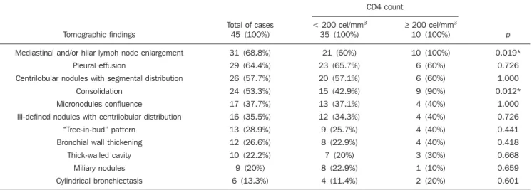
Table 1 Frequency of tomographic findings in 45 cases of pulmonary tuberculosis in adult HIV-positive patients, with identification of CD4 count associated with the findings and respective frequencies.
CD4 count
Tomographic findings
Mediastinal and/or hilar lymph node enlargement
Pleural effusion
Centrilobular nodules with segmental distribution
Consolidation
Micronodules confluence
Ill-defined nodules with centrilobular distribution
“Tree-in-bud” pattern
Bronchial wall thickening
Thick-walled cavity
Miliary nodules
Cylindrical bronchiectasis
Total of cases 45 (100%)
31 (68.8%)
29 (64.4%)
26 (57.7%)
24 (53.3%)
17 (37.7%)
16 (35.5%)
13 (28.9%)
12 (26.6%)
10 (22.2%)
9 (20%)
6 (13.3%)
< 200 cel/mm3
35 (100%)
21 (60%)
23 (65.7%)
20 (57.1%)
15 (42.9%)
13 (37.1%)
12 (34.3%)
9 (25.7%)
8 (22.9%)
7 (20%)
8 (22.9%)
4 (11.4%)
≥ 200 cel/mm3
10 (100%)
10 (100%)
6 (60%)
6 (60%)
9 (90%)
4 (40%)
4 (40%)
4 (40%)
4 (40%)
3 (30%)
1 (10%)
2 (20%)
p
0.019*
0.726
1.000
0.012*
1.000
0.726
0.441
0.418
0.668
0.659
0.601
*p < 0.05 was considered as statistically significant.
Figure 1.Homogeneous lymph node enlargement – Chest CT. Homogeneous right inferior paratracheal, retrotracheal, and para-aortic, lymph node enlarge-ments, associated with opacity with air bronchogram in a view of the apicoposterior segment of the left upper lobe.
Figure 2. Hypodense lymph node enlargement – Chest CT. Right lower paratracheal, confluent retrotracheal lymph node enlargement, with tomo-graphic signs of necrosis in a view of para-aortic ganglionar chain, associated with small bilateral pleural effusion and upper left pulmonary consolidation.
Surprisingly, none of the patients in the present study had undergone regular treat-ments for tuberculosis or AIDS.
Tomographic findings
Table 1 shows the frequency of tomo-graphic findings in decreasing order and respective association with CD4 counts of the 45 cases.
Mediastinal and/or hilar lymph node enlargement (Figures 1 and 2)
Thirty-one (68.8%) of the patients pre-sented mediastinal and/or hilar lymph node enlargement. Among these patients, 21 (67.7%) presented CD4 count < 200 cel/ mm3, while 10 (32.3%) presented CD4
count ≥ 200 cel/mm3.
Based on the total number of patients with CD4 count< 200 cel/mm3 (35) and
with CD4 count ≥ 200 cel/mm3 (10), the
authors observed that, respectively, 21 (60%) of the 35 patients presented medi-astinal and/ or hilar lymph node enlarge-ment, while 10 (100%) of 10 patients pre-sented such tomographic finding.
Pleural effusion
Twenty nine (64.4%) patients presented pleural effusion. Of these patients, 23 (79.3%) presented CD4 count < 200 cel/ mm3, while 6 (20.7%) presented CD4 count
≥ 200 cel/mm3.
Based on the total number of patients with CD4 count< 200 cel/mm3 (35) and with
CD4 count ≥ 200 cel/mm3 (10), the authors
observed that, respectively, 23 (65.7%) of the 35 (60.0%) patients presented pleural effusion and 6 (60%) of the 10 patients presented such tomographic finding.
Among the 29 cases with pleural effu-sion, 20 (68.9%) were bilateral and with small volume.
In none of the cases was pleural effusion the sole radiological finding. The CD4 level ranged from 6 cel/mm3 to 540 cel/mm3
(mean = 165 cel/mm3).
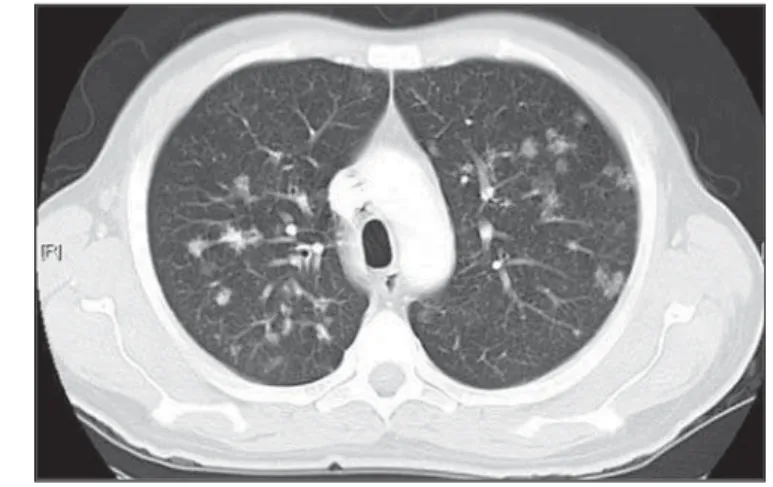
Centrilobular nodules with segmental distribution (Figure 3)
Twenty-six (57.7%) patients presented centrilobular nodules with segmental dis-tribution. Of these patients, 20 (76.9%) presented CD4 count < 200 cel/mm3, while
6 (23.1%) presented CD4 count ≥ 200 cel/ mm3.
Based on the total number of patients with CD4 count< 200 cel/mm3 (35) and
with CD4 count ≥ 200 cel/mm3 (10), the
(57.1%) of the 35 patients presented centrilobular nodules with segmental dis-tribution and 6 (60%) of the 10 patients presented such tomographic finding.
It is important to note that, in spite of the absolute number of cases with centrilobular nodules of segmental distribution being smaller in patients with CD4 count ≥ 200 cel/mm3, the percentage of patients with
this tomographic finding is higher in this group (60% of the cases).
Parenchymal consolidation (Figure 4)
Twenty-four (53.3%) patients presented parenchymal consolidation. Of these pa-tients, 15 (62.5%) presented CD4 count < 200 cel/mm3, while 9 (37.5%) presented
CD4 count ≥ 200 cel/mm3.
Based on the total number of patients with CD4 count< 200 cel/mm3 (35) and
with CD4 count ≥ 200 cel/mm3 (10), the
authors observed that, respectively, 15 (42.5%) of the 35 patients presented
paren-chymal consolidations, and 9 (90%) of the 10 patients presented such tomographic finding.
As regards topographic distribution of the parenchymal consolidations, predomi-nance in the upper lobes was observed.
It can be perceived that, although the absolute number of cases with parenchy-mal consolidation is sparenchy-maller in patients with CD4 count ≥ 200 cel/mm3, the
per-centage of patients with this tomographic finding in higher in this group (90% of the cases).
Micronodular confluence
Seventeen (37.7%) patients presented micronodular confluence. Of these pa-tients, 13 (76.5%) presented CD4 count < 200 cel/mm3 and 4 (23.5%) presented CD4
count ≥ 200 cel/mm3.
Based on the total number of patients with CD4 count< 200 cel/mm3 (35) and
with CD4 count ≥ 200 cel/mm3 (10), the
authors observed that, respectively, 13 (37.1%) of the 35 patients presented micro-nodular confluence, and 4 (40%) of the 10 patients presented such tomographic find-ing.
It can be perceived that, although the absolute number of cases with micronod-ular confluence is smaller in patients with CD4 count ≥ 200 cel/mm3, the percentage
of patients with this tomographic finding in higher in this group (40% of the cases).
Ill defined nodules with centrilobular distribution (Figure 5)
Sixteen (35.5%) patients presented ill defined nodules with centrilobular distribu-tion. Of these patients, 12 (75%) presented CD4 count < 200 cel/mm3 and 4 (25%),
CD4 ≥ 200 cel/mm3.
Based on the total number of patients with CD4 count< 200 cel/mm3 (35) and
with CD4 count ≥ 200 cel/mm3 (10), the
authors observed that, respectively, 12
Figure 3.Centrilobular nodules with segmental distribution – Chest CT. Centrilobular nodules with segmental distribution, confluent in the posterior segment of the right upper lobe. Extensive consolidation in the upper left lobe is observed.
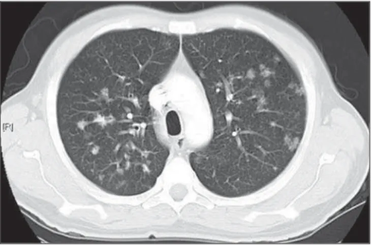
Figure 4.Segmental consolidation in the anterior segment of the right upper lobe – Chest CT. Segmental consolidation in the anterior segment of the right upper lobe caused by the confluence of multiple centrilobular nodules. No-tice centrilobular nodules of segmental distribution in the posterior segment of the right inferior lobe and inferior lingular segment of the left lung.
Figure 5.Ill defined nodules with centrilobular distribution – Chest CT. Bilat-eral, ill defined nodules with centrilobular distribution.
Figure 5 Figure 3
(34.3%) of the 35 patients presented ill defined nodules with centrilobular distribu-tion, and 4 (40%) of the 10 patients pre-sented such tomographic finding.
It can be perceived that, although the absolute number of cases with nodules is lower in patients with CD4 count ≥ 200 cel/ mm3, the percentage of patients with this
tomographic finding is higher in this group (40% of the cases).
“Tree-in-bud” pattern (Figure 6)
Thirteen (28.9%) of the patients pre-sented the “tree-in-bud” pattern. Of these patients, 9 (69.2%) presented CD4 count < 200 cel/mm3, while 4 (30.8%) presented
CD4 count ≥ 200 cel/mm3.
Based on the total number of patients with CD4 count< 200 cel/mm3 (35) and
with CD4 count ≥ 200 cel/mm3 (10), the
authors observed that, respectively, 9 (25.7) of the 35 patients presented the “tree-in-bud” pattern, and 4 (40%) of the 10 patients presented such tomographic finding.
It can be perceived that, although the absolute number of cases with the “tree-in-bud” pattern is smaller in patients with CD4 count ≥ 200 cel/mm3, the percentage
of patients with this tomographic finding is higher in this group (40% of the cases).
Bronchial wall thickening
Twelve (26.6%) patients presented bronchial wall thickening. Of these pa-tients, 8 (66.72%) presented CD4 count < 200 cel/mm3, while 4 (33.3%) presented
with CD4 count ≥ 200 cel/mm3.
Based on the total number of patients with CD4 count< 200 cel/mm3 (35) and
with CD4 count ≥ 200 cel/mm3 (10), the
authors observed that, respectively, 8 (22.9%) of the 35 patients presented bron-chial wall thickening, and 4 (40%) of the 10 patients presented such tomographic finding.
It can be perceived that, although the absolute number of cases with bronchial wall thickening is smaller in patients with CD4 count ≥ 200 cel/mm3, the percentage
of patients with this tomographic finding is higher in this group (40% of the cases).
Thick-walled cavity (Figure 7)
Ten (22.2%) patients presented thick-walled cavity. Among them, 7 (70%) pre-sented CD4 count < 200 cel/mm3 and 3
(30%), CD4 ≥ 200 cel/mm3.
Based on the total number of patients with CD4 count< 200 cel/mm3 (35) and
with CD4 count ≥ 200 cel/mm3 (10), the
authors observed that, respectively, 7 (20%) of the 35 patients presented thick-walled cavity, and 3 (30%) of the 10 pa-tients presented such tomographic find-ing.
The upper right lobe, with four cases (40%), was the most frequent site with this finding.
It can be perceived that, although the absolute number of cases with thick walled cavity is smaller in patients with CD4 count ≥ 200 cel/mm3, the percentage of patients
with this tomographic finding is higher in this group (30% of the cases).
Miliary nodules (randomly distributed small nodules)
Nine (20%) patients presented miliary nodules. Of these patients, 8 (88.9%) pre-sented CD4 count < 200 cel/mm3, while 1
(11.1%) presented CD4 ≥ 200 cel/mm3.
Based on the total number of patients with CD4 count< 200 cel/mm3 (35) and
with CD4 count ≥ 200 cel/mm3 (10), the
authors observed that, respectively, 8 (22.9%) of the 35 patients presented mil-iary nodules, and 1 (10%) of the 10 patients presented such tomographic finding.
Cylindrical bronchiectasis
Six (13.3%) patients presented cylindri-cal bronchiectasis. Of these patients, four (66.7%) presented CD4 count < 200 cel/ mm3 and two (33.3%), CD4 count ≥ 200
cel/mm3.
Based on the total number of patients with CD4 count< 200 cel/mm3 (35) and with
CD4 count ≥ 200 cel/mm3 (10), the authors
observed that, respectively, 4 (11.4%) of the 35 patients presented cylindrical bron-chiectasis, and 2 (20%) of the 10 patients presented such tomographic finding.
As regards topographic distribution of the cylindrical bronchiectasis, one ob-served a predominance of this finding in the lower lobes (four cases - 66.6%).
It can be perceived that, although the absolute number of cases with cylindrical bronchiectasis is smaller in patients with CD4 count ≥ 200 cel/mm3, the percentage
of patients with this tomographic finding is higher in this group (20% of the cases).
Figure 7.Thick walled cavity in the upper right lobe – Chest CT. Thick walled cavity in the posterior segment of the right upper lobe. Centrilobular nodules also suggestive of disease activity are observed.
DISCUSSION
According to Castañer et al.(22), the ra-diological manifestations of tuberculosis in AIDS reflect the level of immunosuppres-sion. Patients with tuberculosis and CD4 count ≥ 200 cel/mm3 present radiological
findings similar to those observed in HIV-negative patients. HIV-positive patients with CD4 count < 200 cel/mm3 tend to
present radiological findings similar to the ones of primary pulmonary tuberculosis(22). Lymph node enlargement is the radio-logical hallmark of primary tuberculosis, and its prevalence decreases with age, be-ing reported with a much lower frequency in adults(16). On the other hand, lymph node enlargement is commonly seen in tubercu-losis in AIDS patients. More than 60% of the AIDS patients with tuberculosis present hilar and mediastinal lymph node enlarge-ment(22). Laissy et al.(18), in a comparative study approaching the use of HRCT among HIV-positive and HIV-negative patients, have demonstrated that the most frequent abnormality in HIV-positive patients was lymph node enlargement. In HIV-positive patients, lymph node enlargement may present peripheral contrast medium uptake, which is radiologically translated into a hypodense center and peripheral enhance-ment(21,24). The study developed by Im et al.(15) reports that the hypodense area cor-responds to necrosis, and the peripheral enhancement of the lymph node enlarge-ment limits results from the hypervascu-larization secondary to the inflammatory process. In the present 45-case study, 68.8% of the patients presented mediasti-nal or mediastimediasti-nal/hilar lymph node en-largement, while hypodensity in lymph node areas after intravenous contrast injec-tion was observed in seven cases (15%). All these patients presented CD4 count < 200 cel/mm3. Based on such observations, it is
possible to infer that this lymph node en-largement pattern is not pathognomonic of tuberculosis, but, the presence of this find-ing indicates severe immunosuppression. In the study developed by Hulnick et al.(25) with HIV-negative patients, cavities
were the parenchymal changes most com-monly associated with pleural effusion. In none of the cases was pleural effusion a sole tomographic manifestation. Of the 45
cases in the present study, 64.4% patients presented bilateral and small pleural effu-sion in association with other tomographic findings, such as parenchymal consolida-tion, bronchogenic dissemination and me-diastinal lymph node enlargement.
Centrilobular nodules with segmental distribution representative of bronchogenic dissemination of tuberculosis are the most frequent tomographic findings in the active phase of the disease, and is present in up to 82% of the cases(12). Such finding was the third most frequently observed in the present study (57.7%) but with no statisti-cally significant difference between patients with CD4 count < 200 cel/mm3 or CD4
count ≥ 200 cel/mm3. Im et al.(15), in a study with 41 immunosuppressed patients with pulmonary tuberculosis, have documented that the centrilobular lesion results from bronchogenic dissemination and that this was the most common finding in active disease, occurring in 95% of their patients. In the present study, the authors could ob-serve that the frequency of parenchymal consolidation was significantly higher in patients with CD4 count ≥ 200 cel/mm3.
The study developed by Leung et al.(26) suggests that, in HIV-positive patients, atypical opacities with involvement of the middle and lower lobes may be secondary to the bronchogenic dissemination of the disease. According to these authors, atypi-cal opacities were the most frequent find-ing in the HIV/tuberculosis association, with bronchogenic dissemination in 57% and miliary dissemination, in 17% of pa-tients. According to McGuinness et al.(27), miliary nodules are observed in more than 13% of HIV-positive patients who devel-oped tuberculosis. In the present study, nine cases (20%) presented miliary nodules.
Among the 45 cases in the present study, 10 (22.2%) presented with cavities, whose location frequency was slightly greater in the upper right lobe (four cases; 40%). Similarly, the low prevalence of cavities was observed in HIV-positive and negative patients (Leung et al.(26); Laissy et al.(18)). According to Andreu et al.(28), cavities can be either single or multiple and, usually, present thickened and irregular walls, with a small amount of fluid inside with air-fluid level. However, according to Aviram et al.(29), cavitation was not frequent in AIDS,
even in severely immunocompromised patients, except in cases of multiple-drug resistance.
Among the 45 cases in the present study, six (13,%) presented radiological signs of bronchiectasis, predominantly in the lower lobes. In the study developed by Laissy et al.(18), bronchiectasis was more frequently found in HIV-negative patients than in HIV-positive patients. According to McAdams et al.(30), bronchiectasis is a common complication of endobronchial tuberculosis, associated with bronchos-tenosis, being radiologically translated, among other signs, into obstructive pneu-monia and mucoid impaction.
According to Laissy et al.(18) and Gutiérrez et al.(31), atypical manifestations of tuberculosis are more frequently ob-served in positive patient than in HIV-negative patients, i.e., lymph node enlarge-ment and pleural effusion are most fre-quently found in AIDS patients. Such find-ings were similar to those observed in the present study that also demonstrated agree-ment with studies developed by Leung et al.(26), which have demonstrated that HIV-positive patients present low prevalence of cavities and high prevalence of lymph node enlargement.
CONCLUSION
In the present study, by order of fre-quency, the main findings in tuberculosis in AIDS patients were the following: me-diastinal and/or hilar lymph node enlarge-ment, pleural effusion, centrilobular nod-ules with segmental distribution and con-solidation.
In this study, differently from reports in the literature, mediastinal/hilar lymph node enlargement and consolidation were sig-nificantly more frequent in patients with CD4 count ≥ 200 cel/mm3. It is important
to highlight that lymph nodes with hypo-dense centers were most frequently ob-served in severely immunosuppressed pa-tients (CD4 count < 200 cel/mm3).
Bilateral pleural effusion in tuberculo-sis in AIDS patients is a frequent finding, differently from what occurs with immuno-competent patients.
does not rule out the diagnosis of tubercu-losis, particularly in AIDS patients and, for that reason, CT scans should be performed in all patients with clinical suspicion of this disease.
REFERENCES
1. WHO. Global tuberculosis control: surveillance, planning, financing. Geneva, Switzerland: World Health Organization; 2006. [acessado em 4 de janeiro de 2008]. Disponível em: htpp://www. who.int/tb/publications/global_report/en/ index.html
2. Batungwanayo J, Taelman H, Dhote R, et al. Pul-monary tuberculosis in Kigali, Rwanda. Impact of human immunodeficiency virus infection in clinical and radiographic presentation. Am Rev Respir Dis. 1992;146:53–6.
3. WHO. Guidelines for HIV: surveillance among tuberculosis patients (Second edition). Geneva, Switzerland; 2004. [acessado em 4 de janeiro de 2008]. Disponível em: http://www.who.int/tb/ publications/global_report/en/index.html 4. Bombarda S, Figueiredo CM, Funari MBG, et al.
Imagem em tuberculose pulmonar. J Pneumol. 2001;27:329–40.
5. Palmieri F, Girardi E, Pellicelli AM, et al. Pulmo-nary tuberculosis in HIV-infected patients pre-senting with normal chest radiograph and nega-tive sputum smear. Infection. 2002;30:68–74. 6. Krysl J, Korzeniewska-Kosela M, Müller NL, et
al. Radiologic features of pulmonary tuberculo-sis: an assessment of 188 cases. Can Assoc Radiol J. 1994;45:101–7.
7. Khan MA, Kovnat DM, Bachus B, et al. Clinical and roentgenographic spectrum of pulmonary tu-berculosis in the adult. Am J Med. 1977;62:31– 8.
8. Hadlock FP, Park SK, Awe RJ, et al. Unusual ra-diographic findings in adult pulmonary tubercu-losis. AJR Am J Roentgenol. 1980;134:1015–8. 9. Berger HW, Samortin TG. Miliary tuberculosis:
diagnostic methods with emphasis on the chest roentgenogram. Chest. 1970;58:586-9. 10. Jones BE, Ryu R, Yang Z, et al. Chest
radio-graphic findings in patients with tuberculosis with recent or remote infection. Am J Respir Crit Care Med. 1997;156(4 Pt 1):1270–3. 11. Greenberg SD, Frager D, Suster B, et al. Active
pulmonary tuberculosis in patients with AIDS: spectrum of radiographic findings (including a normal appearance). Radiology. 1994;193:115–9. 12. Lee KS, Hwang JW, Chung MP, et al. Utility of CT in the evaluation of pulmonary tuberculosis in patients without AIDS. Chest. 1996;110:977– 84.
13. Pereira BAF, Macêdo SGD, Nogueira RA, et al. Aspectos tomográficos da consolidação lobar na tuberculose pulmonar primária. Radiol Bras. 2009;42:109–13.
14. Hatipo—lu ON, Osma E, Manisali M, et al. High resolution computed tomographic findings in pul-monary tuberculosis. Thorax. 1996;51:397–402. 15. Im JG, Itoh H, Shim YS, et al. Pulmonary tuber-culosis: CT findings – early active disease and se-quential change with antituberculous therapy. Ra-diology. 1993;186:653–60.
16. Leung AN. Pulmonary tuberculosis: the essen-tials. Radiology. 1999;210:307–22.
17. Bombarda S, Figueiredo CM, Seiscento M, et al. Estudo comparativo entre a radiografia e a tomo-grafia computadorizada do tórax na forma ativa da tuberculose pulmonar. J Pneumol. 2000;26: S18.
18. Laissy JP, Cadi M, Boudiaf ZE, et al. Pulmonary tuberculosis: computed tomography and high-resolution computed tomography patterns in pa-tients who are either HIV-negative or HIV-serop-ositive. J Thorac Imaging. 1998;13:58–64. 19. Haramati LB, Jenny-Avital ER, Alterman DD.
Effect of HIV status on chest radiographic and CT findings in patients with tuberculosis. Clin Radiol. 1997;52:31–5.
20. Goodman PC. Tuberculosis and AIDS. Radiol Clin North Am. 1995;33:707–17.
21. Saurborn DP, Fishman JE, Boiselle PM. The im-aging spectrum of pulmonary tuberculosis in AIDS. J Thorac Imaging. 2002;17:28–33. 22. Castañer E, Gallardo X, Mata JM, et al.
Radio-logic approach to the diagnosis of infectious pul-monary diseases in patients infected with the human immunodeficiency virus. Eur J Radiol. 2004;51:114–29.
23. Pereira-Silva JL, Kavakama J, Terra Filho M, et al. Consenso brasileiro sobre a terminologia dos descritores de tomografia computadorizada do tórax. J Bras Pneumol. 2005;32:149–56. 24. Jasmer RM, Gotway MB, Creasman JM, et al.
Clinical and radiographic predictors of the etiol-ogy of computed tomography-diagnosed intratho-racic lymphadenopathy in HIV-infected patients. J Acquir Immune Defic Syndr. 2002;31:291–8. 25. Hulnick DH, Naidich DP, McCauley DI. Pleural
tuberculosis evaluated by computed tomography. Radiology. 1983;149:759–65.
26. Leung AN, Brauner MW, Gamsu G, et al. Pulmo-nary tuberculosis: comparison of CT findings in HIV-seropositive and HIV-seronegative patients. Radiology. 1996;198:687–91.
27. McGuinness G, Naidich DP, Jagirdar J, et al. High-resolution CT findings in miliary lung dis-ease. J Comput Assist Tomogr. 1992;16:384–90. 28. Andreu J, Cáceres J, Pallisa E, et al. Radiologi-cal manifestations of pulmonary tuberculosis. Eur J Radiol. 2004;51:139–49.
29. Aviram G, Fishman JE, Sagar M. Cavitary lung disease in AIDS: etiologies and correlation with immune status. AIDS Patient Care STDS. 2001; 15:353–61.
30. McAdams HP, Erasmus J, Winter JA. Radiologic manifestations of pulmonary tuberculosis. Radiol Clin North Am. 1995;33:655–78.