Original article (short paper)
Reliability of electromyography parameters during
stair deambulation in patellofemoral pain syndrome
Marcella Ferraz Pazzinatto Danilo de Oliveira Silva
Universidade Estadual Paulista, Presidente Prudente, Brazil
Ronaldo Valdir Briani
Universidade Estadual do Oeste do Parana, Cascavel, Brazil
Deisi Ferrari
Universidade de São Paulo São Carlos, Brazil
Fernando Amâncio Aragão Carlos Eduardo de Albuquerque
Universidade Estadual do Oeste do Paraná, Cascavel, Brazil
Fábio Mícolis de Azevedo
Universidade Estadual Paulista, Presidente Prudente, Brazil
Abstract—Reliability is essential to all aspects of the measure, as it shows the quality of the information and allows rational conclusions with regard to the data. There has been controversial results regarding the reliability of electromyo-graphic parameters assessed during stair ascent and descent in individuals with patellofemoral pain syndrome (PFPS). Therefore, this study aims to determine the reliability of time and frequency domain electromyographic parameters on both gestures in women with PFPS. Thirty-one women with PFPS were selected to participate in this study. Data from vastus lateralis and medialis were collected during stair deambulation. The selected parameters were: automatic onset, median frequency bands of low, medium and high frequency. Reliability was determined by intraclass correlation
coe-ficient and the standard error of measurement. The frequency domain variables have shown good reliability, with the stair ascent presenting the best rates. On the other hand, onset has proved to be inconsistent in all measures. Our indings
suggest that stair ascent is more reliable than stair descent to evaluate subjects with PFPS in the most cases.
Keywords: electromyography, reproducibility of results, patellofemoral pain syndrome
Resumo—“Reprodutibilidade de parâmetros eletromiográicos durante a subida e descida de escada na síndrome da dor femoropatelar.” A reprodutibilidade é essencial para todos os aspectos da medida, uma vez que mostra a qualidade da informação e permite conclusões racionais no que diz respeito aos dados. Além disso, os resultados são controversos
sobre parâmetros eletromiográicos avaliados durante a subida e descida de escada em indivíduos com síndrome da dor femoropatelar (SDFP). Portanto pretende-se determinar a reprodutibilidade de parâmetros eletromiográicos nos domínios
do tempo e da frequência em ambos os gestos em mulheres com SDFP. Foram selecionadas 31 mulheres com SDFP. Os dados dos músculos vasto lateral e vasto medial foram coletados durante a subida e a descida de escada. Os parâmetros selecionados foram: o onset automático, frequência mediana, bandas de baixa, média e alta frequência. Determinou-se
a reprodutibilidade através do coeiciente de correlação intraclasse e do erro padrão da medida. As variáveis no domínio
da frequência apresentaram boa reprodutibilidade, com a subida apresentando os melhores índices, e o onset mostrou-se
inconsistente. Os resultados sugerem que a subida de escada é mais coniável do que a descida da escada para avaliar
indivíduos com SDPF na maioria dos casos.
Palavras-chave: eletromiograia, reprodutibilidade dos testes, síndrome da dor patelofemoral
Resumén—“Reproducibilidad de los parámetros electromiográicos durante ascenso y descenso de escaleras en la síndrome de dolor patelofemoral.” La reproducibilidad es esencial para todos los aspectos de la medida, ya que muestra la calidad de la información y permite conclusiones racionales con respecto a los datos. Además, los resultados son
controvertidos en parámetros electromiográicos evaluados durante el ascenso y descenso escaleras en las personas
Introduction
Patellofemoral pain syndrome (PFPS) has been responsible for 25 to 40% of all knee problems registered in orthopedic centers (Boling et al., 2010). It is a condition characterized
by non-speciic anterior knee pain, with an insidious onset( -Grenholm, Stensdotter, & Häger-Ross, 2009). Different daily activities such as, ascending and descending stairs, squatting and running, have been pointed as a potential exacerbating factor(de Oliveira Silva et al., 2015; Willson, Sharpee, Mear-don, & Kernozek, 2014).
Stair ascent or descent is a cheap and simple way to simulate a daily life activity in a laboratory environment(Garcia et al., 2010; Riener, Rabuffetti, & Frigo, 2002). As stair deambulation is often used to evaluate the reproducibility of symptoms and to identify abnormal movement patterns indicative of PFPS, knowledge of lower extremity neuromuscular activity during stair deambulation is necessary to better characterize compen-satory behaviors in this population(Salsich, Brechter, & Powers, 2001). The electromyography (EMG) parameters have been frequently investigated in PFPS studies during stair descent (Herrington, Malloy, & Richards, 2005; Saywell, Taylor, & Boocock, 2012) and stair ascent (Kuriki, De Azevedo, Filho, & Alves, 2012; Sung & Lee, 2009), however, data obtained during one gesture may not be applicable to the other due to different motor strategies imposed to their realization. For example, the gesture of descent is more challenging for the neuromuscular coordination system due to the need to generate eccentric forces
to decelerate the movement (Leitner, Schmid, Hiliker, & Ra -dlinger, 2011). On the other hand, during the gesture of ascent the quadriceps muscle is more required and the vastus lateralis (VL) remains active for a longer time compared to the rectus femoris or hamstrings antagonists(McFadyen & Winter, 1988). Therefore, it seems reasonable to investigate the reliability in
both gestures and compare their results to ind the better func -tional activity to study PFPS.
Several studies aim to characterize the activation pattern of the patella stabilizers muscles during stair deambulation (Cavazzuti, Merlo, Orlandi, & Campanini, 2010; Kim & Song, 2012), however, few of them present data related to the quality of these measures (Bolgla, Malone, Umberger, & Uhl, 2010; Cowan, Bennell, & Hodges, 2000; Ferrari, Kuriki, Silva, Alves, & Mícolis de Azevedo, 2014). Thereby, it is unknown which gesture has the best results of precision and reliability,
yet, whether some speciic EMG parameter has better accuracy
depending on the gesture.
The EMG data can be analyzed in two different domains, the time and the frequency domain (Winter, 2009). The delay between vastus medialis (VM) and VL muscle onset is a classical time domain parameter investigated in individuals with PFPS due to its possible relation to the PFP etiology (Kim & Song, 2012; Pal et al., 2010; Patil, Dixon, White, Jones, & Hui, 2011; Uliam Kuriki, Mícolis de Azevedo, de Faria Negrão Filho, & Alves, 2011). On the other hand, frequency domain parameters have been currently used in studies with PFPS and promising results have been shown (Briani et al., 2015; Ferrari et al., 2014). Although some studies have evaluated the reliability of the electromyography data (Bolgla et al., 2010; Briani et al., 2015; Uliam Kuriki et al., 2011), its results are controversial and sometimes the intraclass correlation coeficient (ICC) model chosen for the reliability calculation does not allow the extrapolation of the results to the general population. The comparison with respect to the reliability among these studies
remains quite dificult due to differences in experimental desig -ns. Thus, a study evaluating the reliability of EMG parameters in time and frequency domain during stair ascent and descent, with a standardized experimental design, may shed some light on our understanding about the more reliable gesture to analyze each variable.
The reliability is essential to all aspects of the measure, as it shows the quality of information and allows rational conclusions with regard the data (Weir, 2005), therefore, the aim of this study was to determine the reliability of EMG parameters on the time and frequency domain, during stair ascent and descent in women with PFPS.
Methods
Thirty-one women with PFPS were recruited via advertise-ments placed at universities, gyms and parks around the city. The sample size was calculated based on recommendations of Walter,
Eliasziw, & Donner, (1998) using alpha ≤ .05, p0 = .56 and p1
= .89, the lowest and the highest level of reliability were found in previous studies, respectively (Bolgla et al., 2010; Briani et al., 2015; Ferrari et al., 2014). A minimum of 22 individuals was estimated to be needed to ensure 80% power. All women were between 18 to 30 years. The study was approved by the State University of West Paraná Human Ethics Committee (096/2013), and each participant gave written informed consent prior to participation. The criteria used for the diagnosis of PFPS were based on those used in other PFPS studies (Briani et al.,
parámetros electromiográicos en el tiempo y la frecuencia en ambos gestos en mujeres con SDPF. 31 mujeres com
SDPF fueron seleccionadas. Se recogieron datos del vasto lateral y medial durante ascenso y descenso de escaleras. Los parámetros seleccionados fueron: inicio automático, frecuencia mediana, bandas de baja, media y alta frecuencia.
La reproducibilidad se determinó mediante el coeiciente de correlación intraclase y el error estándar de medición. Las
variables en el dominio de la frecuencia mostraron buena reproducibilidad, el ascenso presentó lós mejores índices,
y el inicio demostró ser inconsistente. Estos resultados sugieren que el ascenso de la escalera es más coniable que el
descenso para evaluar individuos con SDPF en la mayoría de los casos.
2015; de Oliveira Silva et al., 2015; Ferrari et al., 2014): (1) anterior knee pain during at least 2 of the following activities: remaining seated, squatting, kneeling, running, climbing stairs; (2) pain during patellar palpation; (3) symptoms for at least 1 month with an insidious start; (4) pain level up to 3cm on a 10-cm VAS in the previous week; and (5) at least 3 positive clinical signs of the following tests: Noble compression, McConnell, Waldron, Zohler’s sign, Clarke’s sign, Q angle higher than 18º, and patella in the medial or lateral position. These criteria do not indicate the severity of PFPS; they only classify as PFPS
or not. The participants needed to fulill all 5 requirements to
be included.
Any condition besides PFPS was considered an exclusion criterion, such as events of patellar subluxation or dislocation,
lower limb inlammatory process, osteoarthritis, patellar tendon
or meniscus tears, bursitis, ligament tears or the presence of neurological diseases. Those who had knee surgery and knee treatments such as arthroscopy, steroid injections, oral steroids, opiate treatment, acupuncture or physiotherapy during the pre-vious 6 months were excluded from this study.
All the participants were evaluated according to the
exclu-sion and incluexclu-sion criteria by two investigators with ive years
of clinical practice and were only allocated into the PFPS group if these two investigators were in agreement about the criteria.
Instrumentation
The experimental design included a stair designed according to Yu and colleagues (1997) recommendations (Yu, Kienbacher, Growney, Johnson, & An, 1997) with seven steps, each step being 28cm deep, 18cm high and 1m width, with a 2m walkway in front of and at the top of it. These dimensions are according to the Brazilian Regulatory Standards for construction of stairs 9077/2001 (Brazilian Association of Technical Standards).
EMG data were collected using a conditioner module (Lynx®, Sao Paulo, BRA; model 1000-8-4I) with a
four-th-order, zero-lag, Butterworth digital ilter with cutoff fre
-quencies of 20 to 500Hz and an ampliier with a gain of 50. The preampliier circuit on the electrode cable had a gain of
20, a common mode rejection ratio greater than 80dB, and an
impedance of 1012Ω. The raw EMG signal was recorded at a
sampling rate of 4000Hz. Two pairs of bipolar surface-capture
Ag/AgCl electrodes (Kendall, Mansield, MA, USA; model
Medi-Trace) with diameters of 10mm were used to obtain VM and VL EMG data. The data were collected using AqdAnalysis software (Lynx®, Sao Paulo, SP, BRA; model EMG 1000-8-4I). An electrostimulation device (Quark®, Piracicaba, SP,
BRA; model Nemesys 942) was used to ind the VM and VL
motor point in painful lower limb.
To ensure a natural stair climbing pattern, participants were not aware of the force plate (AMTI, OR6, Watertown, MA, USA) which was hidden within the fourth step; only the investigator knew of its existence and position, the force plate was mechanically coupled to the ground (i.e. independent and uncoupled from the stair structure). It was used to obtain ground reaction force data and, thus, to establish the moment when the
subject was passing over the step. The force plate acquisition sampling rate was of 2000Hz.
Procedure
After inding the VM and VL motor point, the skin over the
anterior portion of the thigh was cleaned with rubbing alcohol. The electrodes were placed 2cm below the motor point in the direction of the muscle belly, (Hermens, Freriks, Disselhorst -Klug, & Rau, 2000) with a 20mm interelectrode distance. This motor point method for positioning the electrodes is in accor-dance with the Surface Electromyography for the Non-Invasive Assessment of Muscles (SENIAM) (Uliam Kuriki et al., 2011). The reference electrode was placed over the tibial tubercle.
The laboratory temperature and illumination were control-led. Before data collection, participants were familiarized with the protocol and, once they felt comfortable and the investiga-tors deemed they were ascending and descending stairs with consistent velocity and proper performance, the sEMG data collection commenced.
Each participant was asked to ascent and descent the stairs at
their natural comfortable speed across the staircase and ive suc -cessful trials were collected. The trial was considered valid when the participant touches the force plate with the symptomatic lower limb evaluated by EMG. As demonstrated by Jordan et al, (2007) (Jordan, Challis, & Newell, 2007) controlling the timing of the stair ascent and/or descent can change the sEMG signal for gait in healthy subjects, thus, the speed of stair ascent and descent was not controlled in this study. To ensure a natural stairs negotiation pattern, participants were not made aware of the force plate, which was hidden within the fourth step covered by a rubberized fabric, making it impossible to distinguish the force plate from the other steps. For the reliability analysis, the trials were performed in the same period of the day and in the same manner on 2 separate days, with an interval of 2 to 7 days between the 2 collection periods. The subjects were oriented to keep their daily life activities and routine physical activity between the two days of collect data. Studies have shown that electrode positions may be the cause of measurement variability (Smoliga, Myers, Redfern, & Lephart, 2010) and, as the collection of this study was performed on 2 different days, a template using the participants’ anatomic references was developed (Ferrari et al., 2014), furthermore, the electrodes were placed by the same person in both days.
EMG analysis
The analyzed EMG signals were referenced by the vertical component of ground reaction force measured by the force plate. Therefore, the EMG signal was considered only while the parti-cipant was crossing the fourth step. The vertical component of ground reaction force being a marker of the beginning and the end of the EMG data collection. All processing was performed in MATLAB® (The MathWorks, Inc, Natick, MA).
Smoliga et al., 2010). The iltered EMG data time series was
calculated using the fast Fourier transform. From this calculation the median frequency (Fmed) was extracted, which is when the integral of the left side of the spectrum is equal to that of the right side (Solomonow et al., 1990).
The intensity of the PSD was normalized as follows: (1) calculation of the spectral distribution function, which is the cumulative sum of the power spectrum divided by its maxi-mum value and multiplied by 100, and (2) calculation of the derived spectral distribution function to obtain a PSD with intensity values normalized between 0 and 100. The mean intensity was calculated for each of the 3 frequency bands considered for analysis from the normalized PSD: low (15-45Hz) (B1), medium (45-96Hz) (B2), and high (96-400Hz)
(B3) this bands were deined according to previous PFPS
studies (Briani et al., 2015; Ferrari et al., 2014).
In relation to EMG Onset analysis, automatic algorithm method was utilized. This technique was suggested as the most prevalent onset techniques performed in muscular contraction studies (Cavazzuti et al., 2010; Uliam Kuriki et al., 2011; Williams, Haq, & Lee, 2013). Initially, a linear envelope was
applied to the signal and data were full-wave rectiied and low-pass iltered at 50Hz.
Automatic Onset muscle contraction was quantiied as
more than three standard-deviations of signal alteration for a minimum of 25ms above the baseline level of each muscle by another algorithm (Bolgla et al., 2010; Cowan, Ben-nell, Hodges, Crossley, & McConBen-nell, 2001; Kim & Song, 2012). After identifying the respective values, an algorithm subtracted the VL onset from the VM, where negative diffe-rences indicated previous activation of the VM and positive differences indicated previous activation of the VL (Uliam Kuriki et al., 2011).
Statistical analysis
Statistical analysis was done using SPSS version 18 (SPSS Inc., Chicago IL). Descriptive statistics for sample characteristics were expressed using mean ± standard deviation. Intraclass correlation
coeficient (ICC) (2,k) was used to express relative reliability of
the measures(Weir, 2005). ICC expresses the ratio of between-subject variance to within-between-subject variance and is a unitless value
24. descriptors for reliability coeficients were used to describe
the degree of reliability: .00 to .25 – little, if any correlation; .26 to .49 – low correlation; .50 to .69 – moderate correlation; .70 to .89 – high correlation and .90 to 1.00 – very high correlation (Kellis & Katis, 2008; Mathur, Eng, & MacIntyre, 2005). Standard error of the measurement (SEM) was used to express absolute reliability of the measure (Silva, Silva, Aragão, et al., 2014; Weir, 2005). SEM is calculated from the square root of the error variance (i.e., mean of standard deviations from day 1 and day 2) and has the same unit as the tested variable. In addition, SEM values were normalized by mean to obtain their values on a 0 to 100 percentage scale. Smaller
values of SEM relect more reliable measures (Weir, 2005).
Results
All 31 volunteers have completed the two test days. The data related to the anthropometric measures and lower limbs evaluated are presented in Table 1.
The mean value, standard deviation, SEM and ICC of the variables in the time and frequency domain are described in Tables 2 and 3. The onset does not have to separate the VM and VL muscles values, because it is the delayed onset activation of these muscles. The results were separated according to the gestures of ascent (Table 2) and descent (Table 3).
Data Mean ± SD p-value
Age (years) 21.9 ± 2.72 .457
Body Mass (Kg) 65.72 ± 10.76 .523
Height (m) 1.65 ± 0.05 .225
Lower limbs evaluated (R/L) 25/06
-Table 1. Summary of subject demographics.
Abbreviation: R/L – number of participants with right lower limb pain/ number of participants with left lower limb pain.
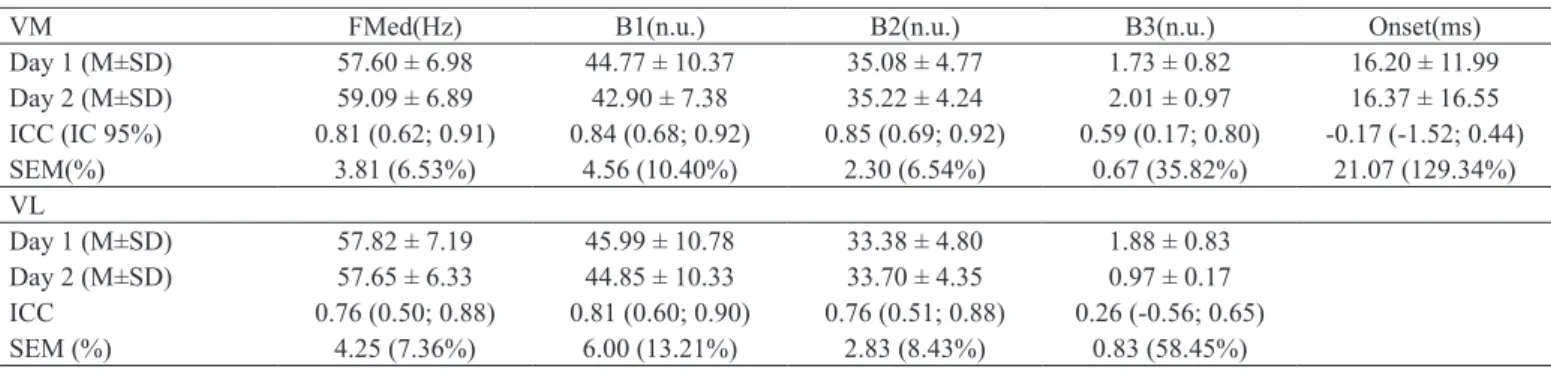
VM FMed(Hz) B1(n.u.) B2(n.u.) B3(n.u.) Onset(ms)
Day 1 (M±SD) 57.60 ± 6.98 44.77 ± 10.37 35.08 ± 4.77 1.73 ± 0.82 16.20 ± 11.99 Day 2 (M±SD) 59.09 ± 6.89 42.90 ± 7.38 35.22 ± 4.24 2.01 ± 0.97 16.37 ± 16.55 ICC (IC 95%) 0.81 (0.62; 0.91) 0.84 (0.68; 0.92) 0.85 (0.69; 0.92) 0.59 (0.17; 0.80) -0.17 (-1.52; 0.44) SEM(%) 3.81 (6.53%) 4.56 (10.40%) 2.30 (6.54%) 0.67 (35.82%) 21.07 (129.34%) VL
Day 1 (M±SD) 57.82 ± 7.19 45.99 ± 10.78 33.38 ± 4.80 1.88 ± 0.83 Day 2 (M±SD) 57.65 ± 6.33 44.85 ± 10.33 33.70 ± 4.35 0.97 ± 0.17 ICC 0.76 (0.50; 0.88) 0.81 (0.60; 0.90) 0.76 (0.51; 0.88) 0.26 (-0.56; 0.65) SEM (%) 4.25 (7.36%) 6.00 (13.21%) 2.83 (8.43%) 0.83 (58.45%)
Table 2. Stair ascent – Mean, standard deviation, ICC and SEM values to time and frequency domain sEMG parameters for VM and VL muscles.
can be extrapolated to other similar equipment (Weir, 2005). Besides, 2.5 ICC model take into account systematic and ran-dom errors, whereas, 3.5 ICC model only gets ranran-dom errors (Weir, 2005).
In general, it has been seen a slight difference between the reliability of the parameters on the frequency domain during ascent and descent stairs. This can be explained by the change in the sequence of recruitment-derecruitment pattern of motor units (Gonzalez-Izal, Cadore, & Izquierdo, 2014; Howell, Fu-glevand, Walsh, & Bigland-Ritchie, 1995). During concentric contraction (stair ascent) the recruitment of motor units occurs from the lower to the higher threshold level stimulation. Firstly,
the low-threshold units are delagrated (fatigue resistant), and
with the overload increasing, the high-threshold units are recrui-ted (low resistance to fatigue) (Kallio et al., 2013). However, during the eccentric contraction can occur activation of stretch receptors, leading to an activation of some polysynaptic afferent pathways, although temporarily, that may inhibit low-threshold motoneurons and excite high-threshold motoneurons, leading to a different activation pattern (Howell et al., 1995; Nardone, Romano, & Schieppati, 1989). Furthermore, the concentric contractions show higher EMG activation, since they require a greater number of active motor units to generate the same force compared to eccentric contractions (Kallio et al., 2013; Yu, 2014).
Considering the results and the paucity of research addres-sing the quality of these measures, we note that in the context of the variables in the frequency domain, either in the ascent or in the descent stair, they are consistent for studying individuals with PFPS. Consequently, it can be explored as assessment and diagnosis method to PFPS. Nonetheless, it is necessary to show the results regarding reliability to show de quality of the measurements. The gesture of ascent is better to determine the pattern of muscle activation in subjects with PFPS and then
compare it with asymptomatic subjects, due to a speciic order
of recruitment (low to high threshold). Therefore, it allows quantify strength physiologically (Silva Jr, 2013) and clearly determine other parameters such as Fmed, for example. On the other hand, stair descent is indicated to check the activity of the quadriceps as a result of higher demands muscle and
VM FMed(Hz) B1(n.u.) B2(n.u.) B3(n.u.) Onset(ms)
Day 1 (M±SD) 55.23 ± 5.80 46.57 ± 8.51 34.12 ± 3.97 1.52 ± 0.65 132.11 ± 133.72
Day 2 (M±SD) 57.74 ± 7.34 43.78 ± 9.21 34.56 ± 4.78 1.77 ± 0.77 149.53 ± 123.04
ICC (IC 95%) 0.56 (0.12; 0.78) 0.72 (0.43; 0.86) 0.79 (0.56; 0.90) 0.71 (0.40; 0.86) 0.59 (-0.2; 0.83)
SEM (%) 5.07 (8.97%) 5.67 (12.55%) 2.59 (7.54%) 0.45 (27.43%) 142.24 (101.01%)
VL
Day 1 (M±SD) 61.47 ± 9.97 41.59 ± 9.78 33.79 ± 3.23 2.23 ± 1.16
Day 2 (M±SD) 60.58 ± 9.50 40.71 ± 10.87 34.99 ± 5.06 2.04 ± 1.09
ICC 0.86 (0.71; 0.93) 0.88 (0.75; 0.94) 0.64 (0.28; 0.82) 0.74 (0.48; 0.87)
SEM (%) 4.86 (7.96%) 4.77 (11.59%) 3.03 (8.81%) 0.71 (33.33%)
Table 3. Stair descent – Mean, standard deviation, ICC and SEM values to time and frequency domain sEMG parameters for VM and VL muscles.
Abbreviation: FMed – median frequency, B1 – low frequency band, B2 – medium frequency band, B3 – high frequency band; n.u. – normalized unit; ms – milliseconds, IC 95% – 95% conidence intervals. As the onset values does not have to separate the VM and VL muscles values, because it is the delayed onset activation of these muscles.
Discussion
The stairs have been placed on experimental designs in order to reproduce a functional gesture. And, in the case of subjects with PFPS, it reproduces a gesture in which the symptoms are exacerbated. However, to date, it is not known if there are and which are the EMG differences between the gestures of ascent and descent stairs. Thus, the aim of this study is to determine the reliability of EMG parameters in these two gestures and report which one presents more reliable results.
Our sample follows the same proile used in studies with
similar experimental design (Bolgla et al., 2010; Ferrari et al., 2014; Kuriki et al., 2012). Studies that evaluated parameters in the frequency domain showed moderate to high reliability during stair deambulation (Briani et al., 2015; Ferrari et al.,
2014), corroborating with our indings. The EMG frequency
parameters demonstrated to be an adequate diagnostic tool for PFPS (Ferrari et al., 2014). Therefore, this experimental design can be implemented to diagnose individuals with PFPS (Yu et al., 1997).
The time domain parameters continue to be investigated, however, few researches have shown the consistency of its measures. The null (-.17) and moderate (.59) reliability during stair descent and ascent, respectively, with high SEM, for the parameters in the time domain reveals that this variable is not consistent and the results are unreliable. In contrast, a research developed with healthy young individuals, revealed very high levels of reliability, being .96 on the descent and .91 on the as-cent (Cowan et al., 2000). Another study obtained an ICC of 0.70 during stair descent in females with PFPS (Bolgla et al., 2010).
mechanical compared to concentric activities, besides being associated with higher rates of pain and joint stress (Powers, 2010). In contrast, the results for the delay in onset proved to be inconsistent and, therefore, should be cautious at the time of extracting the information pertaining to these measures due to its high rates of error.
There are some limitations of the present study that must be acknowledged. Firstly, the sample included only women. Although this subgroup is important to study, as these indivi-duals are the most likely to be committed for PFPS, the results may not be generalizable to the entire population of people with PFPS. Secondly, the speed during stair deambulation was not controlled in any day, which does not allow to determine the
speed inluence on the EMG signal. Therefore, future studies should include men and to control factors that can inluence the
EMG, such as speed during stair deambulation.
Stair ascent has shown EMG characteristics more standard than stair descent. Moreover, it has exhibit moderate to high values of reliability in the frequency domain EMG parameters. Therefore, our results suggest that stair ascent and frequency domain EMG parameters are more reliable to evaluate EMG signals of subjects with PFPS in the most cases.
References
Bolgla, L.A, Malone, T.R., Umberger, B.R., & Uhl, T.L. (2010). Reliability of electromyographic methods used for assessing hip and knee neuromuscular activity in females diagnosed with patellofemoral pain syndrome. Journal of Electromyography and Kinesiology, 20, 142–7. doi:10.1016/j.jelekin.2008.11.008 Boling, M., Padua, D., Marshall, S., Guskiewicz, K., Pyne, S., &
Beutler, A. (2010). Gender differences in the incidence and pre-valence of patellofemoral pain syndrome. Scandinavian Journal of Medicine & Science in Sports, 20, 725–730. doi:10.1111/j. 1600-0838.2009.00996.x.Gender
Briani, R.V., de Oliveira Silva, D., Pazzinatto, M.F., Albuquerque, C.E., Ferrari, D., Aragão, F.A., & Azevedo, F.M. (2015). Comparison of frequency and time domain electromyography parameters in women with patellofemoral pain. Clinical Biomechanics, 30, 302–307. doi:10.1016/j.clinbiomech.2014.12.014
Cavazzuti, L., Merlo, A., Orlandi, F., & Campanini, I. (2010). Delayed onset of electromyographic activity of vastus medialis obliquus re-lative to vastus lateralis in subjects with patellofemoral pain syndro-me. Gait & Posture, 32, 290–5. doi:10.1016/j.gaitpost.2010.06.025 Cowan, S.M., Bennell, K.L., & Hodges, P.W. (2000). The test–retest
reliability of the onset of concentric and eccentric vastus medialis obliquus and vastus lateralis electromyographic activity in a stair stepping task. Physical Therapy in Sport, 1, 129–136. doi:10.1054/ ptsp.2000.0036
Cowan, S.M., Bennell, K.L., Hodges, P.W., Crossley, K.M., & Mc-Connell, J. (2001). Delayed onset of electromyographic activity of vastus medialis obliquus relative to vastus lateralis in subjects with patellofemoral pain syndrome. Archives of Physical Medicine and Rehabilitation, 82, 183–9. doi:10.1053/apmr.2001.19022 De Oliveira Silva, D., Briani, R.V., Pazzinatto, M.F., Ferrari, D.,
Aragão, F.A., de Albuquerque, C.E., … de Azevedo, F.M. (2015). Reliability and differentiation capability of dynamic and static kinematic measurements of rearfoot eversion in patellofemoral pain. Clinical Biomechanics, 30, 144–148.
doi:10.1016/j.clinbio-mech.2014.12.009
Ferrari, D., Kuriki, H.U., Silva, C.R., Alves, N., & Mícolis de Aze-vedo, F. (2014). Diagnostic Accuracy of the Electromyography Parameters Associated With Anterior Knee Pain in the Diagnosis of Patellofemoral Pain Syndrome. Archives of Physical Medicine and Rehabilitation, 95, 1521–6. doi:10.1016/j.apmr.2014.03.028 Garcia, F.R., Azevedo, F.M., Alves, N., Carvalho, A.C., Padovani, C.R.,
& Negrão Filho, R.F. (2010). Effects of electrical stimulation of vastus medialis obliquus muscle in patients with patellofemoral pain syndrome: an electromyographic analysis. Revista Brasileira de Fisioterapia, 14, 477–82. (Retrieved from http://www.ncbi.nlm. nih.gov/pubmed/21340241)
Gonzalez-Izal, M., Cadore, E.L., & Izquierdo, M. (2014). Muscle conduction velocity, surface electromyography variables, and echo intensity during concentric and eccentric fatigue. Muscle & Nerve, 49, 389–397. doi:10.1002/mus.23926
Grenholm, A., Stensdotter, A.-K., & Häger-Ross, C. (2009). Kinematic analyses during stair descent in young women with patellofemoral pain. Clinical Biomechanics, 24, 88–94. doi:10.1016/j.clinbiome-ch.2008.09.004
Hermens, H.J., Freriks, B., Disselhorst-Klug, C., & Rau, G. (2000). Development of recommendations for SEMG sensors and sensor placement procedures. Journal of Electromyography and Kinesio-logy, 10, 361–374. doi:10.1016/S1050-6411(00)00027-4 Herrington, L., Malloy, S., & Richards, J. (2005). The effect of patella
taping on vastus medialis oblique and vastus laterialis EMG ac-tivity and knee kinematic variables during stair descent. Journal of Electromyography and Kinesiology, 15, 604–7. doi:10.1016/j. jelekin.2005.05.002
Howell, J.N., Fuglevand, A.J., Walsh, M.L., & Bigland-Ritchie, B. (1995). Motor Unit Activity During Isometric and Concentric-Ec-centric Contractions of the Human First Dorsal Interosseus Muscle. Journal of Neurophysiology, 74, 0–3.
Jordan, K., Challis, J.H., & Newell, K.M. (2007). Walking speed inluences on gait cycle variability. Gait & Posture, 26, 128–34. doi:10.1016/j.gaitpost.2006.08.010
Kallio, J., Søgaard, K., Avela, J., Komi, P.V, Selänne, H., & Linnamo, V. (2013). Motor unit iring behaviour of soleus muscle in isome -tric and dynamic contractions. PloS One, 8, e53425. doi:10.1371/ journal.pone.0053425
Kellis, E., & Katis, A. (2008). Reliability of EMG power-spectrum and amplitude of the semitendinosus and biceps femoris muscles during ramp isometric contractions. Journal of Electromyography and Kinesiology, 18, 351–8. doi:10.1016/j.jelekin.2006.12.001 Kim, H., & Song, C.H. (2012). Comparison of the VMO/VL EMG
Ratio and Onset Timing of VMO Relative to VL in Subjects with and without Patellofemoral Pain Syndrome. Journal of Physical Therapy Science, 24, 1315–1317. doi:10.1589/jpts.24.1315 Kuriki, H.U., De Azevedo, F.M., Filho, R. de F.N., & Alves, N. (2012).
Onset of quadriceps activation and torque variation during stair ascent in individuals with patellofemoral pain. Conscientiae Saúde, 11, 642–650. doi:10.5585/ConsSaude.v11n4.3810
Leitner, M., Schmid, S., Hiliker, R., & Radlinger, L. (2011). Test-retest reliability of vertical ground reaction forces during stair climbing in the elderly population. Gait & Posture, 34, 421–5. doi:10.1016/j. gaitpost.2011.06.014
Mathur, S., Eng, J.J., & MacIntyre, D.L. (2005). Reliability of surface EMG during sustained contractions of the quadriceps. Journal of Electromyography and Kinesiology, 15, 102–10. doi:10.1016/j. jelekin.2004.06.003
Nardone, A., Romano, C., & Schieppati, M. (1989). Selective re-cruitment of high-threshold human motor units during voluntary isotonic lengthening of active muscles. Journal of Physiology, 409, 451–471.
Pal, S., Draper, C.E., Fredericson, M., Gold, G.E., Delp, S.L., Beau-pre, G.S., & Besier, T.F. (2010). Patellar maltracking correlates with vastus medialis activation delay in patellofemoral pain patients. The American Journal of Sports Medicine, 39, 590–8. doi:10.1177/0363546510384233
Patil, S., Dixon, J., White, L., Jones, A., & Hui, A. (2011). An elec-tromyographic exploratory study comparing the difference in the onset of hamstring and quadriceps contraction in patients with anterior knee pain. The Knee, 18, 329–32. doi:10.1016/j. knee.2010.07.007
Powers, C.M. (2010). The inluence of abnormal hip mechanics on knee injury: a biomechanical perspective. The Journal of Ortho-paedic and Sports Physical Therapy, 40, 42–51. doi:10.2519/ jospt.2010.3337
Riener, R., Rabuffetti, M., & Frigo, C. (2002). Stair ascent and descent at different inclinations. Gait & Posture, 15, 32–44.
Salsich, G.B., Brechter, J.H., & Powers, C.M. (2001). Lower extremity kinetics during stair ambulation in patients with and without patel-lofemoral pain. Clinical Biomechanics, 16, 906–912. doi:10.1016/ S0268-0033(01)00085-7
Saywell, N., Taylor, D., & Boocock, M. (2012). During step descent, older adults exhibit decreased knee range of motion and increa-sed vastus lateralis muscle activity. Gait & Posture, 36, 490–4. doi:10.1016/j.gaitpost.2012.05.007
Silva, C.R., Silva, D.O., Aragão, F.A., Ferrari, D., Alves, N., & Azeve-do, F.M. (2014). Inluence of neuromuscular fatigue on co-contrac -tion between vastus medialis and vastus lateralis during isometric contractions. Kinesiology (Zagreb), 46, 179–185.
Silva, C.R., Silva, D.O., Ferrari, D., Negrão Filho, R.F., Alves, N., & Azevedo, F.M. (2014). Exploratory study of electromyographic behavior of the vastus medialis and vastus lateralis at neuromus-cular fatigue onset. Motriz, 20, 213–220. doi:dx.doi.org/10.1590/ S1980-65742014000200012
Silva Jr, R.A. (2013). Normalização EMG: considerações da literatura para avaliação da função muscular (EMG normalization: conside-rations from literature to assess the muscle function). Conscientiae Saúde, 12, 470–479. doi:10.5585/ConsSaude.v12n3.4362 Smoliga, J.M., Myers, J.B., Redfern, M.S., & Lephart, S.M. (2010).
Reliability and precision of EMG in leg, torso, and arm muscles during running. Journal of Electromyography and Kinesiology :
Oficial Journal of the International Society of Electrophysiological
Kinesiology, 20, e1–9. doi:10.1016/j.jelekin.2009.09.002 Solomonow, M., Baten, C., Smit, J., Baratta, R., Hermens, H.,
D’Am-brosia, R., & Shoji, H. (1990). Electromyogram power spectra frequencies associated with motor unit recruitment strategies. Journal of Applied Physiology, 68, 1177–85.
Sung, P.S., & Lee, D.C. (2009). Gender differences in onset timing and activation of the muscles of the dominant knee during stair climbing. The Knee, 16, 375–80. doi:10.1016/j. knee.2009.02.003
Uliam Kuriki, H., Mícolis de Azevedo, F., de Faria Negrão Filho, R., & Alves, N. (2011). Comparison of different analysis techniques for the determination of muscle onset in individuals with patellofemo-ral pain syndrome. Journal of Electromyography and Kinesiology :
Oficial Journal of the International Society of Electrophysiological
Kinesiology, 21, 982–7. doi:10.1016/j.jelekin.2011.08.002 Walter, S.D., Eliasziw, M., & Donner, A. (1998). Sample Size and
Opti-mal Designs for Reliability Studies. Statistics in Medicine, 17, 101– 110. doi:10.1002/(SICI)1097-0258(19980115)17:1<101::AID-SIM727>3.0.CO;2-E
Weir, J.P. (2005). Quantifying test-retest reliability using the intraclass correlation coeficient and the SEM. Journal of Strength and Con-ditioning Research., 19, 231–240.
Williams, J.M., Haq, I., & Lee, R.Y. (2013). An investigation into the onset, pattern, and effects of pain relief on lumbar extensor electromyography in people with acute and chronic low back pain. Journal of Manipulative and Physiological Therapeutics, 36, 91–100. doi:10.1016/j.jmpt.2012.12.006
Willson, J.D., Sharpee, R., Meardon, S.A., & Kernozek, T.W. (2014). Effects of step length on patellofemoral joint stress in female run-ners with and without patellofemoral pain. Clinical Biomechanics, 29, 243–247. doi:10.1016/j.clinbiomech.2013.12.016
Winter, D.A. (2009). Biomechanics and motor control of human mo-vement (Fourth Edi.). New Jersey.
Yu, B., Kienbacher, T., Growney, E.S., Johnson, M.E., & An, K.N. (1997). Reproducibility of the kinematics and kinetics of the lower extremity during normal stair-climbing. Journal of Orthopaedic Research, 15, 348–52. doi:10.1002/jor.1100150306
Yu, J. (2014). Comparison of Lower Limb Muscle Activity during Eccentric and Concentric Exercises in Runners with Achilles Ten-dinopathy. Journal of Physical Therapy Science, 26, 1351–1353.
Authors’ note
Marcella Ferraz Pazzinatto, Danilo de Oliveira Silva, and Fábio Mícolis de Azevedoare afiliated with the São Paulo State University (Univer -sidade Estadual Paulista), School of Science and Technology, Physical Therapy Department, Presidente Prudente, Brazil.
Ronaldo Valdir Briani, Fernando Amâncio Aragão, and Carlos Edu-ardo de Albuquerque are afiliated with the State University of West Parana (Universidade Estadual do Oeste do Paraná), Physical Therapy Department, Cascavel-PR, Brazil.
Deisi Ferrari is afiliated with the Bioengineering Postgraduate Pro -gram, São Carlos School of Engineering, University of São Paulo (Universidade de São Paulo), São Carlos, SP, Brazil.
Corresponding author
Fábio Mícolis de Azevedo
Department of Physiotherapy FCT/UNESP,
Roberto Simonsen Street, 305, Presidente Prudente, SP 19060-900, Brazil.
Phone: 3229-5820.
E-mail: [email protected]
Manuscript received on February 5, 2015 Manuscript accepted on March 25, 2015