Sodium Chloride Increases A
β
Levels by
Suppressing A
β
Clearance in Cultured Cells
Xiao-Juan Cheng1, Yuan Gao2,3, Yu-Wu Zhao1*, Xiao-Dong Cheng4*
1Department of Neurology, Shanghai Jiao Tong University Affiliated Sixth People’s Hospital, Shanghai, China,2Department of Neurology & Institute of Neurology, Rui Jin Hospital affiliated to Shanghai Jiao Tong University School of Medicine, Shanghai, China,3Department of Neurology, First People’s Hospital of Yunnan Province affiliated to Kunming University of Science and Technology, Kunming, China,4School of Life Sciences and Technology, Tongji University. East Hospital Affiliated To Tongji University, Shanghai, China
*[email protected](YWZ); [email protected](XDC)
Abstract
Recent studies suggest that high-salt diet is associated with cognitive decline in human and mouse. The fact that genetic factors account for less than 50% cases of sporadic Alzhei-mer’s disease (AD) highlights the important contribution of environmental factors, such as high-salt diet, in AD pathogenesis. However, whether and how high-salt diet fits the“ amy-loid cascade”hypothesis remains unexplored. Here, we show sodium chloride (NaCl) could increase Aβlevels in the medium of HEK293 cells overexpressing amyloid precursor pro-tein (APP) or C99 fragment. NaCl treatment dose not affect APP level, gamma secretase level or activity. Instead, NaCl treatment suppresses the capacity of cells to clear Aβand re-duces Apolipoprotein E (ApoE) level. Finally, NaCl treated THP-1 or BV2 cells are inefficient in clearing Aβwhen co-cultured with rat primary neurons. Our study suggests that high-salt diet may increase AD risk by directly modulating Aβlevels.
Introduction
Alzheimer’s disease is a common neurodegenerative disease. Familial AD is caused by APP or presenilin (PS) mutations that lead to abnormal Aβproduction. For sporadic AD which ac-counts for 90% of all AD cases, its underlining mechanism appears to be much more complex and only less than 50% of sporadic AD cases could be attributed to genetic factors [1]. It sug-gests environmental factors may play important roles in the pathogenesis of AD. Nevertheless, the dominant“amyloid cascade”hypothesis believes that Aβplays a vital role in driving the pathogenesis of AD [2].
Several independent epidemiological investigations have shown that high-salt diet is associ-ated with cognitive decline in human [3,4,5]. Recently, it’s found that high-salt diet could im-pair cognitive function in wild-type mice [6]. As cognitive decline is the core symptom of AD, these studies suggest that high-salt diet might be a risk factor for AD. In fact, high-salt diet would lead to hypertension, and then greatly increase the risk of vascular dementia (VaD) [7,8]. Interestingly, autopsy shows that many VaD patients also have Aβplaques, the AD-like a11111
OPEN ACCESS
Citation:Cheng X-J, Gao Y, Zhao Y-W, Cheng X-D (2015) Sodium Chloride Increases AβLevels by Suppressing AβClearance in Cultured Cells. PLoS ONE 10(6): e0130432. doi:10.1371/journal. pone.0130432
Academic Editor:Madepalli K. Lakshmana, Torrey Pines Institute for Molecular Studies, UNITED STATES
Received:March 3, 2015
Accepted:May 20, 2015
Published:June 15, 2015
Copyright:© 2015 Cheng et al. This is an open access article distributed under the terms of the
Creative Commons Attribution License, which permits unrestricted use, distribution, and reproduction in any medium, provided the original author and source are credited.
Data Availability Statement:All relevant data are within the paper.
Funding:The authors received no specific funding for this work.
pathology, in their brains [9,10]. Therefore, the possible direct effects of high-salt diet on Aβ homeostasis and therefore the AD risk could not be underestimated and deserve further investigation.
Studies have shown high-salt diet has profound effects on peripheral blood immune cells in mice and these cells, although far away from the central nervous system (CNS), could play im-portant roles in some nervous system diseases after they pass the blood-brabarriers and in-filtrate into CNS [11,12]. More specifically, macrophages in the peripheral blood could enter brain to affect Aβlevel in AD mouse models [13,14,15]. The possible effects of high-salt diet on these immune cells and their contributions to AD were unclear. Based on the above studies, it’s possible that high-salt diet could affect the function of macrophages in mice and these macro-phages could further modulate Aβlevel in the brain. In the present study, we sought to investi-gate whether and how high-salt treatment could affect Aβhomeostasis in cell models of AD.
Materials and Methods
Cell cultures
HEK293 cell (human embryonic kidney cell), BV2 (murine microglial cell line) and THP-1 (human acute monocytic leukemia derived cell line) were from ATCC (American Type Culture Collection). HEK293-APP stable cell and HEK293-C99 stable cell were established as follows. HEK293 cells were transfected with pcDNA 3.1 vector overexpressing APP or C99. Stable clones were obtained after addition of G418 (final concentration is 500ug/ml) for 2 weeks. To maintain the stable cell lines, 500ug/ml of G418 was used. HEK293 cell, HEK293-APP stable cell, HEK293-C99 stable cell and BV2 (murine microglial cell line) were maintained in Dulbec-co’s Modified Essential Medium (DMEM, GIBCO) with 10% Fetal bovine serum (FBS, GIBCO). THP-1 (human acute monocytic leukemia derived cell line) was maintained in RPMI1640 (GIBCO) with 10% FBS. Primary cortical neurons were dissected from embryonic day 17 (E17) brains of Sprague-Dawley rat or APP/PS1 mouse (#004462, Jackson laboratory) and cultured in Neurobasal (Invitrogen, 21103–049). All animal experiments were in accor-dance with the Institutional Animal Care and Use Committee of Shanghai Jiao Tong Universi-ty, China. The study and protocols were approved by the Institutional Animal Care and Use Committee of Shanghai Jiao Tong University (Permit Number: SYXK 2011–0128). All efforts were made to minimize suffering of the animals.
Co-culture system
The co-culture system consists of lower and upper chambers which are separated by a selec-tively permeable membrane with 0.4um-diameter pores (Corning, Transwell 3450). Rat prima-ry neurons were plated in the lower chamber and maintained to 10 days in vitro (DIV10). Cell lines were plated in the upper chamber and were co-cultured with neurons after treated with normal or NaCl medium for indicated time.
Reagents and antibodies
The following antibodies were used: APP from Invitrogen (13–0200, clone LN27), PS1 from Chemicon (MAB5232), nicastrin from Sigma (MAB5232), Pen2 from Invitrogen (36–7100) and ApoE from Calbiochem (178479). L685,458 (L1790) was from Sigma.
A
β
ELISA
from APP/PS1 mice, they were cultured in 12-well plate for 10 days in vitro (DIV 10) and incu-bated with normal or NaCl medium for 24h. To preclude that the effects of additional 40mM NaCl are unspecific results of increased ion concentration or osmotic pressure, additional 26.7mM MgCl2 (with equal ion concentration) or 80mM Mannitol (with equal osmotic pres-sure) were used as control solutions. After that, the medium was replaced by normal medium (600 ul for each well) for 12h before collection. The collected medium was centrifuged at 13200 rpm/min for 10 min to avoid cells and debris and stored at -80°C until ELISA experiments. Human Aβ42 and Aβ40 levels in the medium were quantified by ELISA kits (Invitrogen, KHB3482 and KHB3544). Rat Aβderived from cultured neurons were quantified by ELISA kit (IBL, 27720). The Aβlevels in the culture medium are normalized to the total protein amount of the cells and then presented as fold changes relative to the control group.
Western blot
Proteins were extracted from cell lines or cultured medium using sodium dodecyl sulfate lysis buffer (2% sodium dodecyl sulfate, 10% glycerol, 0.1 mM dithiothreitol, and 0.2 M Tris—HCl, pH 6.8). Protein samples were resolved by SDS—PAGE and analyzed by immunoblots.
Gamma secretase activity assay
The human coding sequence of C99 was cloned into PET28 vector and expressed as fusion pro-tein with His tag. C99-His recombinant propro-tein was expressed by 1mM IPTG induction for 8 hours at 37°C in BL21 E. Coli. The recombinant protein was purified by Ni-column (QIAGEN) and serves as substrate. HEK293 cell line was homogenized and centrifuged at 13200 rpm/min for 15 min, and gamma secretases was reconstituted by resolving the pellet in 50 mM TrisHCL (pH 6.8), 2 mM EDTA and 0.25% CHAPSO (w/v). Twenty ug of protein was incubated with 2ug C99-His recombinant protein at 37°C for 4h. Then Aβ40 and Aβ42 levels were measured by ELISA.
A
β
clearance assay
Cell lines were plated in 12-well plate and reached 80% confluence 24h later. Cells were treated with normal or NaCl medium for 24h. Then the culture medium was replaced by normal medi-um with 1ug/ml Aβ42. After 8h incubation, remaining Aβ42 in the medium was measured by ELISA.
Annexin V-FITC Apoptosis assay
HEK293 cells were treated with normal or NaCl medium for indicated time and stained with Annexin V-FITC Apoptosis kit (abcam, ab14085) according to the manufacturer’s instruction. Then, the stained cells were analyzed by MoFlo XDP (Beckman Coulter, Inc).
Statistical analysis
Results
NaCl increases A
β
levels in HEK293 cells and primary neurons from
APP/PS1 mouse
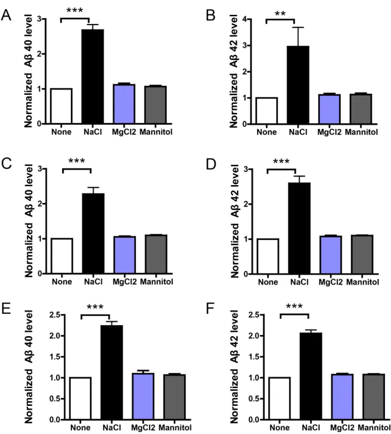
To explore the possible effects of high-salt treatment, we define the condition of high-salt treat-ment as normal medium (DMEM for HEK293 and BV2, RPMI 1640 for THP-1, Neurobasal for primary neurons) with additional 40mM increase in NaCl concentration. HEK293 stable cells overexpressing APP or C99, and DIV10 primary neuron cultures from APP/PS1 mouse were incubated with normal or NaCl medium for 24h. To preclude that the effects of additional 40mM NaCl are unspecific results of increased ion concentration (the total ion concentration is increased by 80mM), we choose the normal medium with additional 26.7mM MgCl2 (the total ion concentration is also increased by 80mM) as a control solution for total ion concentra-tion. Similarly, additional 40mM NaCl also increases osmotic pressure. To see if the effects of additional 40mM NaCl are simply resulted from increased osmotic pressure, we use normal medium with additional 80mM Mannitol (with the same osmotic pressure of 40mM NaCl) as a control solution for osmotic pressure. Then the medium was replaced by normal medium for 12h and Aβlevels in the medium were measured by ELISA. Results show that NaCl treatment greatly increased Aβ40 and 42 levels in the medium (Fig 1). Importantly, the medium contain-ing additional 26.7mM MgCl2 or 80mM mannitol could not affect Aβlevels in the medium. It indicates increased sodium, instead of increased chloride or osmolarity, is responsible for the effect of NaCl medium on Aβlevels. As Aβlevels are determined by the balance between Aβ production and clearance, the above results imply NaCl treatment might affect Aβproduction or clearance. Further, given that C99 is the product ofβ-secretase cleavage of APP and serves as the direct substrate ofγsecretase, our results suggest NaCl treatment might affect the γ-secretase cleavage of APP or Aβclearance.
NaCl dose not affect cell viability or
γ
secretase
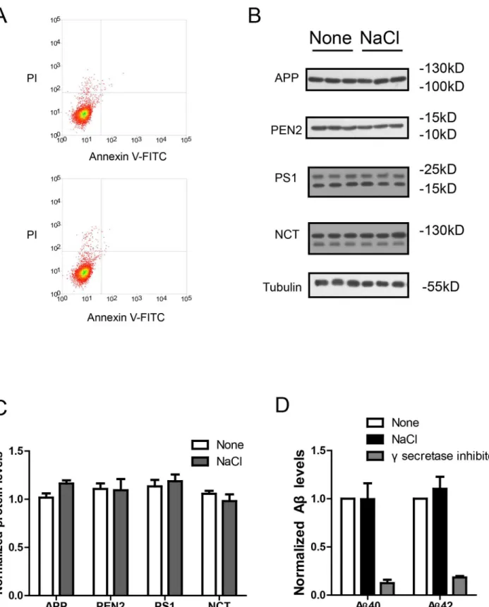
Next, we measured the possible cellular damage resulted from NaCl medium with the Annexin V-FITC apoptosis assay. The results show NaCl treatment for 24h in HEK293 cells did not re-sult in cellular damage (Fig 2A). To explore the possible effects of NaCl treatment on γ-secre-tase cleavage of APP, we investigated whether the expression levels of APP orγ-secretase components were affected by NaCl treatment. Western blots (Fig2Band2C) show that NaCl treatment did not affect the protein levels of APP orγ-secretase components (PS1, NCT, PEN2). Moreover, we measured the enzyme activity ofγsecretase under NaCl treatment in an in-vitro C99 assay [16]. The recombinant C99-His protein was used as the direct substrate ofγ secretase and the membrane fractions from HEK293 cells treated with normal or NaCl medi-um were used asγsecretase. After incubation of the membrane fractions with C99-His protein, the Aβlevels were measured by ELISA and normalized to total protein amount. The normal-ized Aβlevels could reflect theγ-secretase activity. We found NaCl treatment did not affect γ-secretase activity (Fig 2D). In contrast, treatment with L685,458, a potentγ-secretase inhibitor, could abolish theγ-secretase activity. Theses results suggest NaCl treatment could increase Aβ levels without affecting cell viability, APP orγsecretase.
NaCl suppresses A
β
clearance in HEK293, THP-1 and BV2 cells
Fig 1. NaCl treatment increased Aβlevels in culture medium.HEK293 stable cells overexpressing APP (A and B) or C99 (C and D), or primary neurons from APP/PS1 mouse (E and F) were incubated with normal or NaCl medium for 24h and the medium were replaced by normal medium for another 12h. Then Aβlevels in the medium were measured by ELISA (n = 3). Data were presented as means±s.e.m. of indicated numbers of independent experiments.
**P<0.01,***P<0.001.
Fig 2. NaCl treatment did not affect gamma secretase.(A) FACS analysis showed the Annexin V-FITC/ PI staining of HEK293 cells treated with normal (up) or NaCl (low) medium. Western blots (B) and their quantification (C) showed the protein levels of APP and gamma secretase components (PS1, NCT, PEN2) in the lysate of HEK293 cells overexpressing APP after normal or NaCl treatment (n = 3). (D) Enzyme activity assay showed the gamma secretase activity of HEK293 cell lysate after treatment with normal, NaCl medium or gamma secretase inhibitor-L685,458 (n = 3). Data were presented as means±s.e. m. of indicated numbers of independent experiments.
of NaCl treated cell was significantly higher than that of control cells (Fig 3A), indicating that NaCl treated cells was inefficient in Aβclearance. In contrast, MgCl2 or Mannitol has no ef-fects in the Aβclearance assay.
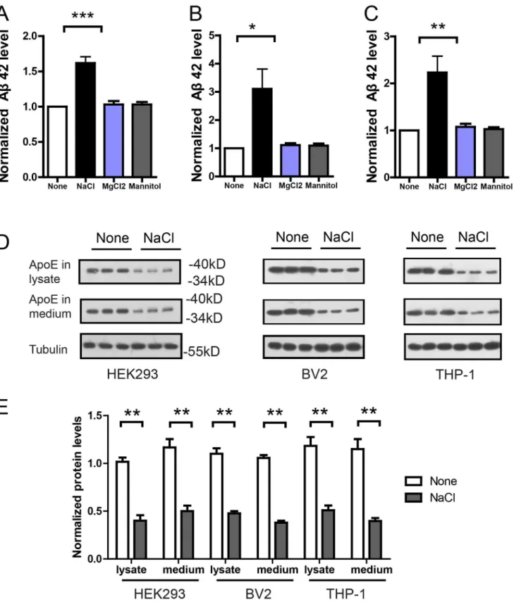
In in-vivo condition, Aβwas cleared mainly by macrophages. Thus, we performed the same Aβclearance assay in microglia cell line BV2 representing macrophages in the brain, and blood macrophage cell line THP-1. Similarly, Aβ42 levels in the medium of NaCl treated BV2 (Fig 3B) or THP-1 (Fig 3C) cells were significantly higher, indicating that NaCl treated BV2 or THP-1 cells were inefficient in Aβclearance. We measured the viability of NaCl treated BV2 and THP-1 cells and found no significant effects of additional NaCl on cell viability with the Annexin V-FITC apoptosis assay (data not shown). In contrast, 26.7mM MgCl2 or 80mM Mannitol treated cells remained efficient in Aβclearance. These results indicate increased sodi-um could suppress Aβclearance. As ApoE plays an important role in Aβclearance [17], we in-vestigated whether ApoE level was affected by NaCl treatment. Indeed, in the lysates and medium of NaCl treated HEK293, BV2 or THP-1 cells, western blots show that ApoE level was significantly reduced (Fig3Dand3E). Together, these results suggest that NaCl treatment could suppress Aβclearance in HEK293, THP-1 and BV2 cells, possibly through down-regulat-ing ApoE level.
NaCl treated THP-1 or BV2 cells are inefficient in clearing A
β
derived
from co-cultured neurons
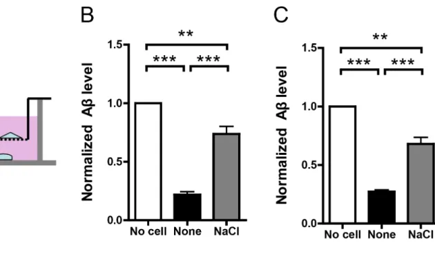
In in-vivo condition, neurons produce Aβwhile macrophages clear Aβ. According to“Aβ cas-cade”hypothesis, dyshomeostasis of this tightly regulated balance would lead to AD. To reca-pitulate this complex process, we adopted an in-vitro co-culture system [18]. In this system, rat primary neurons (DIV10) were cultured in the lower chamber while normal or NaCl medium treated BV2 or THP-1 cells were cultured in the upper chamber (Fig 4A). There was a selec-tively permeable membrane with 0.4 um diameter between the lower and upper chambers, al-lowing free movement of substance smaller than 0.4um, such as Aβ. After 12h co-culture, Aβ level in the medium of the lower chamber was assayed by ELISA. As the results show, cultured rat neurons produced large amount of Aβand its level was significantly reduced when co-cul-tured with normal medium treated BV2 or THP-1 cells. It indicates BV2 or THP-1 cells in the upper chamber could effectively clear Aβin the lower chamber. However, when co-cultured with NaCl treated BV2 or THP-1 cells, Aβlevel in the lower chamber was significantly higher (Fig4Band4C), indicating NaCl treated BV2 or THP-1 cells in the upper chamber were ineffi-cient in Aβclearance.
Discussion
AD is an aging-related disease without effective therapy so far. The comprehensive under-standing of its underlying mechanism is crucial for its prevention and treatment. The familial AD is caused by harmful mutations in APP or PS. For the sporadic AD, however, its mecha-nism of pathogenesis is much more complex. Genetic polymorphism in ApoE gene is the great-est risk factor in sporadic AD and ApoE4 genotype greatly increases AD risk [19]. But less than half of all sporadic AD patients were ApoE4 carriers. Thus, considerable efforts have been made to explore more potential genetic risk factors. Even through, the carriers of new identified risky single nucleotide polymorphism (SNP) are much scarce [1]. These studies suggest that nearly half of sporadic AD patients could not be attributed to genetic factors and potential en-vironmental factors have to be taken into consideration.
Fig 3. NaCl treatment suppressed Aβclearance and reduced ApoE level.Aβclearance assays were performed in HEK293 (A), BV2 (B) or THP-1 (C) cells. Normal or NaCl treated cells were incubated with 1ug/ml Aβ42 for 8h and the remaining Aβ42 in the medium was measured by ELISA (n = 3–5). Western blots (D) and their quantification (E) showed the protein level of ApoE in the lysates and medium of HEK293, BV2 or THP-1 cells after normal or NaCl treatment (n = 3). Data were presented as means±s.e.m. of indicated numbers of independent experiments.*P<0.05,**P<0.01,***P<0.001.
decline [3,4,5]. Moreover, high-salt diet impairs cognitive function in mouse models [6]. These results point to a possibility that high-salt diet might increase AD risk. According to the dominant“amyloid cascade”hypothesis, AD is caused harmful Aβaccumulation. Thus we investigated whether and how could high-salt diet fit into the amyloid cascade. To mimic the effects of high-salt diet, we use DMEM or RPMI 1640 with additional 40mM NaCl to incubate cultured cells. Since the NaCl concentration in the normal DMEM or RPMI 1640 is about 140mM, the final NaCl concentration under our high-salt treatment is about 180mM. We Fig 4. NaCl treatment suppressed Aβclearance in co-culture system.(A) Rat primary neurons were plated in the lower chamber and treated cell lines were plated in the upper chamber. The lower and upper chambers were separated by a selectively permeable membrane with 0.4um-diameter pores. Normal or NaCl treated BV2 (B) or THP-1 (C) cells were co-cultured with neurons for 12h and the remaining Aβin the lower chamber was measured by ELISA (n = 3). (D) Flow chart explains the potential involvement of high-salt diet in AD risk. Data were presented as means±s.e.m. of indicated numbers of independent experiments.**P<0.01,***P<0.001.
choose this concentration because in certain tissues harbouring immune cells such as macro-phages, NaCl concentration could be higher than 180mM in high-salt diet fed mice [20,21]. Also, recent studies suggest that additional 40mM NaCl could produce profound effects on cul-tured peripheral blood immune cells [11,12]. Thus, we choose this in-vitro condition of addi-tional 40mM NaCl to mimic the in-vivo condition of high-salt diet fed mice. Our results show that NaCl treatment could increase Aβlevels in cells overexpressing APP or C99, indicating Aβ production or clearance was affected. APP is cleaved by beta secretase into C99 and then C99 is cleaved by gamma secretase to release Aβ. The results that substrate level (APP) and gamma secretase level or activity remained unchanged imply that Aβclearance, but not Aβproduction, was affected by high-salt treatment. Indeed, in the Aβclearance assay, NaCl treated HEK293, BV2 or THP-1 cells became inefficient in Aβclearance. Finally, to test the effects of NaCl treat-ment in a more physiological and in-vivo condition, we adopted a co-culture system consisting of macrophages in the upper chamber and primary neurons in the lower chamber. Similarly, NaCl treated BV2 or THP-1 cells were inefficient in Aβclearance.
Our study provides a link between high-salt diet and Aβin cultured cells. It may contribute to the association of high-salt diet with cognitive decline in human. As Aβplays a key role in driving the pathogenesis of AD and brain Aβlevels are associated with cognitive decline in AD and normal aging, we speculate that high-salt diet might modulate Aβlevels to affect the cogni-tive decline and therefore AD risk (Fig 4D). It widens our understanding of AD pathogenesis and highlights the importance of healthy life style in AD prevention or reducing AD risk. But there are some limitations in our study. First, the direct association of high-salt diet with AD risk has not been established by epidemiological investigation. Second, the effects of high-salt treatment were investigated in cultured cells which might be quite different from the in-vivo condition. In conclusion, we report that high-salt treatment increases Aβlevels by suppressing Aβclearance in cultured cells. To understand the mechanism underlying the association be-tween the high-salt diet and the cognitive decline, it requires further in-vivo investigation in animal models in the future.
Author Contributions
Conceived and designed the experiments: YWZ XDC. Performed the experiments: XJC YG. Analyzed the data: XJC. Contributed reagents/materials/analysis tools: XJC. Wrote the paper: XJC.
References
1. Bertram L, Tanzi RE. Thirty years of Alzheimer's disease genetics: the implications of systematic meta-analyses. Nat Rev Neurosci. 2008; 9(10):768–78. Epub 2008/09/20. nrn2494 [pii] doi:10.1038/ nrn2494PMID:18802446.
2. Hardy J, Selkoe DJ. The amyloid hypothesis of Alzheimer's disease: progress and problems on the road to therapeutics. Science. 2002; 297(5580):353–6. Epub 2002/07/20. doi:10.1126/science. 1072994297/5580/353 [pii]. PMID:12130773.
3. Afsar B. The relationship between cognitive function, depressive behaviour and sleep quality with 24-h urinary sodium excretion in patients with essential hypertension. High Blood Press Cardiovasc Prev. 2013; 20(1):19–24. Epub 2013/03/27. doi:10.1007/s40292-013-0002-7PMID:23529378.
4. Rondanelli M, Solerte SB, Ferrari E. Electrolytes and cognitive function in the elderly: relationship be-tween serum sodium and chloride concentrations and psychometric test scores. Panminerva Med. 1998; 40(3):191–5. Epub 1998/10/24. PMID:9785915.
6. Liu YZ, Chen JK, Li ZP, Zhao T, Ni M, Li DJ, et al. High-salt diet enhances hippocampal oxidative stress and cognitive impairment in mice. Neurobiol Learn Mem. 2014; 114:10–5. Epub 2014/04/23. doi: S1074-7427(14)00071-9 [pii] doi:10.1016/j.nlm.2014.04.010PMID:24752150.
7. Skoog I, Lernfelt B, Landahl S, Palmertz B, Andreasson LA, Nilsson L, et al. 15-year longitudinal study of blood pressure and dementia. Lancet. 1996; 347(9009):1141–5. Epub 1996/04/27. PMID:8609748.
8. Kilander L, Nyman H, Boberg M, Hansson L, Lithell H. Hypertension is related to cognitive impairment: a 20-year follow-up of 999 men. Hypertension. 1998; 31(3):780–6. Epub 1998/03/12. PMID:9495261.
9. Jellinger KA, Attems J. Neuropathological evaluation of mixed dementia. J Neurol Sci. 2007; 257(1–
2):80–7. Epub 2007/02/28. S0022-510X(07)00078-0 [pii] doi:10.1016/j.jns.2007.01.045PMID: 17324442.
10. Schneider JA, Arvanitakis Z, Bang W, Bennett DA. Mixed brain pathologies account for most dementia cases in community-dwelling older persons. Neurology. 2007; 69(24):2197–204. Epub 2007/06/15. 01. wnl.0000271090.28148.24 [pii] doi:10.1212/01.wnl.0000271090.28148.24PMID:17568013.
11. Wu C, Yosef N, Thalhamer T, Zhu C, Xiao S, Kishi Y, et al. Induction of pathogenic TH17 cells by induc-ible salt-sensing kinase SGK1. Nature. 2013; 496(7446):513–7. Epub 2013/03/08. nature11984 [pii] doi:10.1038/nature11984PMID:23467085; PubMed Central PMCID: PMC3637879.
12. Kleinewietfeld M, Manzel A, Titze J, Kvakan H, Yosef N, Linker RA, et al. Sodium chloride drives auto-immune disease by the induction of pathogenic TH17 cells. Nature. 2013; 496(7446):518–22. Epub 2013/03/08. nature11868 [pii] doi:10.1038/nature11868PMID:23467095; PubMed Central PMCID: PMC3746493.
13. Simard AR, Soulet D, Gowing G, Julien JP, Rivest S. Bone marrow-derived microglia play a critical role in restricting senile plaque formation in Alzheimer's disease. Neuron. 2006; 49(4):489–502. Epub 2006/ 02/16. doi: S0896-6273(06)00075-4 [pii] doi:10.1016/j.neuron.2006.01.022PMID:16476660.
14. Stalder AK, Ermini F, Bondolfi L, Krenger W, Burbach GJ, Deller T, et al. Invasion of hematopoietic cells into the brain of amyloid precursor protein transgenic mice. J Neurosci. 2005; 25(48):11125–32. Epub 2005/12/02. 25/48/11125 [pii] doi:10.1523/JNEUROSCI.2545-05.2005PMID:16319312.
15. Town T, Laouar Y, Pittenger C, Mori T, Szekely CA, Tan J, et al. Blocking TGF-beta-Smad2/3 innate im-mune signaling mitigates Alzheimer-like pathology. Nat Med. 2008; 14(6):681–7. Epub 2008/06/03. nm1781 [pii] doi:10.1038/nm1781PMID:18516051; PubMed Central PMCID: PMC2649699.
16. Edbauer D, Winkler E, Regula JT, Pesold B, Steiner H, Haass C. Reconstitution of gamma-secretase activity. Nat Cell Biol. 2003; 5(5):486–8. Epub 2003/04/08. doi:10.1038/ncb960ncb960 [pii]. PMID: 12679784.
17. Zhao L, Lin S, Bales KR, Gelfanova V, Koger D, Delong C, et al. Macrophage-mediated degradation of beta-amyloid via an apolipoprotein E isoform-dependent mechanism. J Neurosci. 2009; 29(11):3603–
12. Epub 2009/03/20. 29/11/3603 [pii] doi:10.1523/JNEUROSCI.5302-08.2009PMID:19295164.
18. Neher JJ, Neniskyte U, Zhao JW, Bal-Price A, Tolkovsky AM, Brown GC. Inhibition of microglial phago-cytosis is sufficient to prevent inflammatory neuronal death. J Immunol. 2011; 186(8):4973–83. Epub 2011/03/16. jimmunol.1003600 [pii] doi:10.4049/jimmunol.1003600PMID:21402900.
19. Raber J, Huang Y, Ashford JW. ApoE genotype accounts for the vast majority of AD risk and AD pathol-ogy. Neurobiol Aging. 2004; 25(5):641–50. Epub 2004/06/03. doi:10.1016/j.neurobiolaging.2003.12. 023S0197458004001009 [pii]. PMID:15172743.
20. Machnik A, Neuhofer W, Jantsch J, Dahlmann A, Tammela T, Machura K, et al. Macrophages regulate salt-dependent volume and blood pressure by a vascular endothelial growth factor-C-dependent buffer-ing mechanism. Nat Med. 2009; 15(5):545–52. Epub 2009/05/05. nm.1960 [pii] doi:10.1038/nm.1960 PMID:19412173.