Molecular and Cellular Characterization of
a Zebrafish Optic Pathway Tumor Line
Implicates Glia-Derived Progenitors in
Tumorigenesis
Staci L. Solin1, Ying Wang1, Joshua Mauldin1, Laura E. Schultz1, Deborah E. Lincow1, Pavel A. Brodskiy1, Crystal A. Jones1, Judith Syrkin-Nikolau1, Jasmine M. Linn1, Jeffrey J. Essner1, Jesse M. Hostetter2, Elizabeth M. Whitley2,
J. Douglas Cameron3, Hui-Hsien Chou1, Andrew J. Severin4, Donald S. Sakaguchi1, Maura McGrail1*
1.Department of Genetics, Development and Cell Biology, Iowa State University, Ames, Iowa, United States of America,2.Department of Veterinary Pathology, Iowa State University, Ames, Iowa, United States of America,3.Department of Ophthalmology and Visual Neurosciences, University of Minnesota, Minneapolis, Minnesota, United States of America,4.Genome Informatics Facility, Office of Biotechnology, Iowa State University, Ames, Iowa, United States of America
Abstract
In this study we describe the molecular and cellular characterization of a zebrafish mutant that develops tumors in the optic pathway. HeterozygousTg(flk1:RFP)is18
transgenic adults develop tumors of the retina, optic nerve and optic tract. Molecular and genetic mapping demonstrate the tumor phenotype is linked to a high copy number transgene array integrated in the lincRNA genelincRNAis18/ Zv9_00007276on chromosome 3. TALENs were used to isolate a 147kb deletion allele that removes exons 2–5 of the lincRNAis18gene. Deletion allele
homozygotes are viable and do not develop tumors, indicating loss of function of thelincRNAis18locus is not the trigger for tumor onset. Optic pathway tumors in the
Tg(flk1:RFP)is18mutant occur with a penetrance of 80–100% by 1 year of age. The retinal tumors are highly vascularized and composed of rosettes of various sizes embedded in a fibrous matrix. Immunohistochemical analysis showed increased expression of the glial markers GFAP and BLBP throughout retinal tumors and in dysplastic optic nerve. We performed transcriptome analysis of pre-tumorous retina and retinal tumor tissue and found changes in gene expression signatures of radial glia and astrocytes (slc1a3), activated glia (atf3, blbp, apoeb), proliferating neural progenitors (foxd3, nestin, cdh2, her9/hes1), and glioma markers (S100b, vim). The transcriptome also revealed activation of cAMP, Stat3 and Wnt signal transduction pathways. qRT-PCR confirmed .10-fold overexpression of the Wnt pathway OPEN ACCESS
Citation:Solin SL, Wang Y, Mauldin J, Schultz LE, Lincow DE, et al. (2014) Molecular and Cellular Characterization of a Zebrafish Optic Pathway Tumor Line Implicates Glia-Derived Progenitors in Tumorigenesis. PLoS ONE 9(12): e114888. doi:10. 1371/journal.pone.0114888
Editor:Joseph Charles Glorioso, University of Pittsburgh School of Medicine, United States of America
Received:September 23, 2014
Accepted:November 14, 2014
Published:December 8, 2014
Copyright:ß2014 Solin et al. This is an
open-access article distributed under the terms of the
Creative Commons Attribution License, which permits unrestricted use, distribution, and repro-duction in any medium, provided the original author and source are credited.
Data Availability:The authors confirm that all data underlying the findings are fully available without restriction. All RNA-Seq Illumina files are available in the ArrayExpress database (www.ebi.ac.uk/ arrayexpress) under accession number E-MTAB-2886.
Funding:This work was supported by the Roy J. Carver Charitable Trust [14-14418 MM], Research to Prevent Blindness [DJC], the Iowa State University Center for Integrated Animal Genomics [JJE, MM, DSS], Iowa State University DOE/SULI and CC Research Experience for Undergraduate Programs [DEL, PAB], Iowa State University IINspire-LSAMP program [CAJ], and Iowa State University Startup funds [MM]. Dr. Cameron is supported in part by an unrestricted grant from Research to Prevent Blindness to the Department of Ophthalmology and Visual Neurosciences of the University of Minnesota. The funders had no role in study design, data collection and analysis, decision to publish, or preparation of the manuscript.
componentshbegfa,ascl1a, and insm1a. Together the data indicate Mu¨ller glia and/or astrocyte-derived progenitors could contribute to the zebrafish
Tg(flk1:RFP)is18 optic pathway tumors.
Introduction
Glia play critical roles in the function and maintenance of the nervous system. They are involved in neuronal homeostasis and repair, but can also undergo reprogramming in response to injury to generate progenitors that repopulate missing neurons and glia [1]. In the retinas of mice, frog and fish one population of cells that can be reprogrammed in response to injury is Mu¨ller glia [2]. In normal retina Mu¨ller glia have stem-like behaviors, dividing asymmetrically to produce progenitors of the rod photoreceptor lineage [3]. After photoreceptor or retinal neuron damage, Mu¨ller glia can dedifferentiate and produce progenitors that give rise to the major neural retinal cell types. The zebrafish retina has been used extensively as a model system to investigate the molecular mechanisms required for this process [4–6]. Major signal transduction pathways activated in reprogrammed Mu¨ller glia in zebrafish include EGF [7], Stat3 [8–10] and Wnt [11,12]. Understanding how these signaling pathways promote glia reprogram-ming and neural regeneration is important for advancing treatments of central nervous system injury and disease.
In this study we present the characterization and molecular cloning of a zebrafish transgenic line Tg(flk1:RFP)is18 that develops highly penetrant tumors in the retina and optic tract with features of retinoblastoma and fibrous glioma. The tumor phenotype is linked to a high copy number array of an RFP expressing reporter transgene in line Tg(flk1:RFP)is18. The transgene integrated in a long intergenic noncoding RNA gene lincRNAis18, which was previously identified in the zebrafish embryonic transcriptome as Zv9_00007276 and Zv9_00007274[13]. Isolation of a second targeted deletion allele of thelincRNAis18gene did not result in tumor formation, suggesting loss of function of the locus is not the initiating event that triggers tumor onset. Histological, cytological, and transcriptome analyses in pre-tumorous retina and tumor tissue reveal gene signatures of radial glia, neural progenitors, and injury induced activation of glia and astrocytes. The
Tg(flk1:RFP)is18 tumors are similar to the zebrafish model of optic pathway glioma driven by activated Sonic hedgehog signaling in neural progenitors [14]. Our analyses indicate the Tg(flk1:RFP)is18 tumors originate from neural
Results
Isolation and molecular mapping of the optic pathway tumor line
Tg(flk1:RFP)is18
We isolated a transgenic zebrafish line in which heterozygous adults develop neoplasia of the retina, optic nerve and optic tract. Transgenic line
Tg(Tol2,flk1:RFP-CAAX.)is18 (abbreviated as Tg(flk1:RFP)is18)was generated using a Tol2 transposon reporter construct that expresses membrane-targeted RFP-CAAX throughout vascular endothelial cells (Fig. 1A, B). Heterozygous
Tg(flk1:RFP)is18adults developed large ocular tumors that first became evident at approximately 5 months of age (Fig. 1C) with a penetrance of .80%.
Multigenerational genetic analysis demonstrated that the tumor phenotype was linked to inheritance of the Tg(flk1:RFP)is18 transgene, as determined by RFP expression (Fig. 1D). In each generation non-transgenic siblings were healthy and showed no evidence of tumor formation. These results indicated that the tumor phenotype was due either to the location of theTg(flk1:RFP)is18transgene in the genome or the presence of the transgene itself. Attempts to map the
Tg(flk1:RFP)is18 transgene integration site by standard ligation mediated PCR and inverse PCR methods suggested that during isolation of the line, multiple copies of the entire pTol2,flk1:RFP. construct, including the vector backbone, had integrated as a concatemer array. We confirmed by genomic Southern blot that the Tg(flk1:RFP)is18 line contains an array with ,100 copies of the Tol2,flk1:RFP. construct (Fig. 1E). Genomic DNA from Tg(flk1:RFP)is18
individuals from 3 consecutive generations hybridized with an RFP-specific probe revealed multiple bands, including the expected 1.3 kb EcoRI-ScaI fragment from the Tol2,flk1:RFP. construct. In each generation non-transgenic siblings that lacked RFP expression in vascular tissue did not inherit the concatemer (Fig. 1E). Together these results provided strong evidence that the transgene in line
Tg(flk1:RFP)is18 is a stable, high-copy number concatemer that displays Mendelian transmission from one generation to the next.
To clone the genomic DNA flanking theTg(flk1:RFP)is18 concatemer integration site and map its location in the zebrafish genome, we designed a custom SureSelect Target Enrichment kit (Agilent Technologies) with single stranded biotinylated RNA probes that tile across the entire pTol2,flk1:RFP.
vector (S1 A Figure;Table 1). Random sheared Tg(flk1:RFP)is18 genomic DNA was hybridized to the biotinylated bait library in order to capture all of the concatemer transgene sequences. Since the genomic DNA was randomly sheared, we predicted a percentage of captured sequences would span the junction of the ends of the concatemer and the genomic DNA. Five independent genomic DNA samples isolated from muscle or tumor tissue, representing 3 Tg(flk1:RFP)is18
one end and zebrafish genomic sequences at the other. Four candidate integration sites that contained 9 or more reads from multiple samples (S3 Table) were chosen for further analysis. Three of the four sites mapped to regions with highly
Figure 1. Genetic linkage of zebrafish ocular tumor model in transgenic lineTg(flk1:RFP)is18.(A) Endothelial-specific membrane targeted RFP expression in live 3 dpf zebrafish larva from transgenic lineTg(flk1:RFP)is18. (B) Endothelial-specific membrane targeted RFP expression and nuclear eGFP expression in live 3 dpfTg(flk1:RFP)is18; Tg(fli1:eGFP)y1zebrafish larva. (C) Gross morphology of ocular tumors in heterozygousis18/+adults at 5 months and 1 year of age. (D) Pedigree of one family showing trans-generational linkage analysis of ocular tumor phenotype with inheritance of the
Tol2,flk1:RFP.transgene in heterozygousis18/+individuals. Two families, A and B, were established from the F4 generation. Marked individuals were
used in subsequent STR/STS linkage (L/S) and Southern blot analyses (#). (E) Diagram of endothelial-specific RFP reporter constructpTol2, flk1:RFP-CAAX.with positions ofEcoRI andSacI sites. Genomic Southern blot hybridized with RFP probe detects 1.3 kbEcoRI -SacI fragment (arrows) of the
Tol2,flk1:RFP-CAAX.transgene. The high copy-numberTol2,flk1:RFP.transgene array is transmitted specifically to RFP expressing, tumor positive
Tg(flk1:RFP)is18progeny through the F6 generation.
repetitive DNA, indicating non-specific capture of during hybridization. The fourth site had 80 reads that aligned to either side of position 24.212 Mb on chromosome 3 of the v9 zebrafish genome (S1 B Figure). This location maps in between the HoxBacluster and the Hp1 heterochromatin binding protein family member cbx1a (Fig. 2A). The integration site was confirmed by sequencing of PCR amplification products that span the Tg(flk1:RFP)is18 transgene-genomic DNA junction (S1 C, D Figure). An 8 bp duplication was present at the integration site, which indicated a simple Tol2 transposon integration (S1 C Figure). However, additional molecular analyses confirmed that the high copy array detected by genomic southern (Fig. 1E) is also tightly linked to the integration site on chromosome 3.
Genomic Southern blot analysis of restriction fragment length polymorphism caused by the transgene integration confirmed the location of theTg(flk1:RFP)is18
concatemer on chromosome 3 (S2 andS3 Figures). Further confirmation of the location of the transgene concatemer was obtained by PCR linkage analysis with chromosome 3 sequence tagged site (STS) and short tandem repeat (STR) markers (S4 Figure). We identified a haplotype segregating on the transgene-containing chromosome 3 for 4 STS markers: Z7419, G39247, Z5197, and an STR
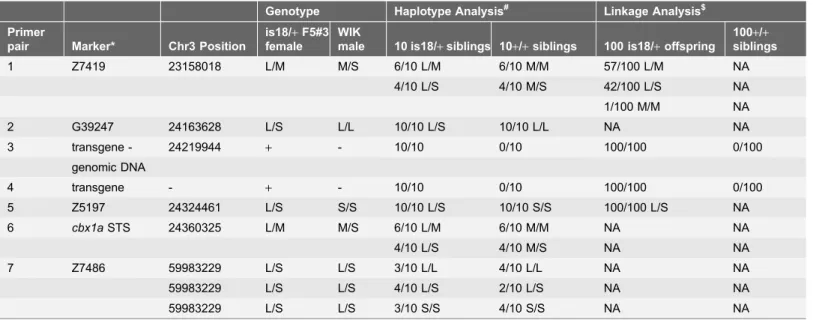
Table 1.Linkage ofTg(flk1:RFP)is18transgene integration site to position 24.219 Mb Chromosome 3.
Genotype Haplotype Analysis# Linkage Analysis$
Primer
pair Marker* Chr3 Position
is18/+F5#3 female
WIK
male 10 is18/+siblings 10+/+siblings 100 is18/+offspring
100+/+ siblings
1 Z7419 23158018 L/M M/S 6/10 L/M 6/10 M/M 57/100 L/M NA
4/10 L/S 4/10 M/S 42/100 L/S NA
1/100 M/M NA
2 G39247 24163628 L/S L/L 10/10 L/S 10/10 L/L NA NA
3 transgene - 24219944 + - 10/10 0/10 100/100 0/100
genomic DNA
4 transgene - + - 10/10 0/10 100/100 0/100
5 Z5197 24324461 L/S S/S 10/10 L/S 10/10 S/S 100/100 L/S NA
6 cbx1aSTS 24360325 L/M M/S 6/10 L/M 6/10 M/M NA NA
4/10 L/S 4/10 M/S NA NA
7 Z7486 59983229 L/S L/S 3/10 L/L 4/10 L/L NA NA
59983229 L/S L/S 4/10 L/S 2/10 L/S NA NA
59983229 L/S L/S 3/10 S/S 4/10 S/S NA NA
*Microsatellite STS markers Z7419, G39247, Z5197, Z7486.cbx1a STSis a Short Simple Repeat located in the 5th intron of thecbx1agene. Genotype determined by size of PCR amplification products after gel electrophoresis. L, long; M, middle; S, short allele PCR products.is18transgenic genotype (+or 2) confirmed by PCR with primers that amplify across the genomic DNA-transgene junction (3) or amplify an internal fragment of the transgene concatemer (4).
#Genotype ofis18/
+female and WIK male was determined. Segregation of STS markers in 10 is18/+and 10+/+siblings was used to determine the
haplotype of the chromosome containing the is18 transgene integration. TheTg(RFP)is18chromosome haplotype in the region surrounding the integration site is Z7419-L, G39247-S, Z5197-L,cbx1aSTS-L.
$100is18/
+and 100+/+siblings from a cross between aTg(flk1:RFP)is18/+female and WIK wild type male were genotyped by PCR. 1/100
Tg(flk1:RFP)is18/+progeny was homozygous Z7419 M/M and heterozygous G39247 L/S. This indicates a single recombination event between markers
Z7419 and G39247 and genetic map distance of 1 centimorgan.
Figure 2. TheTg(flk1:RFP)is18transgene integration site on chromosome 3 disrupts expression of lincRNAis18.v2.(A) Integration site of the transgene array at 24.2 Mb on chromosome 3. The integration sits downstream of theHoxBacluster and upstream relative to the heterochromatin binding family membercbx1a.
in exon 5 of cbx1a (Table 1). PCR linkage analysis demonstrated that the
Tg(flk1:RFP)is18 chromosome 3 haplotype at marker Z5197 located,105 Kb
from the integration site segregated with the transgene and tumor phenotype in 100 RFP expressing progeny from a tumor-bearing F5 adult Tg(flk1:RFP)is18
female outcrossed to a WIK wild type male (Table 1). As expected, one recombinant was recovered between the transgene and marker Z7419 located
,1.05 Mb 59 to the integration site (Table 1). Together with the genomic
Southern blot analyses, the linkage data provided strong evidence that the optic pathway tumor phenotype in line Tg(flk1:RFP)is18 was due to the presence of a high copy number concatemer that had integrated at position 24.212 MB on chromosome 3.
Zebrafish
Tg(flk1:RFP)is18
transgene integration in the
lincRNAis18
gene
We searched NCBI, Ensembl, and the recent zebrafish embryonic transcriptome study [13] for long intergenic noncoding RNAs (lincRNAs) that map to the region between theHoxBacluster andcbx1a on chromosome 3 and identified two. One of the genes, si:ch211-84g22.1 ENSDARG00000097724, was located,14 kb 39to the integration site (Fig. 2A). The second lincRNA was identified by a ,930 bp
transcript FDR202-P00026-DEPE-F_N10 FDR202 EH545544/Zv9_00007276 containing 8 exons that map to a genomic region spanning ,350 kb (Fig. 2A). The 59region and first exon overlaps thecbx1a gene, and downstream exons 6, 7, and 8 map in the intergenic regions between genes in theHoxBacluster (Fig. 2A). A shorter transcript of ,480 bp (si:ch211-246i5.4 ENSDARG00000097621/
FDR202-P00032-DEPE-R EH568666.1/Zv9_00007274) terminates in an alter-native third exon. Neither transcript is predicted to encode a polypeptide longer than 73 amino acids (reading frame 3). We named the locus encoding these transcripts lincRNAis18. The shorter transcript is designated lincRNAis18.v1, the longer transcript is designated lincRNAis18.v2(Fig. 2A). The Tg(flk1:RFP)is18
concatemer integration site is in the second intron of lincRNAis18.v2 (Fig. 2A). Since the concatemer contains many copies of the SV40 bidirectional
polyadenylation sequence, the transgene is predicted to cause premature
transcriptional termination oflincRNAis18.v2. Together, the data showed that the optic pathway tumor phenotype was linked to the integration of the
Tg(flk1:RFP)is18 concatemer in the second intron of lincRNAis18.v2.
We performed a number of experiments to determine whetherlincRNAis18 is involved in formation of ocular tumors in Tg(flk1:RFP)is18 heterozygotes. We
analyses with primers in exon 1 and exon 8 oflincRNAis18.v2showing expression oflincRNAis1.v28is disrupted in 6 dpf homozygousTg(flk1:RFP)is18larvae. 5 individual larvae of each genotype are shown. The genotype of each larva was confirmed (Figure S6). Control, expression of ribosomal protein S6 kinase b, polypeptide 1,rps6kb1. (C) TALENs targeting exons 2 and 5 oflincRNAis18. (D) Predicted structure of the
lincRNAis18e2e5deldeletion allele. Sequence of amplicon spanning exon 2 – exon 5 junction from F1
lincRNAis18e2e5del/
+adult genomic DNA.
examined the expression pattern of lincRNAis18 byin situhybridization and RT-PCR to determine whether it is expressed in the zebrafish retina. in situ
hybridization on adult retina detected a low level oflincRNAis18expression in the inner nuclear layer and the ganglion cell layer (S5 A Figure), indicating it is not highly expressed in the retina. RT-PCR on an adult tissue panel revealed high levels of lincRNAis18.v2 transcripts in the male and female germline and lower levels in muscle and retina (S5 B Figure). High levels of maternally supplied
lincRNAis18.v2 were observed in the developing embryo before the onset of zygotic transcription (S5 C Figure) then rapidly declined later stages. Multiple alternatively spliced forms oflincRNAis18were cloned from wild type adult retina, ovary and embryonic and larval stages (S5 Figure). Exons 1, 2 and 8 were consistently present in all isoforms, indicating a possible functional role of these exons. Although a low level of lincRNAis18.v2 expression was detected in the retina, the specificity of expression in this tissue, and its absence from other adult tissues except muscle, suggested it may have a role in retina function.
To further examine the coding potential oflincRNAis18, we searched the recently published ribosome profiling datasets of expressed transcripts from 8 early developmental stages of zebrafish [15]. The datasets included 2–4 cell, 256 cell and 1000 cell embryos in which a high level of maternally supplied
lincRNAis18 is present. None of the,900 million 35 bp sequence reads fully
aligned with the lincRNAis18 sequence, providing further evidence that
lincRNAis18 represents a novel lincRNA that most likely is not translated into a functional protein. Database searches showed lincRNAis18 has no significant homology with lincRNAs from other species. Despite the lack of homologous sequences in other vertebrate species, the high levels oflincRNAis18 expression in the embryo suggests it could play an important role in early zebrafish
development.
To determine whether the transgene integration in Tg(flk1:RFP)is18 disrupts expression oflincRNAis18.v2, we examinedlincRNAis18.v2expression by RT-PCR in homozygous Tg(flk1:RFP)is18/Tg(flk1:RFP)is18 individuals. Progeny from an intercross of Tg(flk1:RFP)is18/+ heterozygous adults were sorted into
RFP-expressing and RFP-negative classes. Genomic DNA and total RNA were isolated from each individual for genotyping and RT-PCR. Expression of lincRNAis18.v2
was undetectable in 5/5 homozygous is18/is18 larvae (Fig. 2B). The genotype of each individual was confirmed by PCR (S6 Figure). These data demonstrate that integration of theTg(flk1:RFP)is18transgene disrupts expression of the long form of lincRNAis18, lincRNAis18.v2.
To determine whether homozygosity results in additional phenotypes, all surviving progeny from the is18/+ heterozygous intercross were raised and
between 7 dpf and 4 weeks of age. These data indicate that homozygous
Tg(flk1:RFP)is18 larvae lack expression oflincRNAis18.v2 (Fig. 2B), due to premature transcription in the transgene, as predicted by the location of the transgene integration in intron 2. However, this does not rule out the possibility that the onset of edema and homozygous lethal phenotype might be caused by the presence of the Tg(flk1:RFP)is18 transgene concatemer or its effect on nearby genes.
To test the hypothesis that disruption of expression of lincRNAis18.v2 is homozygous lethal and involved in tumor formation, we isolated a second allele using TALEN genome editing to create a deletion in the lincRNAis18 gene (Fig. 2C). TALENs targeting exons two and five (S7 Figure) were co-injected to create a deletion that removes 147 kb of genomic DNA and fuses exon two to exon five. Exons further upstream or downstream were not targeted in order to avoid affecting the genomic region surrounding the cbx1a and HoxBa cluster genes. We recovered 1 founder out of 27 that transmitted the deletion allele to F1 progeny. Sequencing of the exon 2 - exon 5 junction fragment and genotyping with STR markers confirmed the identity of F1 individuals carrying the deletion allele lincRNAis18e2e5del (Fig. 2D). To test whether the deletion allele was
homozygous lethal, heterozygous lincRNAis18e2e5delF1 adults were intercrossed and the progeny were raised to adulthood. Genotyping identified homozygous F2 adults (S8 Figure), demonstrating that deletion of the genomic sequences between exons two and five oflincRNAis18did not result in lethality. By 8 months neither heterozygous nor homozygouslincRNAis18e2e5deladults developed ocular tumors. Together, these data suggest that tumor onset in Tg(flk1:RFP)is18 individuals is most likely not due to a loss of function of thelincRNAis18gene, or disruption of expression of lincRNAis18.v2. However, since the deletion allele that was
generated is predicted to produce a transcript containing exons 1, 2/5, 6, 7 and 8, it does not represent a null allele, and may retain some function that prevents homozygous lethality and tumor onset. Alternatively, because the
Tg(flk1:RFP)is18 transgene mutation segregates as a dominant allele, it might create a dominant effect on the expression of other lincRNAis18 transcript isoforms. It is also possible that the presence of the high copy number transgene in
Tg(flk1:RFP)is18 adults induces ocular tumor formation through an oncogenic
mechanism that induces overexpression of nearby genes. Transcriptome analyses presented below do not support the latter mechanism.
Histopathology of the zebrafish
Tg(flk1:RFP)is18
optic pathway
tumors reveals features of retinoblastoma and fibrous glioma
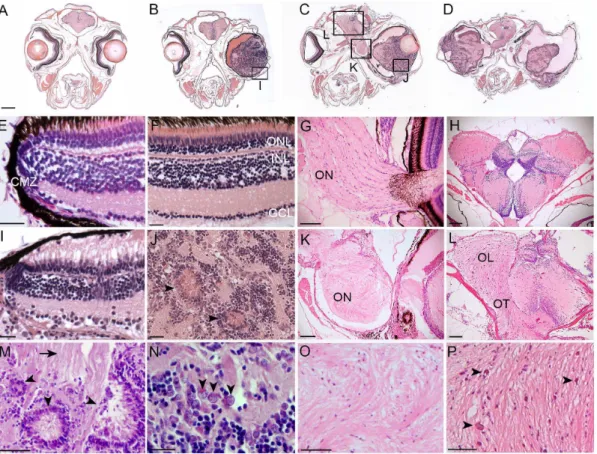
vessels were present throughout the tumors, indicating neovascularization in the advanced tumors. There were extensive glial fibrillar structures within the tumors (Fig. 3J, Marrow) and the optic nerve and tract (Fig. 3K, L). In advanced tumors the optic nerves and tract were completely replaced by neoplastic tissue, and the architecture of the brain lobes was highly disrupted (Fig. 3C, D, K, L). Cells having a ‘‘salt and pepper’’ chromatin dispersion pattern were present, again consistent with neuro-ectodermal tumor cell cytomorphology (Fig. 3N, arrow-heads). Areas of necrosis were present throughout the optic lobe (Fig. 3Parrows). Examination of tumors in younger adults revealed relatively normal organization of the retina at the ciliary marginal zone. In a 5-month old adult with a large ocular mass (Fig. 3B), the neural retinal layers were observed to be intact at the
Figure 3. Histological analysis ofTg(flk1:RFP)is18tumors reveals similarities with retinoblastoma and glioma.Coronal sections through heads of wild type (A, E–H) andTg(flk1:RFP)is18/+adults (B–D, I–L). (B) 5-month-oldTg(flk1:RFP)is18/+adult with retinal tumor filling the vitreous. 1-year-old Tg(flk1:RFP)is18/+adults with unilateral (C) or bilateral tumors (D). Tumor cells extend through the optic pathway to the tectum. (E,F) Ciliary marginal zone
and mature retina in wild type. (G) Fibrillar ribbon-like structure of a wild type optic nerve exiting the eye. (H) Section through forebrain shows the sacus dorsalis and left and right lobes of the anterior region of the optic tectum. (I) Intact ciliary marginal zone in the tumorous retina fromTg(flk1:RFP)is18/+adult shown in B. Degeneration of the retinal pigment epithelium and photoreceptor outer segments is evident. Disorganization of the retinal layers adjacent to the ciliary marginal zone is present. Streaking across the inner plexiform layer appears similar to reactive Mu¨ller glia. (J) Advanced tumors contain rosettes and blood vessels extending throughout the tumor tissue. (K) Disorganization and dysplasia in the optic nerve of theTg(flk1:RFP)is18/+adult in C. (L) Brain from
the adult in panel D showing dysplasia of the optic tract with infiltration and disruption of normal brain structures. (M) Dysplastic areas of advanced tumor with rosettes of various sizes (arrowheads) and extensive glial fibrillar proliferation (arrow). (N) ‘‘Salt and pepper’’ chromatin dispersion pattern (arrowheads) consistent with a neuroectodermal tumor cell. (O) Disorganization of the optic nerve with absence of organized fibrils. (P) Expansion of the optic lobe with possible areas of necrosis (arrowheads). CMZ, ciliary marginal zone; GCL, ganglion cell layer; INL, inner nuclear layer; OL, optic lobe; ON, optic nerve; ONL, outer nuclear layer; OT, optic tract. Scale bars A, B, C, D 500mm; E, F, G, I, J, N 20mm; H, K, L 100mm; M, O, P 50mm.
ciliary marginal zone (Fig. 3I). These initial examinations indicated the ocular masses likely originated from cells located in the differentiated tissue of the neural retina, not from the retinal stem cell population that resides at the ciliary marginal zone. Overall, the retinal tumors appeared similar to fibrous glioma with features present in human retinoblastoma.
Evidence for involvement of glia in
Tg(flk1:RFP)is18
tumor
proliferation
To gain further insight into the identity of theTg(flk1:RFP)is18retinal tumors we examined the expression of retinal neural markers in cryosections of tumor tissue. The calcium binding protein Recoverin is normally expressed in all photo-receptors in the retina (Fig. 4A), while the antibody RT97, which broadly recognizes neuronal intermediate filament proteins, also labels photoreceptor outer segments (Fig. 4A). In tumors Recoverin was detected in the cell bodies and RT97 labeled structures projecting into the center of the rosettes (Fig. 4E–H). RT97 also strongly labeled the fibrous stroma of the tumor (Fig. 4E–F), indicating the presence of neuronal processes in this matrix. The synaptic vesicle marker SV2 was also detected throughout the fibrous stroma (G). These analyses were consistent with the histopathology suggesting the tumors likely originate from the neural retina.
To examine the proliferative cell populations in theTg(flk1:RFP)is18 retinal tumors, we labeled adult fish with BrdU for 2 hours, followed by a 4 hour recovery period. In wild type retina BrdU incorporation was detected at the ciliary marginal zone where progenitor cells reside (Fig. 4D, arrow). Very rarely BrdU was detected in single cells in more central retina, consistent with the slow cycling of the Mu¨ller glia-rod photoreceptor lineage. We examined BrdU incorporation in advanced tumors and found robust labeling throughout the tumor mass with some overlap with rosettes (Fig. 4H). BrdU did not consistently co-label cells expressing a specific retinal neural cell type marker, making it difficult to conclusively determine the identity of proliferating cells in very advanced tumors. Moreover, histopathology did not indicate high mitotic activity in the advanced tumors (Fig. 3). This suggests the possibility that the large number of cells that incorporated BrdU was not due to DNA synthesis during S phase but was the result of the activity of global DNA repair processes.
Numerous studies have demonstrated that the Mu¨ller glia can be activated in response to retinal injury and reprogrammed to produce progenitors that repopulate all retinal neural cell types [16]. To determine if the Tg(flk1:RFP)is18
glial cell type. Increased expression was also evident in the dysplastic optic nerve at the lamina cribrosa of an eye with an advanced tumor (Fig. 4O), indicating expansion of the astroglia of the optic nerve. To visualize activated Mu¨ller glia, we examined immunolocalization of Brain Lipid Binding Protein/fatty acid binding protein (BLBP/fabp). In wild type retinas BLBP is expressed in progenitor cells at the ciliary marginal zone (Fig. 4L) but is absent from Mu¨ller glia. In the
Tg(flk1:RFP)is18 retinal tumors an increase in BLBP was detected throughout the tumor and did not appear to co-localize with GFAP positive Mu¨ller glia cells (Fig. 4L, P). This suggested BLPB was labeling a population of cells distinct from mature Mu¨ller glia. Together these results indicateTg(flk1:RFP)is18tumors might
Figure 4. Characterization oflincRNAis18tumors indicates a glial cell origin.Immunolabeling and in situ hybridization of cryosections from wild type retina (A–D, I, J, M–P) and advancedlincRNAis18tumors (E–H, K, L, Q–T). Cells in rosettes in the tumors label with neurofilament marker RT-97 (red) and photoreceptor marker recoverin (green) (E, F). The synaptic vesicle marker SV2 (green), which is enriched in the retinal plexiform layers (C), was distributed throughout the fibrous tumor mass (G). (D) BrdU (green) incorporated into proliferating progenitor cells at the ciliary marginal zone of the wild type retina (arrow). (H) Intense labeling of BrdU incorporation was detected in cells forming rosettes and throughout the tumor mass. In wild type the glial marker GFAP is most evident in the Mu¨ller glia end feet that sit in the retinal ganglion cell layer (I–L). GFAP expression is absent in the oligodendrocytes and astrocytes of the optic nerve (I, K arrow). In contrast, GFAP was readily detected in long streaks throughout the tumor tissue (M, N, P), and was expressed by cells in the mutant optic nerve (O, arrow). BLBP is present at the ciliary marginal zone of wild type retina (L) and appeared present throughout the tumor mass (P). A, E, I, M, Differential interference contrast (DIC) overlay on immunofluorescence labeling images. CMZ, ciliary marginal zone; GCL, ganglion cell layer; INL, inner nuclear layer; ON, optic nerve; ONH, optic nerve head; ONL, outer nuclear layer. All scale bars represent 50mm,
except in panels K and O scale bars represent 100mm.
be the result of abnormal levels of glial proliferation, with features similar to activated Mu¨ller glia and the neural progenitors derived from them.
Differential gene expression analysis of
Tg(flk1:RFP)is18
retinal
tumor progression
To identify the molecular signatures that correlate with onset and tumorigenesis of Tg(flk1:RFP)is18tumors we performed differential gene expression analysis by RNA-Seq. Libraries were prepared from 6 month old age-matched wild type retina (Wild Type), pretumor retina from heterozygous Tg(flk1:RFP)is18 adults
(Pretumor), and advanced tumor tissue from heterozygous Tg(flk1:RFP)is18
adults (Tumor) (S6 Table). The data was mapped to the zebrafish v9 genome, gene model assembly version 71 (S7 Table). The gene models included 1133 recently identified lincRNAs expressed during early zebrafish development [13]. Genes with FPKM (Fragments Per Kilobase per Million sequenced reads) value of
.51 in Wild Type retina were examined for significant changes in expression level in Pretumor and Tumor samples (S7 Table). GO term analysis (Table 2) indicated translational activity was elevated in Pretumor and Tumor tissue. In Pretumor and Tumor tissue cellular respiration was decreased; photoreception and ion transport processes were also decreased in tumor. Consistent with the GO term analysis, the gene set that showed the greatest down-regulation between Wild Type and Tumor was enriched for genes that function in photoreceptor
maintenance and photoreception (S8 Table), indicating that in the
Tg(flk1:RFP)is18 tumors, normal photoreception and synaptic transmission are disrupted. As expected for proliferating tumor, mitosis and cell division processes were increased inTg(flk1:RFP)is18tumors. 252 of the 1133 lincRNAs identified in the zebrafish embryonic transcriptome were expressed in wild type retina (S7 Table). 11 of the 252 lincRNAs were significantly increased or decreased in expression level in Pretumor and/or Tumor tissue (S9 Table).
We examined the expression levels of genes that map in the region of the
Tg(flk1:RFP)is18 transgene integration in order to determine if the array influenced expression of nearby genes. Only 5 reads in the wild type retina transcriptome mapped to lincRNAis18, indicating the majority of lincRNAis18
mRNA was lost during isolation of polyadenylated RNA for RNA-Seq library construction. We confirmed this by RT-PCR using the input RNA for library construction and samples of the resulting libraries. The presence of lincRNAis18
mRNA was detected by RT-PCR in the total RNA input sample, but was only detectable in the wild type RNA-Seq library after nested RT-PCR (S9 Figure). The lincRNA si:ch211-84g22.1, located in intron 2 of lincRNAis18 and positioned 14 kb 39to the Tg(flk1:RFP)is18 transgene integration site, was only represented by 4 reads in wild type retina. Expression levels of si:ch211-84g22.1 in
Tg(flk1:RFP)is18 Pre-Tumor and Tumor tissue were increased (98 and 132 reads
lincRNA si:ch211-84g22.1. Other genes located in the region of the
Tg(flk1:RFP)is18 transgene integration were examined for altered expression. No significant change in expression level was detected for genes that map within 1 Mb upstream or downstream of the transgene integration, including the HoxBa
cluster and cbx1a genes. Overall, the analyses indicate theTg(flk1:RFP)is18
transgene integration is responsible for inducing tumor formation, but the mechanism does not involve altering the expression level of genes in cis other than the lincRNA si:ch211-84g22.1.
Human Mutation-Driver and Glioma Marker Genes
We examined the Tg(flk1:RFP)is18 tumor differentially expressed gene set for changes in tumor suppressors and oncogenes that identify disrupted signal transduction pathways in cancer. Although the ocular tumors showed features of retinoblastoma tumors, the expression level of the rb1 tumor suppressor was unchanged in pretumor and elevated ,3 fold in tumor tissue (S7 Table),
indicating the tumors do not arise due to deletion of the rb1locus. 62 of the 138 designated Mutation-Driver human cancer genes [17] showed at least a 2-fold change in expression (Fig. 5A). In the Wnt signaling pathway, two direct molecular targets of ß-catenin transcriptional activation, the myconcogene [18] and cell cycle regulatorcyclinD1[19], showed significant increases in expression in
Tg(flk1:RFP)is18 tumor tissue. Two tumor suppressors in the PI3K-mTOR
pathway altered in the majority of human gliomas [20–23], PI3K regulatory subunitpi3kr1and thetsc1repressor of mTOR, were decreased in expression level in the Tg(flk1:RFP)is18 tumors (Fig. 5A and S7 Table). The tumors showed a significant increase in expression of glioma diagnostic markers [24,25] [22]
vimentin (vim), S100ß(s100ß) and the matrix metalloprotease mmp9 (Table 3).
Table 2.Categories of biological processes altered inTg(flk:RFP)is18/+Pre-tumor and Tumor tissues.
DGE Group* GO Process p-value
Increased in Pretumor translation 4.50e-13
ribonucleoprotein complex biogenesis 9.59e-8
regulation of translational initiation 5.62e-5
ribosome biogenesis 9.28e-5
Decreased in Pretumor cellular respiration 9.38e-7
energy derivation by oxidation of organic compounds 4.53e-5
ATP metabolic process 9.01e-5
Increased in Tumor ribonucleoprotein complex biogenesis 1.99e-13
translation 3.86e-12
mitotic cell cycle 9.48e-9
cell division 5.02e-7
Decreased in Tumor sensory perception of light stimulus 2.00e-4
cellular respiration 3.11e-3
ion transport 3.67e-3
*DGE Group: Differential Gene Expression Group. Genes were grouped according to direction of change in expression level in Pretumor and Tumor tissue.
The zebrafish homolog of the putative tumor suppressor ajap1[26] was also significantly decreased in expression in Tg(flk1:RFP)is18 tumor tissue (Table 3). qRT-PCR confirmed ajap1decreased in expression 4-fold (Fig. 5C). These data are consistent with the histological and immunohistochemical data that suggests
Tg(flk1:RFP)is18 retinal tumors express a glial gene signature.
Glial activation and Neuronal Progenitors
Two populations of glia cells in the retina may contribute progenitor cells that give rise to the Tg(flk1:RFP)is18 tumors. The Mu¨ller glia reside within the neural retina and in the mature retina function as late retinal progenitors that produce the rod photoreceptor lineage. The nerve fiber layer contains astrocytes that migrate into the retina from the optic nerve during embryogenesis. We examined the transcriptome data for changes in gene expression of markers for glia, glial progenitors, reactive astrocytes and activated Mu¨ller glia. Glutamate transporter GLAST/SLC1A3 has been identified as a marker of radial glia, glial progenitor cells, and glial progenitor-derived astrocytes [27]. Zebrafish slc1a3a and-b were both significantly increased in expression in the Tg(flk1:RFP)is18 tumors
(Table 3). Activating transcription factor 3 (ATF3) is a member of the cAMP-response element binding protein family of transcriptional activators that bind the CRE (cAMP response element) enhancer and are activated in response to a rise in cytosolic cAMP levels. atf3 expression increases in the retinal ganglion and nerve fiber layers of the retina and the optic nerve after injury to the optic nerve in adult zebrafish [28]. In theTg(flk1:RFP)is18 transcriptome atf3expression levels increased significantly in Pretumor and Tumor tissue (Table 3). In contrast,bystin
(bysl), whose expression increases substantially after optic nerve injury and marks reactive astrocytes [29], remained unchanged (S7 Table). Changes in expression levels ofatf3andbyslwere confirmed by qRT-PCR (Fig. 5C). These data indicate reactive gliosis was not solely responsible for the increased expression of glial progenitor markers in the Tg(flk1:RFP)is18 ocular tumors.
Many of the genes required for proliferation of neuroglial progenitor cells were increased in expression level in the Tg(flk1:RFP)is18 tumor tissue. This included nerve growth associated factor gap43, neural stem cell intermediate filament protein nestin, and neural cadherin 2cdh2 (Table 3). Transcription factors required in neural stem cells (foxd3,sox21b,sox4a,sox4b,sox11aandsox11b) were also increased inTg(flk1:RFP)is18Tumor tissue (Fig. 5B;Table 2). In response to photoreceptor degeneration after retinal injury, the Mu¨ller glia become activated to reenter the cell cycle and produce retinal progenitors. The gene expression pattern that defines proliferating Mu¨ller glia (apoeb,blbp,cdh2,hes1,rlbp1aand–
Figure 5. Differential gene expression analysis of retina RNA-Seq from wild type, pre-tumor, and retinal tumor tissues.A, B Heat map
representations of differentially regulated transcripts associated with tumor initiation and progression. (A) Differential gene expression of 62 Mutation-Driver genes. Tumor suppressors are in green, oncogenes are in red. (B) Differential gene expression of genes required for photoreceptor and neural function, neural progenitor proliferation, and injury induced regeneration. Scale represents log2 fold change in gene expression. (C) Comparison of absolute fold change in gene expression levels forajap1,ascl1a,atf3,bysl,hbegfa, andinsm1ameasured by RNA-Seq (upper panel) and qRT-PCR (lower panel).
Tg(flk1:RFP)is18/+Pretumor (red bars) andTg(flk1:RFP)is18/+Tumor (blue bars).
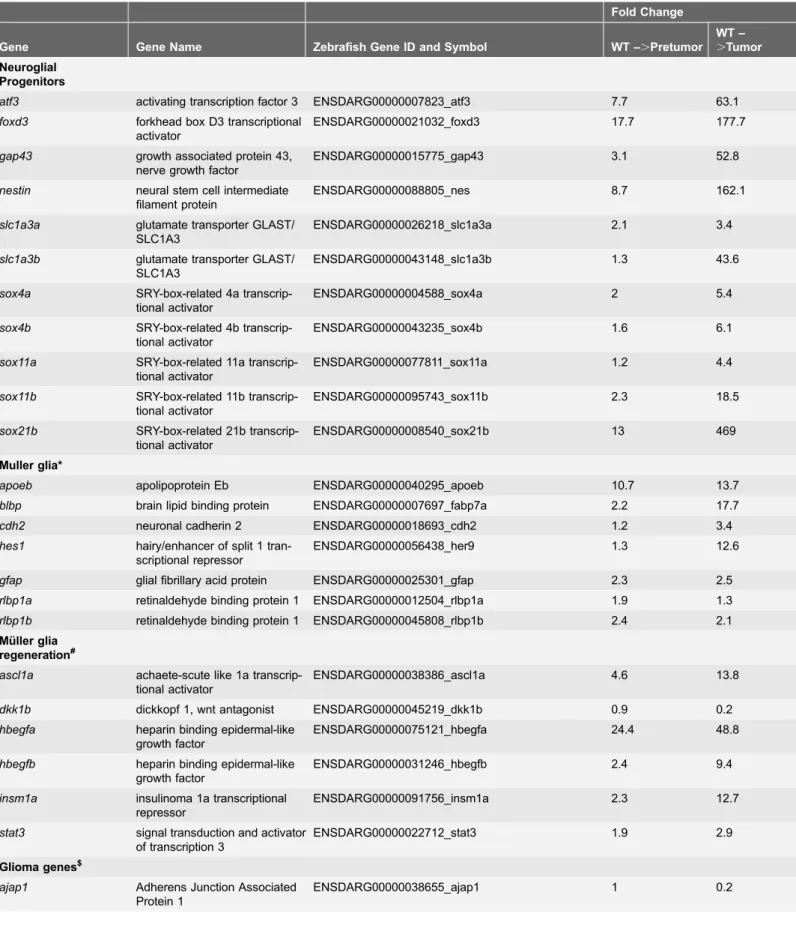
Table 3.Differential Expression of Neural Progenitor, Mu¨ller glia Regeneration, and Glioma Genes inTg(flk1:RFP)is18/+tumor progression.
Fold Change
Gene Gene Name Zebrafish Gene ID and Symbol WT –.Pretumor
WT –
.Tumor
Neuroglial Progenitors
atf3 activating transcription factor 3 ENSDARG00000007823_atf3 7.7 63.1
foxd3 forkhead box D3 transcriptional
activator
ENSDARG00000021032_foxd3 17.7 177.7
gap43 growth associated protein 43,
nerve growth factor
ENSDARG00000015775_gap43 3.1 52.8
nestin neural stem cell intermediate filament protein
ENSDARG00000088805_nes 8.7 162.1
slc1a3a glutamate transporter GLAST/ SLC1A3
ENSDARG00000026218_slc1a3a 2.1 3.4
slc1a3b glutamate transporter GLAST/ SLC1A3
ENSDARG00000043148_slc1a3b 1.3 43.6
sox4a SRY-box-related 4a
transcrip-tional activator
ENSDARG00000004588_sox4a 2 5.4
sox4b SRY-box-related 4b
transcrip-tional activator
ENSDARG00000043235_sox4b 1.6 6.1
sox11a SRY-box-related 11a transcrip-tional activator
ENSDARG00000077811_sox11a 1.2 4.4
sox11b SRY-box-related 11b transcrip-tional activator
ENSDARG00000095743_sox11b 2.3 18.5
sox21b SRY-box-related 21b transcrip-tional activator
ENSDARG00000008540_sox21b 13 469
Muller glia*
apoeb apolipoprotein Eb ENSDARG00000040295_apoeb 10.7 13.7
blbp brain lipid binding protein ENSDARG00000007697_fabp7a 2.2 17.7
cdh2 neuronal cadherin 2 ENSDARG00000018693_cdh2 1.2 3.4
hes1 hairy/enhancer of split 1 tran-scriptional repressor
ENSDARG00000056438_her9 1.3 12.6
gfap glial fibrillary acid protein ENSDARG00000025301_gfap 2.3 2.5
rlbp1a retinaldehyde binding protein 1 ENSDARG00000012504_rlbp1a 1.9 1.3
rlbp1b retinaldehyde binding protein 1 ENSDARG00000045808_rlbp1b 2.4 2.1
Mu¨ller glia regeneration#
ascl1a achaete-scute like 1a transcrip-tional activator
ENSDARG00000038386_ascl1a 4.6 13.8
dkk1b dickkopf 1, wnt antagonist ENSDARG00000045219_dkk1b 0.9 0.2
hbegfa heparin binding epidermal-like growth factor
ENSDARG00000075121_hbegfa 24.4 48.8
hbegfb heparin binding epidermal-like growth factor
ENSDARG00000031246_hbegfb 2.4 9.4
insm1a insulinoma 1a transcriptional repressor
ENSDARG00000091756_insm1a 2.3 12.7
stat3 signal transduction and activator of transcription 3
ENSDARG00000022712_stat3 1.9 2.9
Glioma genes$
ajap1 Adherens Junction Associated
Protein 1
b) also showed increased expression in the Tg(flk1:RFP)is18 tumors. Reprogramming of Mu¨ller glia is mediated by activation of multiple signal transduction pathways [4]. The JAK/Stat cytokine signal transduction pathway transcription factorstat3, which is required for proliferation after light dependent retinal injury and regeneration [9,10], was elevated nearly 3-fold in the tumor (Table 3).Wnt-ß-catenin signal transduction is activated in Mu¨ller glia derived progenitors via a network involving heparin binding epidermal growth factor
hbegf, the transcriptional activatorascl1a, and the transcriptional repressorinsm1a
[7,12]. Each of these genes was significantly increased in theTg(flk1:RFP)is18
Tumor, while the wntantagonist dkk1bwas significantly decreased in expression (Table 3). We confirmed the differential gene expression ofhbegfa, ascl1a, and
insm1a by qRT-PCR (Fig. 5C). Together, these data are consistent with the hypothesis that Tg(flk1:RFP)is18 retinal tumors might arise from neural progenitors derived from Mu¨ller glia and/or astroglia.
Discussion
In this report we describe the isolation and characterization of a zebrafish optic pathway tumor line that is linked to integration of a Tol2,flk1:RFP-CAAX.
transgene concatemer in line Tg(flk1:RFP)is18.By 1 year, greater than 80% of heterozygous Tg(flk1:RFP)is18 adults develop tumors in the retina, optic nerve and optic tract with features of retinoblastoma and fibrous glioma. Histological, immunohistochemical, and transcriptomic analyses of Tg(flk1:RFP)is18 ocular tumors are consistent with the tumor originating in part from a glial cell population in the retina that includes the Mu¨ller glia. Astrocytes residing within the optic nerve fiber layer may also contribute to the retinal tumor cell progenitor population. The dominant pattern of inheritance indicates the presence of the transgene results in an oncogenic mechanism that induces tumor onset. The
Tg(flk1:RFP)is18 transgene integration is homozygous lethal, and animals die between 1 and 4 weeks of age. The transgene disrupts expression of the lincRNA gene,lincRNAis18 (Zv9_00007276) but elevates expression of the opposite strand
lincRNA si:ch211-84g22.1 ,25 fold. A second deletion allele generated using
Table 3.Cont.
Fold Change
Gene Gene Name Zebrafish Gene ID and Symbol WT –.Pretumor
WT –
.Tumor
mmp9 matrix metalloprotease 9 ENSDARG00000042816_mmp9 2.0 7.4
S100ß neural calcium binding protein ENSDARG00000057598_s100b 1.6 5.0
vim vimentin ENSDARG00000010008_vim 2.8 9.6
*Raymond et al., 2006.
#Kassen el al., 2009; Nelson et al., 2012; Ramachandran et al., 2011; Ramachandran et al., 2012; Wan et al., 2012. $
Zhao et al., 2010; Lin et al., 2012.
TALEN genome editing that removes exons 2–5 of lincRNAis18 and the entire lincRNAsi:ch211-84g22.1locus did not result in a tumor or lethal phenotype. This suggests that in lineTg(flk1:RFP)is18 the presence of the Tol2,flk1:RFP-CAAX.
array is responsible tumor formation and lethality, however, the mechanism underlying tumor onset is not known.
Our study provides several pieces of evidence that the Tg(flk1:RFP)is18 retinal tumors likely originate from glial-derived neural progenitors in the retina. The
Tg(flk1:RFP)is18 phenotype is very similar to the previously reported zebrafish optic pathway glioma model in which activation of Sonic hedgehog signaling in neural progenitors induces tumors [14]. Radial glia in the CNS are the source of neural stem cells and glial progenitors during development and in adult
neurogenesis [30]. The dramatic increase in expression of the radial glial marker
GLAST/slc1a3b [27] and the neural stem cell markers foxd3, nestin, sox4, sox11, and sox21 in Tg(flk1:RFP)is18 tumor tissue supports a neuroglial progenitor population. The CREB transcriptional activator ATF3 and the reactive astrocyte marker Bystin are associated with reactive gliosis and upregulated in the optic nerve in response to injury [28,29]. While atf3 was dramatically increased in expression in the Tg(flk1:RFP)is18 retinal tumors, zebrafish bystin-like (bysl) showed no change in expression level. Consequently, the activation of glia in the
Tg(flk1:RFP)is18 model is not merely a result of reactive gliosis due to insult or injury in the retina. ATF3 is a member of the cAMP-response element binding protein family of transcriptional activators that bind the CRE (cAMP response element) enhancer and are activated in response to a rise in cAMP levels. In fetal brain synergism between the cytokine LIF and BMP signal transduction pathways is mediated by CREB, which acts as a bridge between STAT3 and smad1 to promote astrocyte formation from neural progenitors [31]. Consistent with increase in CREB family member ATF3, both zebrafishsmad1andstat3expression levels were elevated inTg(flk1:RFP)is18 tumors, indicating a role for cAMP signal transduction in tumor growth.
Activation of multiple signal transduction pathways required for inducing Mu¨ller glia proliferation and progenitor production was observed in the
Tg(flk1:RFP)is18 ocular tumors. Mu¨ller glia markers, such as apolipoprotein Eb,
apoeb, brain lipid binding protein, blbp, and hairy/enhancer of split 1
transcriptional repressor, hes1 [32], were significantly increased in the tumors. The JAK/Stat signal transduction and transcriptional activator stat3, which is required to stimulate Mu¨ller glia proliferation in the injured zebrafish retina [9,10], was elevated as well in tumor tissue. Theinsm1a, ascl1a/dkk1b, andhbegf
pathways that active wnt signaling to induce Mu¨ller glia proliferation [7,11,12] were also significantly altered. Together these data support the hypothesis that the Mu¨ller glia in the Tg(flk1:RFP)is18 retinas dedifferentiate and produce
transformed neuroglial progenitors. Retinal tumor progenitors might also arise from astrocytes located in the nerve fiber layer. Overall, the transcriptome data, histological analyses, and immunohistochemical labeling of tumor tissue provides significant support for the conclusion that transformed glia give rise to the
proliferation in the Tg(flk1:RFP)is18 retina will require additional studies. The zebrafish Tg(flk1:RFP)is18 line presents a highly penetrant and consistent retinal tumor model that will be useful for investigating the mechanisms driving glia activation and reprogramming in the vertebrate central nervous system.
Materials and Methods
Zebrafish husbandry and genetics
Zebrafish were reared in an Aquatic Habitat system (Aquatic Ecosystems, Inc., Apopka, FL). Fish were maintained on a 14-hr light/dark cycle at 27
˚
C. Transgenic lines were established in a WIK wild type strain obtained from the Zebrafish International Research Center (http://zebrafish.org/zirc/home/guide.php). For in situ hybridization experiments, embryos were collected and maintained at 28.5˚
C in fish water (60.5 mg ocean salts/l) containing 0.003% 1-phenyl-2-thiourea (PTU) until harvesting. Embryos were staged according to published guidelines [33]. All experimental protocols were approved by the Iowa State University Institutional Animal Care and Use Committee (Log # 11-06-6252-I) and are in compliance with American Veterinary Medical Association and the National Institutes of Health guidelines for the humane use of laboratory animals in research. Adult fish were anesthetized and euthanized in MS-222 Tricaine Methanesulfonate prior to sacrifice and tissue dissection for histopathology and immunolabeling.Animal Care and Humane Endpoint Establishment
Transgenic fish predisposed to tumor formation were raised side by side with non-transgenic siblings. Heterozygous and homozygous transgenic fish and sibling fish were monitored daily during routine feeding for viability and
morbidity, and monitored bi-weekly for gross presentation of ocular tumors. 50% of each generation of transgenic fish developed pericardial edema. All
homozygous transgenic fish presented with pericardial edema beginning at 2– 7 dpf and developing through 4 weeks of age. Fish presenting with edema were sacrificed before swimming and feeding behavior were adversely affected. Juvenile and adult fish were anesthetized and euthanized in MS-222 Tricaine
Methanesulfonate according to experimental protocols approved by the Iowa State University Institutional Animal Care and Use Committee (Log # 11-06-6252-I) in compliance with the American Veterinary Medical Association and the National Institutes of Health guidelines for the humane use of laboratory animals in research.
for size and length relative to non-transgenic siblings. Fish were sacrificed before tumor burden reached 3 mm in size/25 mg in weight, constituting less than 10% of the total body weight of an adult fish (300–500 mg), as outlined for mouse and rat studies [34]. No adverse affect on growth rate, feeding behavior or fertility was detected in Tg(flk1:RFP)is18 fish with a tumor burden less than 3 mm/10% of body weight. For transcriptome studies age matched 6 month old fish with tumor size ranging from undetectable to 2 mm in size were anesthetized and euthanized in MS-222 Tricaine Methanesulfonate. Retinal and tumor tissue was dissected for isolation of total RNA. In each generation of Tg(flk1:RFP)is18 fish, individuals presenting with ocular tumors were sacrificed when tumor burden reached 3 mm/ 25 mg of total body weight, or by 1 year of age, whichever endpoint was first reached.
Isolation of transgenic line
Tg(flk1:RFP)is18
The endothelial specific membrane targeted RFP reporter construct flk1:RFP-CAAX was assembled using standard PCR cloning methods. The zebrafish promoter for theflk1gene was amplified from WIK genomic DNA. The construct was cloned into the minipTol2transgenesis vector [35]. Transgenics were isolated by co-injection of in vitro transcribed, capped, polyadenylatedTol2 transposase mRNA [35] and the pTol2,flk1:RFP-CAAX.construct into 1 cell zebrafish WIK embryos, as described previously [36]. Three independent lines expressing RFP in the endothelial cells of the vasculature were isolated. Line Tg(flk1:RFP)is18
contains a high copy number array Tol2,flk1:RFP-CAAX.. The
Tg(flk1:RFP)is18 line is available upon request.
Tol2
,
flk1:RFP
.
is18
transgene integration site cloning
A custom RNA bait library (Agilent) was used for capture of theTol2,flk1:RFP.
transgene concatemer and flanking genomic DNA sequences following the Agilent SureSelect Targeted Capture protocol. Briefly, a panel of overlapping biotin-labeled RNA baits (S1 Table) complementary to the pTol2,flk1:RFP. construct was synthesized. Genomic DNA from 5 samples was subjected to shearing, hybridization capture, library amplification, and index barcoding as outlined in the Custom SureSelect Target Enrichment protocol (Agilent). Genomic DNA was isolated from muscle tissue from one Tol2,flk1:RFP.is18/+ heterozygous adult
from the F2, F3 and F4 generation of the is18 pedigree was isolated. Genomic DNA was also isolated from the tumor tissue from the F3 and F4 individuals. The genomic DNA was isolated with the Agilent SureSelect gDNA Extraction kit (Agilent). 3 mg of genomic DNA from each of the 5 samples was sheared to
,250 bp (Covaris, Inc., Woburn, MA). The SureSelect Captured Libraries were
75 bp paired end multiplex sequenced in one lane on an Illumina GA II
GSNAP [37] to theDanio rerio reference genome (Zv9 64) amended with the
pTol2,flk1:RFP.construct sequence as a separate scaffold. Reads were filtered to remove those that failed to map to both the transgene and a unique location in the zebrafish genome. The remaining reads were used to identify 1000 bp intervals in the genome that had a higher level of mapped reads than expected by random chance using a modification of a previously published bootstrap method written in the R programming language [38]. 4 locations were identified in which paired end sequences had one end mapped to the pTol2,flk1:RFP. construct and the other end mapped to a unique, non-repetitive sequence in the genome (S3 Table). Each site was tested for confirmation by PCR amplification of the predicted transgene-chromosome junction fragment with primers complementary to the
pTol2,flk1:RFP.construct and the flanking genomic DNA (S4 Table). Only one of the 4 sites, chromosome 3:24,212,813–24,212,885, was validated by amplifica-tion in all 5 samples (S1 B, C Figure). A total of 81 sequences in the 5 samples mapped to the transgene and to genomic DNA flanking either the 59or 39sides of the integration site at 24,212,944 on chromosome 3 (S1 D Figure).
Genomic Southern blot analyses
Genomic DNA was isolated from adult zebrafish using a Qiagen midiprep kit and chemiluminescent non-radioactive Southern blot analyses performed as described previously [39]. Sequences of primers for probes for genomic Southern analyses: 450 bp probe complementary to chr 3 position 24,214,849–24,215,303; forward 59
CTCATTCTGTCCATGTGTTCACAG 39, reverse 59 CTTCTTGCCTGACT-TTCACAGCC 39: 450 bp probe complementary to chr 3 positions 24,211,859– 24,212,364; forward 59 CTGACAAGCAGCTGACAGATTGG 39, reverse 59
GGAAGTTGCTCTCATAATTCACG 39: 450 bp probe complementary to RFP cDNA; forward 59CTTCAGGGCCATGTCGCTTCTG 39, reverse 59 CATGG-AGGGCACCGTGAACAA 39: 477 bp probe complementary to ßlactamase cDNA in pTol2 vector backbone; forward 59 ATCAGTGAGGCACCTATCTCAGC 39, reverse 59 CATAACCATGAGTGATAACACTGC 39.
Imaging, histopathology and immunohistochemistry
For immunohistochemistry heads were removed from anesthetized adults and fixed in 4% paraformaldehyde overnight at 4
˚
C, decalcified in Cal-Ex for 2 days at 4˚
C, then processed for embedding in optimal cutting temperature (OCT) medium (Fisher). Heads were serial sectioned at 12 mm on a Microm HM 550cryotstat at 25
˚
C. For BrdU labeling experiments, adults were incubated in 5 mMBrdU (Sigma) in fish water (60.5 mg ocean salts/l) for 2 hours, placed in fresh fish water for 4 hours, then sacrificed and processed as above for immunohisto-chemical labeling experiments. Antibody labeling of cryosectioned tissue was as described previously [9,40]. Dilutions and primary antibodies used for labeling sectioned tissue were as follows: rabbit polyclonal anti-recoverin 1:1000
(Millipore); mouse monoclonal anti-glial fibrillary acid protein GFAP 1:1000 obtained from the Zebrafish International Research Center (ZIRC); rabbit polyclonal anti-Brain Lipid Binding Protein BLBP 1:200 (Abcam); mouse monoclonal anti-SV2 1:100 (Developmental Studies Hybridoma Bank); mouse monoclonal RT97 1:250 (Developmental Studies Hybridoma Bank); anti-BrdU 1:500 (Bio-Rad). Alexa-594 and Alexa-697 conjugated secondary antibodies (Invitrogen) and Cy3 and Cy5 conjugated secondary antibodies (Jackson
Immunoresearch Labs) were used at a dilution of 1:500. Tissues were counter-stained with 5 mg/ml DAPI (Sigma) and mounted in Fluorogel (EMS). To aid
antigen retrieval tissues were pretreated with 2 M HCl (for anti-BrdU labeling). Immunofluorescent labeling of cryosections was imaged on a Nikon Microphot-FXA microscope and captured using a QImaging Retiga 2000R Fast 1394 camera and QCapture software. All images were edited and assembled in Adobe
Photoshop CS2.
RT-PCR and in situ hybridization
Total RNA for staged developmental series, tissue panel, and analysis of
homozygous embryos, was isolated with a Qiagen RNeasy Isolation Kit. RT-PCR was carried out using a One-Step RT-PCR Kit (Invitrogen). cDNA for
lincRNAis18 was amplified by RT-PCR out of total RNA isolated from wild type
48 hpf embryos or adult retina. cDNAs were cloned into pBluescript or the pCR4-TOPO vector (Invitrogen). Primers for amplification were as follows:lincRNAis18
forward1 59TCACTGTCTGCTGCTGACGATC 39,lincRNAis18nested forward 59
GACAGACTCTGGCACAATCTCTG 39, lincRNAis18 forward2 59 CAACAG-TTTCCTGAACACGC 39, lincRNAis18 reverse 59 CAACGCTTTAACAGAACAG-ACTTC 39,lincRNAis18nested reverse 59TGACATACTCACATAAACTCCACGC
39; cbx1aforward 59TCGATGAGCATGAGCCAACC 39,cbx1a reverse 59
CCAA-GCCTAGTTCTTGTCATCTTTC 39.
Primers complementary to theribosomal protein S6 kinase b,rps6kb1gene were used as control for RT-PCR reactions. forward rps6kb1 forward 59
was performed as described [41]. Probes were purified over Biorad Biospin or Qiagen RNeasy MinElute Cleanup Kit columns following the manufacturers instructions and stored in 50% formamide at 220
˚
C.TALEN directed isolation of
lincRNAis18
deletion allele
TALEN pairs targeting sequences in exon 2 and exon 5 of lincRNAis18 were designed using TAL Effector-Nucleotide Targeter 2.0 [42] and synthesized using the modified GoldyTALEN scaffold [43]. 1-cell stage WIK embryos were co-injected with 30 or 60 pg in vitrotranscribed TALEN mRNA targeting sequences in exons two and exons five of lincRNAis18. Individual injected embryos were assayed for mutation efficiency by disruption of restriction enzyme sites in an amplicon spanning the targeted region. Genomic DNA was extracted from embryos and adult fin clips by placing tissue in 50 ml 50 mM NaOH and heating
at 95
˚
C for 10 minutes. Primers to amplifylincRNAis18 exon two: exon2F 59GGTCATGTCCTTGTGTTTTG 39; exon2R9CTCCAGCTCCTGTGTATTCATTG 39. Primers to amplify lincRNAis18exon five: exon5F 59 CCACAAGTTTCATGT-GGCTCT 39; exon5R 59TGGATTACTCGTAACTGAGGAAAAAC 39. Founders were raised to adulthood and screened for germline transmission of the deletion allele. 20 individual embryos from 27 F0 adults were tested by PCR amplification across the exon two – exon five junction using primers exon2F 59 GGTCATG-TCCTTGTGTTTTG 39 and exon5R 59 TGGATTACTCGTAACTGAGGAA-AAAC 39.
RNA-Seq, real time PCR and differential gene expression
analyses
RNA-Seq data are available in the ArrayExpress database (www.ebi.ac.uk/ arrayexpress) under accession number E-MTAB-2886. Tissues for isolation of total RNA for RNA-Seq libraries were dissected from three age-matched genotypes. Total RNA was isolated using Qiagen RNeasy RNA Isolation Kit (Qiagen). The first sample, ‘‘Wild Type’’, contained three pooled retinas from 6-month-old wild type adults. The second sample, ‘‘Pretumor’’, consisted of three pooled retinas from age-matched heterozygousTg(flk1:RFP)is18/+ adults that did
not show obvious gross ocular tumors. The third sample, ‘‘Tumor’’, consisted of tumor tissue dissected from the eyes of two age-matched heterozygous
HTSeq-count (http://www-huber.embl.de/users/anders/HTSeq/) [44] to identify unique reads that mapped within a gene model. Upper quartile normalization was applied to the raw reads across the three samples. The Fisher’s exact test was performed to determine differential expression of a gene between samples. Q-value estimation for false discovery rate was performed in R using open source software qvalue at bioconductor (http://bioconductor.org/biocLite.R; Alan Dabney, John D. Storey and with assistance from Gregory R. Warnes. qvalue: Q-value estimation for false discovery rate control. R package version 1.32.0). Heat maps representing log2(fold change) in gene expression were created in Excel. GO
Term analysis was done at the Gene Ontology (GO) Tools website http://go. princeton.edu.
Two-Step real time PCR was performed on a Roche LightCycler 480 instrument using SYBR green reaction mix (Fisher). RNA isolation from Wild Type, Pre-tumor, and Tumor tissues was as described above. cDNA template was synthesized with SuperScript II (Invitrogen). qRT-PCR reactions were run in triplicate for each tissue template and primer pair. Primers for qRT-PCR are listed in S4 Table.
Supporting Information
S1 Figure. Molecular mapping of theTol2,flk1:RFP.concatemer transgene in zebrafish lineTg(flk1:RFP)is18to chromosome 3.(A) Schematic of Agilent Sure Select Target Enrichment mapping technique. Tg(flk1:RFP)is18 and flanking genomic sequences were captured with complementary biotin-RNA probes followed by Illumina GAIIx sequencing of barcoded libraries and mapping to the zebrafish genome (B) Snapshot of alignment to chromosome 3 in the zebrafish genome of genomic DNA-transgene junction fragments captured with a custom SureSelect Target Enrichment kit. (C) Diagram illustrating integration site of the
Tol2,flk1:RFP. concatemer at position 24, 212, 944 on chromosome 3. The
sequence flanking the transgene, containing an 8 bp duplication at the integration site, is shown below. Primers used for PCR amplification of the junction
fragments at the integration site are shown. Chr3F and chr3R, position of primers on chromosome 3. Tol2-59R and Tol2-39F sit within the left and right inverted repeats of the Tol2,flk1:RFP. transposon. (D) PCR products verify the location of the transgene integration in the 5 genomic samples used for SureSelect Target Enrichment. *, amplification of the 350 bp 59 genomic-transgene junction fragment. **, amplification of the 200 bp 39 genomic-transgene junction fragment.
doi:10.1371/journal.pone.0114888.S001 (TIF)
S2 Figure. RFLP caused by integration of Tol2,flk1:RFP. concatemer at position 24, 212, 944 on chromosome 3. (A) Diagram ofTol2,flk1:RFP.
transposon construct with position of probe complementary to RFP cDNA (red box). (B) BamHI/BglII restriction map of region surrounding Tol2,flk1:RFP.
complementary to region on chromosome 3 just 59 to integration site. (C) Genomic Southern blots of BamH1/BglII double digested genomic DNA from wild type WIK, 6thgenerationTg(flk1:RFP)is18, and 6thgeneration non-transgenic
+/+ siblings. BamHI cuts once within theTol2,flk1:RFP. transposon, releasing
each copy from the concatemer. Left panel shows chromosome 3 RFLP due to transgene integration (blue asterisk) present only in Tg(flk1:RFP)is18 transgenic fish. Right panel shows an intense band at the expected size for the
Tol2,flk1:RFP. transposon construct and many other bands of varying sizes. This confirms the identity of the Tg(flk1:RFP)is18 transgenic fish and reveals the complex nature and disorganization of the transgenes in the high copy number array.
doi:10.1371/journal.pone.0114888.S002 (TIF)
S3 Figure. RFLP caused by integration of Tol2,flk1:RFP. concatemer at position 24, 212, 944 on chromosome 3. (A) Diagram ofTol2,flk1:RFP.
transposon construct with position of probe complementary to ßlactamase cDNA (orange box) in the vector backbone. (B) BstEII restriction map of region surrounding Tol2,flk1:RFP. concatemer integration on chromosome 3.BstEIII does not cut in the Tol2,flk1:RFP. concatemer. Blue box shows position of probe complementary to region on chromosome 3 just 59to integration site. (C) Genomic Southern blots of BstEIII digested genomic DNA from wild type WIK, 5thgeneration Tg(flk1:RFP)is18, 6th generationTg(flk1:RFP)is18, and 6th
generation non-transgenic +/+ siblings. Left panel shows blot of linear digested
plasmids of known size for comparison. Panel second from left shows high molecular weight band (blue asterisk) corresponding to chromosome 3 RFLP caused by concatemer integration. Right panels show blots hybridized with a probe specific to the transgene construct in the concatemer. The intense band (orange asterisk) corresponds to the concatemer integrated in chromosome 3. The band runs at the same position as the band recognized by the chromosome 3 probe. Far right panel represents longer exposure of blot shown in second panel from right.
doi:10.1371/journal.pone.0114888.S003 (TIF)
S4 Figure. STR marker linkage analysis of theTol2,flk1:RFP.concatemer in line Tg(flk1:RFP)is18. Upper panel. Genomic position and name of Short Tandem Repeat markers in the region of the transgene integration site on chromosome 3. Representative images of marker PCR products show genotype of an F5 generation Tg(flk1:RFP)is18 and a wild type WIK fish used for linkage analysis. Lower panel. Analysis in 20 offspring from a cross between the genotyped
Tg(flk1:RFP)is18 and wild type WIK adults shows linkage of the chromosome to
the long allele of Z7419, the short allele of G39247, the long allele of Z5197, and the long allele of cbx1aSTS. Further analyses of 200 progeny (Table 1) confirmed tight linkage of the concatemer integration site to the Z7419, G3927, Z5197 and cbx1aSTS markers.
S5 Figure. Examination of zebrafish lincRNAis18 expression in early development and adult tissues. (A) Nested RT-PCR showing expression of
lincRNAis18within the adult zebrafish retina. (B) In situ hybridization using non-radioactive DIG-labeled lincRNAis18 probes on adult zebrafish retina cryosec-tions. lincRNAis18 expression is detected in the ganglion cell layer (GCL) and a subset of cells at the vitreal side of the inner nuclear layer (INL) (left, middle). Negative control, lincRNAis18 sense DIG-labeled probe (right). (C, D) RT-PCR showing relative levels oflincRNAis18expression throughout development and in adult tissues. Control, expression of ribosomal protein S6 kinase b, polypeptide 1,
rps6kb1. Blue bracket and asterisks indicate lincRNAis18 cloned and sequence confirmed products. Red bracket and asterisks indicate nonspecific products cloned and confirmed by sequencing. GCL, ganglion cell layer; INL, inner nuclear layer; ONL, outer nuclear layer; RPE, retinal pigmented epithelium. Scale bars 100 mm.
doi:10.1371/journal.pone.0114888.S005 (TIF)
S6 Figure. Genotyping of progeny from Tg(flk1:RFP)is18 incross. (A–C) Five individual larvae from each progeny class were genotype confirmed by PCR. Primer pair 1, chr3F and chr3R, amplify a fragment of the wild type chromosome 3 spanning the concatemer integration site. Primer pair 2, chr3F and Tol2R, amplify a genomic DNA-transgene junction fragment. Primer pair 3, control primers for amplification of a fragment of the flhgene. (A) Wild type +/+ sibling
larvae. As expected primer pair 1 amplifies the wild type fragment of chromosome 3, while the concatemer genomic junction fragment that would be amplified by primer pair 2 is absent. (B) Homozygous mutant Tg(flk1:RFP)is18/
Tg(flk1:RFP)is18 larvae. As expected, the wild type fragment of chromosome 3 is absent, while the concatemer genomic DNA junction fragment is present. (C) Heterozygous Tg(flk1:RFP)is18/+ genotyped larvae. Both the wild type
chromo-some 3 fragment and the concatemer genomic DNA junction fragment amplify.
doi:10.1371/journal.pone.0114888.S006 (TIF)
S7 Figure. TALEN directed mutagenesis oflincRNAis18exons two and five.(A) TALEN pairs targeting exon 2 and exon 5 of lincRNAis18. TALEN spacers are shown in red./marks location of FOK1 endonuclease cut site. HincII restriction enzyme site (exon 2) andMseI restriction enzyme site (exon 5) are underlined. (B) Individual embryos injected with TALENs targeting lincRNAis18 exon 2 (left panel) or exon 5 (right panel). The presence ofHincII andMseI digestion resistant amplicons demonstrates mutation of the targeted site.
doi:10.1371/journal.pone.0114888.S007 (TIF)
S8 Figure. The lincRNAis18e2e5deldeletion allele is homozygous viable. (A) Diagram oflincRNAis18gene structure and exon 2- exon 5 deletion allele. Primers e2F and e2R flank exon 2; primers e5F and e5R amplify exon 5. The genetic marker G38247 is located between exons 2 and 3. The genetic marker Z7419 is located 1 Mb downstream of the 39 end of lincRNAis18. (B) Genotyping of fin clips from 10 adult progeny of alincRNAis18e2e5del/+ incross. Genomic DNA was