Their Impact on T-Cell Proliferation
Esmaeil Mortaz1,2,3*, Aletta D. Kraneveld1, Joost J. Smit4, Mirjam Kool5,6, Bart N. Lambrecht5,6, Steven L. Kunkel7, Nicholas W. Lukacs7, Frans P. Nijkamp1, Gert Folkerts1
1Division of Pharmacology and Pathophysiology, Utrecht Institute for Pharmaceutical Sciences, Utrecht University, Utrecht, The Netherlands, 2Department of Biochemistry, Faculty of Medical Sciences, Tarbiat Modarres University, Tehran, Iran,3Department of Basic Science, Section of Biochemistry, Faculty of Veterinary Medicine, Urmia University, Urmia, Iran,4Institute for Risk Assessment Sciences (IRAS), Utrecht University, Utrecht, The Netherlands,5Department of Pulmonary Medicine, Erasmus University Medical Center, Rotterdam, The Netherlands,6Laboratory of Immunoregulation and Mucosal Immunity, Department of Pulmonary Medicine, Ghent University, Ghent, Belgium,7Department of Pathology, University of Michigan Medical School, Ann Arbor, Michigan, United States of America
Abstract
Chronic obstructive pulmonary disease (COPD) is characterized by chronic airway inflammation. Cigarette smoke has been considered a major player in the pathogenesis of COPD. The inflamed airways of COPD patients contain several inflammatory cells including neutrophils, macrophages,T lymphocytes, and dendritic cells (DCs). The relative contributions of these various inflammatory cells to airway injury and remodeling are not well documented. In particular, the potential role of DCs as mediators of inflammation in the smoker’s airways and COPD patients is poorly understood. In the current study we analyzed the effects of cigarette smoke extract on mouse bone marrow derived DC and the production of chemokines and cytokines were studied. In addition, we assessed CSE-induced changes in cDC function in the mixed lymphocyte reaction (MLR) examining CD4+and CD8+T cell proliferation. Cigarette smoke extract induces the release of the chemokines CCL3 and CXCL2 (but not cytokines), via the generation of reactive oxygen species (ROS). In a mixed-leukocyte reaction assay, cigarette smoke-primed DCs potentiate CD8+T cell proliferation via CCL3. In contrast, proliferation
of CD4+T cells is suppressed via an unknown mechanism. The cigarette smoke-induced release of CCL3 and CXCL2 by DCs
may contribute to the influx of CD8+T cells and neutrophils into the airways, respectively.
Citation:Mortaz E, Kraneveld AD, Smit JJ, Kool M, Lambrecht BN, et al. (2009) Effect of Cigarette Smoke Extract on Dendritic Cells and Their Impact on T-Cell Proliferation. PLoS ONE 4(3): e4946. doi:10.1371/journal.pone.0004946
Editor:Olivier Neyrolles, Institut de Pharmacologie et de Biologie Structurale, France ReceivedNovember 4, 2008;AcceptedJanuary 20, 2009;PublishedMarch 18, 2009
Copyright:ß2009 Mortaz et al. This is an open-access article distributed under the terms of the Creative Commons Attribution License, which permits unrestricted use, distribution, and reproduction in any medium, provided the original author and source are credited.
Funding:This study was performed within the framework of Dutch Top Institute Pharma (project numbers T1-103 and D1.101). The funders had no role in study design, data collection and analysis, decision to publish, or preparation of the manuscript.
Competing Interests:The authors have declared that no competing interests exist. * E-mail: [email protected]
Introduction
Chronic Obstructive Pulmonary Disease (COPD) is a multi-component disease characterize by emphysema and/or chronic bronchitis [1]. The pulmonary component is characterized by airflow limitation that is not fully reversible. The airflow limitation is usually progressive and associated with an abnormal inflamma-tory response of the lung to noxious particles or gases [2].
COPD is mostly associated with cigarette smoking and thereby cigarette smoke is defined as a major risk factor [3]. Several inflammatory cells and their mediators, both of the innate and adaptive immune system, participate in the inflammatory response in COPD., Macrophages, neutrophils and CD8+ T cells are the cells usually considered the prime effector cells in pathogenesis of COPD [4], but recently DCs have been suggested to be a potentially important new player/orchestrator of the pattern of inflammation that characterizes of COPD [5].
In both humans and mice there are several subtypes of DCs, as characterized by surface markers and function. Generally, DCs can be distinguished into conventional DCs (cDCs) and plasma-cytoid DCs (pDCs) [6–8] . cDCs are crucial antigen-presenting cells (APCs) for primary T-cell responses. They arise from bone
marrow (BM) precursors that colonize peripheral tissues through the blood or lymph [9]. In vitro studies using bone marrow and monocyte-derived DCs exposed to varying doses of nicotine [10,11] and cigarette smoke extract (CSE) [11] have yielded contrasting results with respect to their effect on DC function.
cDCs might play a central role in bridging innate and adaptive immunity via direct cell-cell interactions and/or cytokine produc-tion [12,13]. These interacproduc-tions may influence the activaproduc-tion status of cells from the adaptive immune system such as CD4+T cells and
CD8+
T cells [5,7,13–15] CD8+
T cells could be essential for the development of cigarette smoke-induced COPD [12]. In this context, it has been reported that cigarette smoke in humans reduces DC maturation and function. Changes that favor repeated infection, increased exacerbation frequency, and the altered (CD8+T-cell predominant) pattern of inflammation associated
with this progressive chronic disease [15]. Moreover, Robbins et al provided evidence that cigarette smoke exposure causes specific defects in DC maturation and suppresses the proliferation of CD4+
studied. In addition, we assessed CSE-induced changes in cDC function in the mixed lymphocyte reaction (MLR) examining CD4+and CD8+T cell proliferation.
Materials and Methods
Reagents
GM-CSF was purchased from PeproTech (London, UK). Trizol and SuperScript II were purchased from Invitrogen (CA, USA). Sybrgreen Universal PCR Master Mix was obtained from ABgene (Hamburg, Germany). LPS, propidium ionide (PI), N-acetylcysteine (NAC), SB 239063, and curcumin were obtained from Sigma-Aldrich (Zwijndrecht, The Netherlands). The CCL3, CXCL2, MCP-1, KC ELISA kits, neutralizing antibodies for CCL3 and CXCL2 were purchased from R&D systems (Oxon, UK). Mouse inflammatory and Th1/Th2 cytokine beads array (CBA) kits, annexin V, 7-AAD were purchased from BD (Alphen, The Netherlands). Rabbit polyclonal antibody against IkB-a and p65 were obtained from Santa Cruz Biotechnology (Heerhugowaard, The Netherlands). Mouse monoclonal antibodies specific for JNK/ SAPK, phospho-Erk1/2,b-actin, phospho p38, p38, phospho c-jun and c-jun were obtained from Cell Signaling (Leiden, The Netherlands). Functional Grade Purified anti-mouse Toll-like receptor 4 (TLR4)/MD-2 (Clone: MTS510 0) and isotype control (Rat IgG2a,k) were purchased from ebioscience (San Diego, CA, USA). ATF-2 and c-fos and lamin C were obtained from Stressgen (Uden, The Netherlands). Horseradish peroxidase (HRP)-conjugat-ed rabbit-anti mouse IgG, mouse anti-rabbit and goat anti-rabbit IgG were purchased from Dako (Heverlee, Belgium). A nuclear and cytoplasmic extraction kit, super blocking buffer and bicinchoninic acid (BCA) protein assay kit were purchased from Pierce (Amsterdam, The Netherlands). CFSE dye and miniTM protease inhibitors were obtained from Molecular Probes (Eugene, OR, USA) and Roche (Almere, The Netherlands), respectively.
Experimental animals
Ten- to 12-week-old Balb/c or C57BL/6 and MyD88 knockout mice (kindly provided by Dr. S. Kunkel) were purchased from The Jackson Laboratory (ME, USA) and maintained in the pathogen-free Central Animal Facility of the University of Utrecht and University of Michigan. All experiments were approved by the University Utrecht and University of Michigan Committee on the Use and Care of Animals.
Preparation of Cigarette Smoke Extract (CSE)
CSE was produced following the method as described before [16]. Nontoxic concentrations of CSE were assayed performing toxicological assays (lactase dehydrogenase) and flow cytometery analysis (annexin-V and 7-AAD staining). We also performed a dose–response to establish the effect of different CSE concentra-tions on chemokine and cytokine release of cDCs. No toxic effects of up to 1.5% concentration of CSE was found since viability was consistently established to be.95% (trypan blue exclusion).
Generation of bone marrow dendritic cells culture with GM-CSF
The method for generating BM-derived cDCs was modified (for higher purity) from that described originally by Inaba and coworkers [17].
Cell activation
Cells at 9 days of culture were washed and pre-incubated with pharmacological inhibitors for 30 min, and then stimulated with
CSE (1.5%) or LPS (100 ng/ml, positive control) for 30 min for protein expression in cytoplasmic and nuclear fraction, for determination of chemokines at mRNA or protein levels by ELISA and Real time-PCR, at 4 and 16 h, respectively. For MLR, cDC were incubated with CSE (1.5%) for 24 h and then washed and co-cultured with CD8+
and CD4+
T cells for 72 h.
Chemokines and cytokine assays
CCL3, CXCL2, MCP-1 and KC at protein concentrations in supernatants of cells were quantified using ELISA kits according to the manufacturer’s instructions. To quantify the inflammatory cytokines (TNF-a, IL-2, IL-6, IL-10, IL-12p70, MCP-1, IFN-c), 50ml of culture medium were subjected to CBA kits by using FACS analysis according to the manufacturer’s instruction.
RNA isolation and real time PCR
Total RNA was extracted from cDCs by using Trizol according to standard protocols. Reverse transcription was performed with SuperScript II. For real-time RT-PCR, cDNA was analyzed for the expression of CCL3, CXCL2 and GAPDH/B2M genes using Sybrgreen using an ABI Prism 7000 Sequence Detection System (Applied Biosystems) under conditions of 50uC for 2 minutes, 95uC for 10 minutes, then 40 cycles of 95uC for 15 seconds and 60uC for 1 minute. The sequences for PCR primers (Eurogentec) were used as described before [18,19].
Measurement of intracellular ROS
Intracellular ROS levels were measured by flow cytometry in cells cultured in serum-free medium and loaded with the redox-sensitive dye DCFH-DA (D399) [20]. Thirty minutes before the end of each incubation period, cells were incubated with 10mM DCFH-DA in dark. Cells were thoroughly and quickly washed with PBS and immediately acquired for analyzed for fluorescence as described before [20,21] by FACSCalibur (BD Bioscienes). The data were plotted and analyzed using CellQuest software. PMA at concentration 0.1mg/ml used as a positive control.
Preparation of cytoplasmic and nuclear extracts
Cells were washed twice with PBS and layzed with cytoplasmic extraction reagent containing protease inhibitors (MiniTM protease inhibitors, cocktail).as described before [22]. Protein concentrations were determined by using a bicinchoninic acid (BCA) protein assay kit (Pierce).
Western blot analysis
After activation, cDC were washed once with PBS and lysed in lysis buffer containing MiniTM protease inhibitors. The lysate (25 or 50mg) was subjected to SDS/PAGE [10% (w/v) gel] and blotteing as described before [22]. After blocking the membranes with blocking buffer, the membranes were probed with antibodies in recommended concentration as described in usage instruction antibodies. After three washes with TBS-T, membranes were treated for 1 h with HRP-conjugated indicated antibodies diluted to 1:20,000 in TBS-T. After three washes with TBS-T, immunoreactive protein bands were revealed with an ECL and ECL Plus (Amersham). Films were scanned and analyzed on a GS7-10 Calibrated Imaging Densitom-eter equipped with Quantity One v. 4.0.3 software (Bio-Rad).
Quantification of AP-1 and NF-kB activities
Mixed lymphocyte reaction (MLR)
cDCs (from Balb/c mice) at day 8 were pretreated with CSE (1.5%) for 24 h and then used to stimulate CD4+
T cells or CD8+
T cells (from C57BL/6 mice) . The MLR was conducted in round-bottom; 96-well micro test plates in 0.2 ml of RPMI with (10%) FCS in the continued presence of the blocking mAb at 20mg/ml. Graded doses of cDCs were added as indicated inResults section. To monitor the MLR, the CD4+T cells or CD8+T cells were isolated
from spleen by using CD4+ and CD8+T cell isolation kits
(Miltenyibiotec) and then cells (56107/ml) loaded with the proliferation-tracking dye CFSE at a concentration of 4mM in phosphate-buffered saline for 15 min at 37uC. Labelled cells were then washed three times. The MLR was assessed by CFSE dilution after 72 h after co-culturing with T cells by FACS analysis. For FACS analysis, cells were washed and labelled with CD3 conjugated with APC, CD4+
and CD8+
conjugated with PE antibodies plus PI for 30 min. Then after 2 times washing with FACS buffer (PBS, 5% FCS, 0.1% sodium azide) the proliferation of T cells were measured by flow cytometery. T To determine the production of IL-2 induced by CSE-conditioned cDCs, superna-tants of T cells were harvested for measurement of IL-2.
Application of neutralizing antibodies
Using neutralizing antibodies directed against CCL3 or CXCL2, we investigated the role of these chemokines in MLR response of CSE-primed cDC. cDC were pretreated with CSE for 24 h and then were washed with PBS and treated with 10mg/mL anti-CCL3 or 0.1mg/ml anti CXCL2 antibodies or control IgG antibodies for 30–60 min at 37uC. Thereafter, cells were subjected to MLR as described before.
Statistical analysis
Experimental results are expressed as mean6S.E.M. Results were tested statistically by an unpaired two-tailed Student’s t-test or one-way ANOVA, followed by Newman–Keuls test for comparing all pairs of groups. Analyses were performed by using GraphPad Prism (version 4). Results were considered statistically significant when p,0.05.
Results
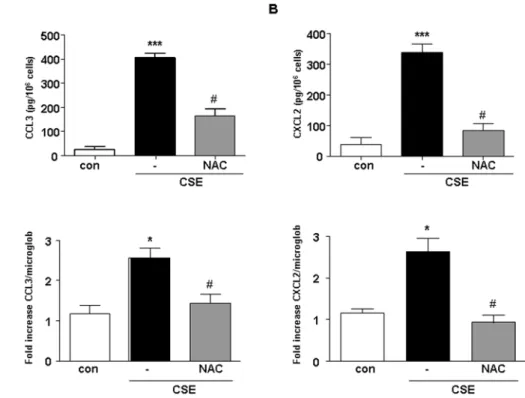
CSE induces CCL3 and CXCL2 production by cDCs CSE dose dependently (0.035–2.5%) induced the release of chemokines (data not shown). The CSE concentration of 1.5% is most effective in inducing chemokines release from cDCs (Fig. 1A and B, upper panels). Therefore, this concentration was used in all subsequent experiments. Stimulation of cells with CSE (1.5%) did not induce significant TNF-a, IL-2, IL-6, IL-10, IL-12p70, MCP-1 and IFN-cproduction (data not shown). CSE-induced CCL3 and CXCL2 production is associated with an increased in mRNA levels for both chemokines (Fig. 1A and B, lower panels). To investigate the involvement of ROS and oxidative stress in the production of CCL3 and CXCL2 by CSE-exposed cDCs, the effect of the antioxidant agent N-Acetyl-Cysteı¨ne (NAC 2.5 mM) was investigated.
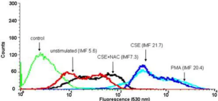
NAC attenuated the production of CCL3 and CXCL2 induced by CSE (Fig. 1A and B). Furthermore, intracellular ROS production after CSE treatment was measured. Exposure of cDCs to CSE or PMA, as a positive control, resulted in the production of ROS (Fig. 2). Pretreatment of cDCs with the antioxidant NAC resulted in an inhibition of CSE-induced ROS production (Fig. 2).
Figure 1. CSE induces the expression of mRNA and the production of chemokines in cDCs.The supernatants of CSE-exposed cDCs were tested for the production and release of CCL3 (A) and CXCL2 (B) ELISA (upper panels) and cell pellets were tested for CCL3 and CXCL2 mRNA levels by real time PCR (upper panels). White bars represent cDCs treated with medium, black bars represent cDCs treated with CSE and gray bars cDCs treated with NAC and CSE. Data are representative of three independent experiments, showing the means6SEM from triplicate cultures. * represents significant differences compared with medium-treated cells (*p,0.05; ***p,0.001).#
indicates significant differences between cells treated with CSE in combination with NAC and cells treated with CSE.
TLR4 and MyD88 are involved in the CSE-induced CCL3 and CXCL2 production of cDC
We and others have demonstrated that CSE activates inflammatory cells via TLRs [16,23]. By using neutralizing antibody against TLR4, the releases of CCL3 and CXCL2 was decreased (Fig. 3A and B).
MyD88 is a critical adapter molecule for the transduction of TLRs signals [24]. Therefore, cDCs lacking MyD88 were investigated. CSE did not induce the release of CCL3 and CXCL2 from cDCs obtained from MyD88 knockout mice (Fig. 3C and D). Similarly, LPS, the positive control did not induce a response in MyD882/2cDCs (Fig. 3C and D).
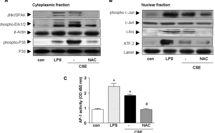
Involvement of MAPKs and NF-kB in CSE-induced CCL3 and CXCL2 release by cDCs
It has been reported that CSE activates MAPK and NF-kB in many inflammatory cells [25–27]. Therefore, in the current study, the involvement of these pathways were investigated. CSE stimulated phosphorylation of the JNK/SAPK, Erk1/2 and p38 pathways in cytoplasm of cDCs (Fig. 4A). In addition, NAC abrogated the phosphorylation of all these molecules. Next, the effect of pharmacological inhibitors were examined on chemokine release after CSE stimulation. Inhibition of p38 MAP kinase by SB 239063 induced a 63%617 and 43%631 reduction of CSE-induced CCL3 and CXCL2 production by cDCs, respectively. Inhibition of Erk1/2 by PD98059 induced a 28.4%62 and 29%616 reduction in CCL3 and CXCL2 production, respec-tively. In the nuclear fraction, CSE increased the phosphorylation of c-jun, c-fos and ATF-2 in cDC (Fig. 4B) and addition of NAC abrogated the phosphorylation of these molecules (Fig. 4B).
CSE and LPS (the positive control) significantly increased the activity of AP-1 compared to control. Pre-incubation of cells with NAC, suppressed the activation of AP-1 induced by CSE (Fig. 4C). Further, the regulation of NF-kB signaling in cDCs by CSE was investigated. To address the mechanism involved in the degrada-tion of IkB-aby CSE, phosphorylation of IkB-aby Western blot analysis was examined. CSE or LPS (the positive control) increased IkB-a phosphorylation (Fig. 5A) and exposure to CSE or LPS resulted in the degradation of IkB-a (Fig. 5A). Pretreat-ment with NAC inhibited the CSE-induced phosphorylation and degradation of IkB-a (Fig. 5A). In the nuclear fraction CSE and LPS increased the nuclear translocation of p65 which was abrogated by NAC (Fig. 5B). Pretreatment of cDC with the pharmacological NF-kB inhibitor (curcumin) resulted in an 80%63 reduction of the chemokine production (data not shown). For determination the activity of NF-kB, nuclear proteins were
Figure 2. CSE increases the production of intracellular ROS in cDC.cDCs were incubated with CSE , with or without NAC or PMA (as a control) and ROS generation was assayed by FACS analysis. The mean fluorescent intensity (MFI) of the following groups are indicated in the figure: control: unlabelled CSE-treated cells (green line), unstimulated: control labeled cells (red line), CSE: CSE-stimulated labeled cells (blue line), CSE+NAC: CSE-stimulated labeled cells treated with NAC (black line), PMA: PMA-stimulated labeled cells (light blue line).
doi:10.1371/journal.pone.0004946.g002
Figure 3. CSE increases the production of CCL3 and CXCL2 by TLR4 and MyD88 dependent manner.cDC were prepared by culturing BM cells from Balb/c mice preincubated with anti-TLR4 antibody (20mg/ml) or isotype control (20mg/ml) for 1 h and then stimulated with CSE for 16 h and amount of CCL3 (A) and CXCL2 ( B) were determined by ELISA. cDCs were prepared by culturing BM cells from Balb/c mice and age- and sex-matched MyD88-deficient mice. CSE or LPS were incubated for 16 h and supernatant were harvested for determination of CCL3 (C) and CXCL2 (D) by ELISA. White bars represent cDCs treated with medium, dotted bars are cDCs treated with LPS and black bars represent cDCs treated with CSE. Data are representative of three independent experiments, showing the means6SEM from triplicate cultures. * represent significant differences compared with medium-treated cells (***p,0.001).
subjected to a reaction containing biotin conjugated-oligonuclo-tides NF-kB. CSE increased the activity of NF-kB in cDCs and pretreatment with NAC suppressed NF-kB activity (Fig. 5C).
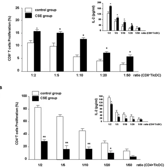
CSE-primed cDCs stimulate the proliferation of CD8+T cells
CSE significantly increased the ability of cDCs to stimulate the proliferation of CD8+T cells compared with untreated cDCs
(Fig. 6A) and decreased proliferation of CD4+T cells (Fig. 6B). In a
reciprocal fashion, the MLR was also determined by using T cells exposed to CSE. CSE did not effect proliferation of T cells (CD4+
or CD8+
T cells) when co-cultured with untreated allogeneic cDCs (data not shown). To further characterize the effect of CSE on the priming capacity of cDCs, the effect of CSE on IL-2 production in the MLR was assessed. As depicted in Fig 6A upper panels, IL-2 production of CD8+T cells was increased and decreased by
CD4+T cells (Fig. 6B, upper panel).
CCL3 antibodies suppress CSE-primed cDC- induced proliferation of CD8+T cells
Finally, the involvement of CCL3 and CXCL2 in MLR reaction were investigated.
cDCs were exposed with CSE for 24 h, treated with neutralizing antibodies directed against CCL3 or CXCL2 and
were then subjected to MLR reaction. Incubation of neutralizing CCL3 antibody in MLR, profoundly suppressed the CSE-primed cDC–induced proliferation of CD8+
T cells in the MLR (Fig. 7). Incubation with CCL3 antibody did not affect the CSE-primed cDCs-induced reduction of CD4+
T cell proliferation (data not shown). Moreover, the effects of CXCL2 neutralizing antibodies on the MLR with CSE-primed cDCs and CD8+
or CD4+
T cells were investigated. CXCL2 antibody had no effect on the proliferation of CD8+
T cells or CD4+
T cells when co-cultured with CSE-primed cDCs (data not shown).
Discussion
In this study, the effects of CSE on cDCs were explored with particular emphasis on the function and cellular immune responses. Among the tested cytokines and chemokines, CSE induced the release of CCL3 and CXCL2 by a ROS dependent manner. Interestingly, CSE did not induce the production of TNF-a, IL-2, IL-6, IL-10, IL-12p70, MCP-1, IFN-c and even suppressed the production of these cytokines induced by LPS (data not shown). Similar data on IL-12 and IL-23 have been published by Kroening et al, [28]. Our findings are consistent with the work of others showing the induction of IL-8 by CSE or cigarette smoke in human pulmonary DCs and in an in vivo model of smoke exposed mice [29].
Figure 4. CSE increases the activity of the MAPK pathway in cDCs.Western blot analysis of the cytoplasmic fraction (A) for JNK/SAPK, p-Erk1/ 2 and p- p38 and p38 from whole cell extracts and of the nuclear fraction (B) ATF-2, p-c-jun, c-jun and c-fos. Representative results of three independent experiments are shown.b-actin and lamin served as loading controls from cytoplasmic and nuclear fractions, respectively. AP-1 activity after stimulation of cells with CSE, LPS or CSE and NAC (C). Values (mean6SEM) are representative data from one of five independent sets of experiments. * indicates significant differences between medium-treated cells and cells treated with LPS or CSE (* p,0.05) and#
represents the significance between cells treated with CSE in combination with NAC and cells treated with CSE (#
Moreover, we show for the first time that CSE modulates cDC-mediated of T cells and specifically augments proliferation of CD8+T cells and inhibits proliferation of CD4+T cells in MLR.
The increase in proliferation of CD8+
T cells is mediated by CCL3, since the increase in proliferation is inhibited by antibodies against CCL3. CXCL2 antibodies did not have an effect (data not shown).
Cigarette smoke contributes to oxidant-induced damage of the cells via oxidants and free radicals [30] and generation of intracellular ROS [31]. We showed, that CSE induces ROS production in cDCs leading to the production and release of CXCL2 and CCL3. Interestingly, the generation of chemokines by cigarette smoke-activated DCs could be mitigated by anti-oxidants, NAC treatment. These data indicate that anti-oxidant therapy with agents like NAC may effect cigarette smoke-induced chemokine release of cDCs.
Next we found that MyD88/TLR4 activation and NF-kB/ MAPK signaling is involved in the induction of chemokines by CSE in cDCs. The first signaling protein to be recognized as oxidative stress-sensitive molecules are transcription factors, such as NF-kB [32] . ROS strongly affects the activation of NF-kB [32]. Besides, the MAPK pathway is an important signaling pathway affected by CSE [26]. In the current study, CSE induces the release of chemokines by both the activation of NF-kB and the MAPK pathways since inhibition of these intracellular signaling pathways suppresses the release of both chemokines.
Chemokines regulate the movement of leukocytes such as neutrophils and lymphocytes [33]. The predominant chemokine for human neutrophils is the CXC chemokine CXCL8. Mice lack CXCL8 but have the neutrophilic CXC chemokine ligand 2, MIP-2 or CXCL2 [34]. The importance of this chemokine in promoting pulmonary inflammation associated with COPD has extensively been investigated in vitro and in vivo [35–37]. Therefore, the CSE-induced release of CXCL2 by cDCs may result in the infiltration and activation of neutrophils in the airways.
Interestingly, in current study we show that CSE-primed cDCs increase the proliferation of CD8+
and suppress CD4+
T cells proliferation. In the supernatants of MLR samples the IL-2 production is elevated in CD8+T cells which is in agreement with
the proliferation of cells. These data could explain the enhanced CD8+
T cell population observed in lungs of smokers and smoke-treated mice [37–39]. Until now, the mechanism for this process is not well documented. Maeno and coworkers, described a critical role for CD8+T cells in inflammatory cell recruitment
and lung destruction in a cigarette smoke-induced murine model for COPD [12]. Earlier evidence reported that CCL3 is involved in CD8+
T cell proliferation [40]. Interestingly, CCL3 production by cDCs after CSE stimulation has a central role in the induction of the proliferation of CD8+T cells since proliferation was
blocked by adding CCL3 neutralizing antibody. Moreover, CSE-primed cDCs suppress CD4+
T cells proliferation which is
Figure 5. CSE increases the activity of the NF-kB pathway in cDCs.Western blot analysis of the cytoplasmic fraction (A) for IkB-aand p-IkB-a from whole cell extracts and of the nuclear fraction (B) p65 were carried out with related antibodies. Representative results of three independent experiments are shown.b-actin and lamin served as loading controls from cytoplasmic and nuclear fractions, respectively. NF-kB activity after stimulation of cells with CSE, LPS or CSE and NAC. (C). Values (mean6SEM) are representative data from one of five independent sets of experiments. * Indicates significant differences between medium-treated cells and cells treated with LPS or CSE (* p,0.05) and#represents the significance between cells treated with CSE in combination with NAC and cells treated with CSE (#
agreement with recentin vivostudies [38]. The role and amounts of CD4+
T cells in COPD is not well documented but early studies reported that cigarette smoke exposure led to a specific decrease in the percentage of activated CD4+
T cells, but not CD8+T cells in the lung [37]. Interestingly, very recently,
Harissison et al reported that the total number of BAL CD4+
and CD8+
T cells is higher in mice exposed to cigarette smoke. Furthermore, CD4+
T cells were proportionally higher than CD8+
T cell [41]. We tested the effects of neutralization antibodies against CCL3 and CXCL2 on the decreased CD4+
T cell proliferation induced by CSE-primed cDCs and did not find any suppressive effects on (data not shown). The reasons for the above mentioned discrepancies are not clear. The decrease in proliferation of CD4+
T cells may indicate suppressive effects of cigarette smoke on immune responses and may account for the higher susceptibility of smokers to viral and bacterial infections [42,43].
The above-mentioned explanation for the regulation of proliferation of T cells by CSE primed-DCs is an over simplification and is mainly used as a working hypothesis. In
Figure 6. CSE increases cDC-induced CD8+T cell but inhibits CD4+T cell proliferation.
cDCs from Balb/c mice were incubated with medium (white bars) or CSE ( black bars) and coincubated with allogenic T cells from C57BL/6 mice [CD8+(A) and CD4+T cells (B)] in a MLR. Presented are pooled
data from eight individual experiments using cDCs generated from eight isolations. Values are represented as mean6SEM. A statistically significant modulation of proliferation of T cells with CSE-primed cDCs occurred (*
p,0.05 and ** p,0.01 when compared to medium-treated cDCs). The supernatants of MLR were collected for the measurement of IL-2 by ELISA (inserted graphs in A & B). Presented are pooled data from eight individual experiments using cDCs generated from eight isolations. Values are represented as mean6SEM. * Indicates significant differences between medium-treated cells (* p,0.05).
doi:10.1371/journal.pone.0004946.g006
Figure 7. CCL3 neutralizing antibodies suppresses CSE-primed cDC-induced proliferation of CD8+T cells.cDCs were incubated
with medium (white bars) or CSE and then incubated without (black bars) or with polyclonal antibodies neutralizing CCL3 (gray bars). The cDCs were co-cultured with CD8+
summary, cigarette smoke induces the release of CXCL2 and CCL3 by cDCs. CXCL2 is considered as a chemokine that can recruite neutrophils. CCL3 results in the proliferation of CD8+T
cells and may be a key factor for increasing this cell in smokers and COPD patients. However, the relevance of above mentioned data should be confirmed in animal model with COPD and human.
Author Contributions
Conceived and designed the experiments: EM JjS MK BNL SLSLK NWL FPN GF. Performed the experiments: EM MK. Analyzed the data: EM ADK JjS MK BNL SLSLK NWL FPN GF. Contributed reagents/ materials/analysis tools: EM ADK MK BNL SLSLK NWL FPN. Wrote the paper: EM GF.
References
1. Barnes PJ, Shapiro SD, Pauwels RA (2003) Chronic obstructive pulmonary disease: molecular and cellular mechanisms. Eur Respir J 22: 672–688. 2. Rabe KF, Hurd S, Anzueto A, Barnes PJ, Buist SA, et al. (2007) Global strategy
for the diagnosis, management, and prevention of chronic obstructive pulmonary disease: GOLD executive summary. Am J Respir Crit Care Med 176: 532–555.
3. Spurzem JR, Rennard SI (2005) Pathogenesis of COPD. Semin Respir Crit Care Med 26: 142–153.
4. Yoshida T, Tuder RM (2007) Pathobiology of cigarette smoke-induced chronic obstructive pulmonary disease. Physiol Rev 87: 1047–1082.
5. Kitamura H, Iwakabe K, Yahata T, Nishimura S, Ohta A, et al. (1999) The natural killer T (NKT) cell ligand alpha-galactosylceramide demonstrates its immunopotentiating effect by inducing interleukin (IL)-12 production by dendritic cells and IL-12 receptor expression on NKT cells. J Exp Med 189: 1121–1128.
6. Bell D, Young JW, Banchereau J (1999) Dendritic cells. Adv Immunol 72: 255–324.
7. Banchereau J, Steinman RM (1998) Dendritic cells and the control of immunity. Nature 392: 245–252.
8. Lambrecht BN, Hammad H (2002) Myeloid dendritic cells make it to the top. Clin Exp Allergy 32: 805–810.
9. Hart DN (1997) Dendritic cells: unique leukocyte populations which control the primary immune response. Blood 90: 3245–3287.
10. Nouri-Shirazi M, Guinet E (2006) A possible mechanism linking cigarette smoke to higher incidence of respiratory infection and asthma. Immunol Lett 103: 167–176.
11. Aicher A, Heeschen C, Mohaupt M, Cooke JP, Zeiher AM, et al. (2003) Nicotine strongly activates dendritic cell-mediated adaptive immunity: potential role for progression of atherosclerotic lesions. Circulation 107: 604–611. 12. Maeno T, Houghton AM, Quintero PA, Grumelli S, Owen CA, et al. (2007)
CD8+T Cells Are Required for Inflammation and Destruction in Cigarette Smoke-Induced Emphysema in Mice. J Immunol 178: 8090–8096.
13. Robbins CS, Franco F, Mouded M, Cernadas M, Shapiro SD (2008) Cigarette smoke exposure impairs dendritic cell maturation and T cell proliferation in thoracic lymph nodes of mice. J Immunol 180: 6623–6628.
14. Palucka K, Banchereau J (1999) Dendritic cells: a link between innate and adaptive immunity. J Clin Immunol 19: 12–25.
15. Tsoumakidou M, Demedts IK, Brusselle GG, Jeffery PK (2008) Dendritic cells in chronic obstructive pulmonary disease: new players in an old game. Am J Respir Crit Care Med 177: 1180–1186.
16. Karimi K, Sarir H, Mortaz E, Smit JJ, Hosseini H, et al. (2006) Toll-like receptor-4 mediates cigarette smoke-induced cytokine production by human macrophages. Respir Res 7: 66.
17. Inaba K, Inaba M, Romani N, Aya H, Deguchi M, et al. (1992) Generation of large numbers of dendritic cells from mouse bone marrow cultures supplement-ed with granulocyte/macrophage colony-stimulating factor. J Exp Msupplement-ed 176: 1693–1702.
18. Kim HY, Kim HS (2007) Upregulation of MIP-2 (CXCL2) expression by 15-deoxy-Delta(12,14)-prostaglandin J(2) in mouse peritoneal macrophages. Im-munol Cell Biol 85: 60–67.
19. Jing H, Vassiliou E, Ganea D (2003) Prostaglandin E2 inhibits production of the inflammatory chemokines CCL3 and CCL4 in dendritic cells. J Leukoc Biol 74: 868–879.
20. Royall JA, Ischiropoulos H (1993) Evaluation of 29,79-dichlorofluorescin and dihydrorhodamine 123 as fluorescent probes for intracellular H2O2 in cultured endothelial cells. Arch Biochem Biophys 302: 348–355.
21. Vowells SJ, Sekhsaria S, Malech HL, Shalit M, Fleisher TA (1995) Flow cytometric analysis of the granulocyte respiratory burst: a comparison study of fluorescent probes. J Immunol Methods 178: 89–97.
22. Mortaz E, Redegeld FA, Nijkamp FP, Engels F (2005) Dual effects of acetylsalicylic acid on mast cell degranulation, expression of cyclooxygenase-2 and release of pro-inflammatory cytokines. Biochem Pharmacol 69: 1049–1057.
23. Doz E, Noulin N, Boichot E, Guenon I, Fick L, et al. (2008) Cigarette smoke-induced pulmonary inflammation is TLR4/MyD88 and IL-1R1/MyD88 signaling dependent. J Immunol 180: 1169–1178.
24. Hemmi H, Takeuchi O, Kawai T, Kaisho T, Sato S, et al. (2000) A Toll-like receptor recognizes bacterial DNA. Nature 408: 740–745.
25. Birrell MA, Wong S, Catley MC, Belvisi MG (2008) Impact of tobacco-smoke on key signaling pathways in the innate immune response in lung macrophages. J Cell Physiol 214: 27–37.
26. Li CJ, Ning W, Matthay MA, Feghali-Bostwick CA, Choi AM (2007) MAPK pathway mediates EGR-1-HSP70-dependent cigarette smoke-induced chemo-kine production. Am J Physiol Lung Cell Mol Physiol 292: L1297–L1303. 27. Liu X, Togo S, Al-Mugotir M, Kim H, Fang Q, et al. (2008) NF-kappaB
mediates the survival of human bronchial epithelial cells exposed to cigarette smoke extract. Respir Res 9: 66.
28. Kroening PR, Barnes TW, Pease L, Limper A, Kita H, et al. (2008) Cigarette smoke-induced oxidative stress suppresses generation of dendritic cell IL-12 and IL-23 through ERK-dependent pathways. J Immunol 181: 1536–1547. 29. Vassallo R, Kroening PR, Parambil J, Kita H (2008) Nicotine and oxidative
cigarette smoke constituents induce immune-modulatory and pro-inflammatory dendritic cell responses. Mol Immunol 45: 3321–3329.
30. Kinnula VL (2005) Production and degradation of oxygen metabolites during inflammatory states in the human lung. Curr Drug Targets Inflamm Allergy 4: 465–470.
31. Wu CC, Hsieh CW, Lai PH, Lin JB, Liu YC, et al. (2006) Upregulation of endothelial heme oxygenase-1 expression through the activation of the JNK pathway by sublethal concentrations of acrolein. Toxicol Appl Pharmacol 214: 244–252.
32. Beaudeux JL, Peynet J, Bonnefont-Rousselot D, Therond P, Delattre J, et al. (2006) Cellular sources of reactive oxygen and nitrogen species. Roles in signal transcription pathways. Ann Pharm Fr 64: 373–381.
33. Lukacs NW, Oliveira SH, Hogaboam CM (1999) Chemokines and asthma: redundancy of function or a coordinated effort?. J Clin Invest 104: 995–999. 34. Ma B, Zhu Z, Homer RJ, Gerard C, Strieter R, et al. (2004) The C10/CCL6
chemokine and CCR1 play critical roles in the pathogenesis of IL-13-induced inflammation and remodeling. J Immunol 172: 1872–1881.
35. Stevenson CS, Coote K, Webster R, Johnston H, Atherton HC, et al. (2005) Characterization of cigarette smoke-induced inflammatory and mucus hyper-secretory changes in rat lung and the role of CXCR2 ligands in mediating this effect. Am J Physiol Lung Cell Mol Physiol 288: L514–L522.
36. Folkerts G, Kraneveld AD, Nijkamp FP (2008) New endogenous CXC chemokine ligands as potential targets in lung emphysema. Trends Pharmacol Sci 29: 181–185.
37. Thatcher TH, McHugh NA, Egan RW, Chapman RW, Hey JA, Turner CK, et al. (2005) Role of CXCR2 in cigarette smoke-induced lung inflammation. Am J Physiol Lung Cell Mol Physiol 289: L322–L328.
38. O’Shaughnessy TC, Ansari TW, Barnes NC, Jeffery PK (1997) Inflammation in bronchial biopsies of subjects with chronic bronchitis: inverse relationship of CD8+T lymphocytes with FEV1. Am J Respir Crit Care Med 155: 852–857. 39. Saetta M, Baraldo S, Corbino L, Turato G, Braccioni F, et al. (1999) CD8+ve cells in the lungs of smokers with chronic obstructive pulmonary disease. Am J Respir Crit Care Med 160: 711–717.
40. Taub DD, Turcovski-Corrales SM, Key ML, Longo DL, Murphy WJ (1996) Chemokines and T lymphocyte activation: I. Beta chemokines costimulate human T lymphocyte activation in vitro. J Immunol 156: 2095–2103. 41. Harrison OJ, Foley J, Bolognese BJ, Long E III, Podolin PL, et al. (2008) Airway
infiltration of CD4+CCR6+Th17 type cells associated with chronic cigarette smoke induced airspace enlargement. Immunol Lett 121: 13–21.
42. Sallusto F, Cella M, Danieli C, Lanzavecchia A (1995) Dendritic cells use macropinocytosis and the mannose receptor to concentrate macromolecules in the major histocompatibility complex class II compartment: downregulation by cytokines and bacterial products. J Exp Med 182: 389–400.