Cardiac Ventricular Weights Recorded at the Autopsy of Healthy
Subjects Who Died of External Causes
Sérgio Lamêgo Rodrigues, Enildo Broetto Pimentel, José Geraldo Mill
Laboratory of Cardiovascular Physiopathology, Post-Graduation Program in Physiological Sciences, Centro de Ciências da Saúde da Ufes, Vitória, ES - Brazil
Summary
Objective: To establish cardiac ventricular weights recorded during the autopsy of healthy individuals who died of external causes, aiming at determining normality patterns in our population.
Methods: A total of 94 hearts were evaluated at the Forensics Department of the city of Vitória, Espírito Santo. After the heart removal and resection of the atria and epicardial fat, the right ventricle (RV) and the left ventricle (LV), including the septum, were separated and weighed and the mass was indexed by the height. The Kolmogorov-Smirnov test was used to test the normality of the distribution. Data are presented as means± SD.
Results: After the exclusion of 12 hearts (possible cardiovascular disease detected post-mortem) 82 hearts were examined (52 males and 30 females, aged 16-68 yrs, mean = 31±12 yrs). The weight of the LV was 181±25 g and 125±15 g, and the weight of the RV was 54±7 g and 38±6 g; the LV mass indexed by height was 105±14 g/m and 78±8 g/m, for males and females, respectively. The P95 of the LV weight was 218 g and 128 g/m in males and 148 g and 88 g/m in females. No significant correlation between ventricular mass and age was observed.
Conclusion: The weight of the LV in the males from our sample was higher than that reported in the contemporary literature. Our results suggest that the presence of LV hypertrophy can be inferred in the presence of LV mass > 218 g or 128 g/m in males and 148 g or 88 g/m in females. (Arq Bras Cardiol 2007;88(5):252-257)
Key words: Heart ventricles/anatomy; organ size; hypertrophy, left ventricular.
Mailing address: José Geraldo Mill •
Programa de Pós-Graduação em Ciências Fisiológicas da Ufes Av. Marechal Campos 1468 – Maruípe - 29042-755 - Vitória, ES - Brazil E-mail: [email protected]
Manuscript received December 13, 2006; revised received March 30, 2007; accepted May 03, 2007.
Introduction
The increase in the left ventricular mass (LVM) is an independent prognostic factor for cardiovascular mortality1,
mortality due to all causes2,3 and sudden death4. Hypertensive
individuals with left ventricular hypertrophy (LVH) present a 2-to-5-fold increase in fatal and non-fatal cardiovascular events when compared to those with hypertension only5.
Therefore, the accurate evaluation of the cardiac mass is fundamental not only for the determination of the degree of hypertrophy as well as for the assessment of its regression. The LIFE study confirmed the decrease in cardiovascular events with the regression of LHV, regardless of the decrease in blood pressure6-8; thus, the LHV regression must be one of the objectives
to be attained during the treatment of arterial hypertension. The electrocardiogram (ECG) was one of the first methods used to identify the LVH. However, this method has low sensitivity9, and, therefore, presents a low power to detect
this condition, notably in the initial phases of cardiac growth. Therefore, M-mode echocardiography, given its higher sensitivity, has been the most frequently type of propedeutics
used, not only in the detection, but also in the regression of LVH after treatment. However, the echocardiography is limited by its low accuracy10 and low interstudy reproducibility11.
Recently, the cardiac magnetic resonance (CMR) has appeared as an image-acquiring method with a higher precision and less variability11, becoming the reference
pattern for the determination of LVH12, after the validation in
animals13 and in the human heart ex-vivo14. Its use has been
recommended in the evaluation of remodeled ventricles and in the detection of small alterations in the cardiac mass during the clinical follow-up of patients or in scientific research15.
The reference values for normality in the determination of LVH and the cutoffs obtained from studies, however, are discordant. Considering well-conducted studies in this area, the means of LVH indexed by body surface area (BSA) for healthy men and women were, respectively, 71 and 58 g/m2, 91 and 79 g/m2,
78 and 61 g/m2 and 94 and 80 g/m2 16-19. The difference
between the highest and the lowest LVH values in these studies was 32% for men and 37% for women, which is unacceptable for a method considered to be a reference pattern.
At the Cardiovascular Monitoring Project (Projeto de Monitoramento Cardiovascular -MONICA Project) developed in Vitória, ES20,21, we measured LVH by CMR in a subsample
of 96 individuals (48 men), with 40 healthy individuals and 56 with LVH detected at the ECG (Sokolow and Lyon index
≥3.5 mV) and/or the echocardiogram (LV mass ≥116 g/m2
ventricles, followed by the detachment of the large base vessels below their respective valves. The ventricles were photographed and measured with a pachimeter at the largest axis and cut into five transversal slices (short axis direction), mimicking the CMR slices. The thickness of the septum and posterior wall was measured above the papillaries and photographed with the pachimeter fixed at 1 cm opening in order to have a reference pattern. The ventricular mass sliced in discs allows a better visualization of the epicardial fat, which was resected. The free wall of the right ventricle was separated with tweezers, detached from the septum and resected following the plane of curvature of the interventricular septum. The trabeculae encrusted in the septum were preserved. The mitral and tricuspid valves were removed. Both ventricles were weighed separately in an electronic scale (Urano, UDC 30000/5) with a 1-gram precision. All the biological material was returned to the corpse.
The data are expressed as mean ± standard deviation (SD), minimum and maximum values and 95% percentile (P95). The Kolmogorov-Smirnov test with significance levels of Lilliefors was used to test the normal distribution in the variables with continuous distribution. The difference between the means was carried out by two-tailed t test for independent samples and the equality of the variances was assessed by Levene’s test. The degree of correlation was determined by Pearson’s coefficient (r) and the regression lines were calculated by the minimum squares. The level of significance was set at p < 0.05. The SPSS 13.0 software was used for the statistical analysis.
Results
The 82 healthy hearts were from 52 men and 30 women, of which 57 were Brazilian Mulatto, 18 were Caucasian and 7 were African-Brazilian, with the Mulatto ethnicity being predominant among men (34/52) as well as among women (23/30), totaling 76.6% of the sample, followed by Caucasians 18/82 (22%) and African-Brazilians7/82 (8.5%). Age varied from 14 to 68 years. As the corpses underwent autopsy while still in rigor mortis status, the time of death was between 24 and 48 hs26. The most frequent cause of death was
firearm-related (60% in men and 50% in women). The accidents due to means of transportation (car, motorcycle, plane) accounted for 21% of the deaths among men and 23% among women. Table 1 shows age, height, ventricular mass and thickness of the left ventricular wall of the examined hearts. All of the variables were adjusted to Gaussian distribution curves, except age, which showed a preponderance of individuals below 40 yrs of age. A significant difference was observed regarding gender between the means of all variables analyzed, except for age the ratio between masses of the left and right ventricles. The P95 values for the LVM also showed important differences between genders.
Table 2 shows the comparison of the values of the LVM and right ventricular mass (RVM) in our study with the literature data. The age was different between the samples. The RVM was similar in all studies. A significant difference was observed in LVM of men in our sample when compared to the data from London28 and probably with the study
carried out in the USA26.
for men and 104 g/m2 for women). The mean LV mass (LVM)
indexed by BSA in the subsample of healthy individuals (20 men and 20 women, mean age = 48 yrs) was 57 and 42 g/m2, respectively, which are quite discordant values in
comparison to the international literature16-19. However, these
measurements are similar to the findings of Ghorayeb et al22,
who reported a mean LVM of 57 g/m2 in a sample of 30 normal
men evaluated by CMR.
The Brazilian literature has scarce data on the cardiac mass of normal individuals. The only available data were published by Tafuri & Chapadeiro23 40 years ago. However, these authors
reported only the total cardiac mass, which is inadequate for estimating the ventricular mass.
Considering the discrepancy between the Brazilian data and the international literature regarding the cardiac mass obtained at the CMR, we aimed at determining the actual ventricular mass of healthy individuals. To do that, we evaluated hearts obtained at autopsies, which is a method that is classically used as an absolute reference pattern, as it directly quantifies the mass of the organ or parts of it24-26. The present study included
hearts of individuals with no apparent signs of cardiac disease that died of external causes. The project was approved by the Ethics Committee in Research of the Center of Health Sciences of Ufes and the study was developed in collaboration with the Forensics Department of Vitória, ES.
Methods
From June to November 2005, 94 hearts obtained at the autopsies of apparently healthy individuals that died of external causes and were referred to the Forensics Department of the city of Vitoria were examined. The hearts of individuals with signs of systemic disease, evidence of cardiac surgery, those of malnourished, obese or pregnant individuals, as well as hearts with external or internal evidence of abnormalities (infarction, valvular heart disease, etc) were excluded. Additionally, within the first month after the death, a telephone contact was made with the person in charge of the removal of the body from the Forensics Department in order to obtain information about the presence of arterial hypertension or other cardiovascular diseases, or whether the deceased regularly used medication for cardiac disease. Based on these observations and information, 12 hearts (8 males and 4 females) were excluded. Therefore, 82 hearts were classified as “normal”.
The length of the corpse was measured in the dorsal decubitus position, using an inelastic measuring tape from the top of the head (vertex) to the heel, taking into account the necessity to correct the height retraction due to the muscular contraction caused by the rigor mortis. Ethnicity-race was evaluated by the forensic physician and researcher, and the corpses were classified as Caucasian, Brazilian Mulatto, African-Brazilian or others.
The heart was removed during the usual necropsy procedures during the rigor mortis, preserving an adequate extension of the base and pulmonary vessels. Next, the fresh heart was dissected using the Bove et al27 method with a
Arq Bras Cardiol XXXX; XX(X) : XXX-XXX
Figure 1 shows a strong correlation (r = 0.66; P<0.01) between LVM and height and this variable accounts for 43% of the total variability of the LVM of the sample (R2 = 0.43).
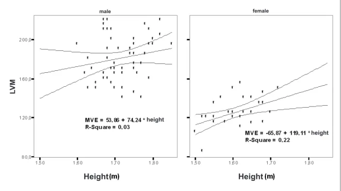
However, when the LVM indexed by height is stratified by sex (Fig. 2), it is observed that the correlation between these variables in men disappears (r = 0.17), with a significant correlation (r = 0.47; p<0.05) being kept among women, data also reported by Hangartner et al28. No correlation between
RVM and age or height was observed.
Discussion
Data on normal cardiac weight in the Brazilian population are scarce. Schvartzman et al29, using echocardiographic
measurements in a random sample of 100 participants from the urban population of the city of Porto Alegre, found mean values of LVM of 72 and 64.8 g/m2 for men and women,
respectively. In a study carried out in Vitoria, we found values of LVM at the ECG of 84 and 74 g/m2 in healthy men and
women, respectively. These constituted the healthy subgroup (N=251) of a sample of 682 adults (30-70 yrs) that participated in the Phase II of the Monitoring Project of Tendencies and Determinants of Cardiovascular Diseases, started in Vitoria in 199930,31. The subjects defined as “healthy” were
normotensive, non-obese, non-diabetic individuals with no signs of cardiac abnormalities at the ECG and echocardiogram and no renal dysfunction. The values found at the Vitoria study31 are closer to those found by Ilercil et al32.
Tafuri & Chapadeiro23 reported the cardiac weight of
normal adults submitted to necropsy using a sample of 230 healthy individuals (20-70 yrs). The mean total cardiac weight was 329.9 g for men and 258.9 g for women. Differences between genders were found only below 40 yrs of age. There were no differences among the ethnicities, which was confirmed in the present study, despite the small number of African-Brazilians in our sample (n=7). They concluded that the cardiac weight of Brazilian men was higher than that
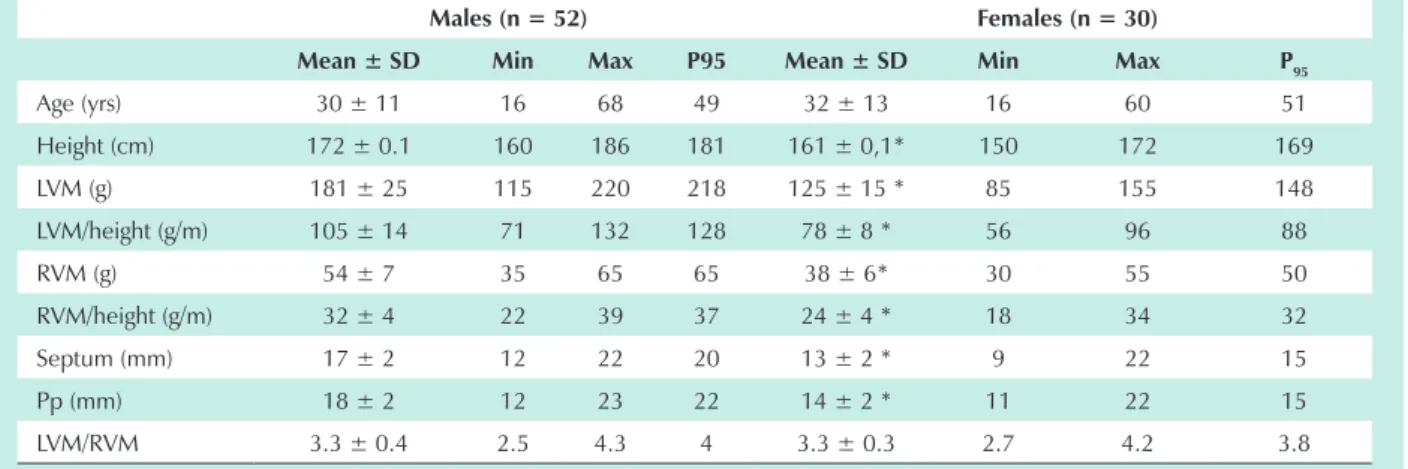
Table 1 – Morphometric characteristics of the hearts of normal individuals who died of external causes
Males (n = 52) Females (n = 30)
Mean ± SD Min Max P95 Mean ± SD Min Max P95
Age (yrs) 30 ± 11 16 68 49 32 ± 13 16 60 51
Height (cm) 172 ± 0.1 160 186 181 161 ± 0,1* 150 172 169
LVM (g) 181 ± 25 115 220 218 125 ± 15 * 85 155 148
LVM/height (g/m) 105 ± 14 71 132 128 78 ± 8 * 56 96 88
RVM (g) 54 ± 7 35 65 65 38 ± 6* 30 55 50
RVM/height (g/m) 32 ± 4 22 39 37 24 ± 4 * 18 34 32
Septum (mm) 17 ± 2 12 22 20 13 ± 2 * 9 22 15
Pp (mm) 18 ± 2 12 23 22 14 ± 2 * 11 22 15
LVM/RVM 3.3 ± 0.4 2.5 4.3 4 3.3 ± 0.3 2.7 4.2 3.8
LVM - left ventricular mass; RVM - right ventricular mass; PW - posterior wall; Min - minimum, Max - maximum; P95 - 95
th percentile. * p < 0.05 vs. males.
Table 2 – Weight of the cardiac ventricles of normal individuals submitted to autopsy in Vitória and those reported by other studies
Vitória Italy 29 London 28 USA 26
N 52 30 13 22 32 24 53
Gender M F M F M F ND
Age (yrs) 30 ± 11 32 ± 13 61 ± 14 55 ± 18 65 ± 9 54 ± 14
Height (cm) 171 ± 6 161 ± 6 ND 170 ± 6 162 ± 9 ND
LVM (g) 181 ± 25 125 ± 15 153 ± 33 164 ± 38* 122 ± 26 135 ± 20
RVM (g) 54 ± 8 38 ± 6 52 ± 15 56 ± 14 40 ± 8 44 ± 9
Data expressed as means ± SD. The data in italics (USA and Italy) represent the means of the measurements of both genders. M - male; F - female; LVM - left ventricular mass; RVM - right ventricular mass; ND - not described. Italy, Corradi et al, (2004); London, Hangartner et al, (1985); USA, Murphy et al, (1988); n - number of individuals (*) - p < 0.05 with LVM Vitória, t test.
weight, presents restrictions regarding the current use of echocardiography or CMR, as it does not accurately reflect the ventricular mass, considering that other components are part of the total heart weight, such as the epicardial fat and atrial weight, which are not negligible33.
The normality standard for organ weights must be established in a specific reference sample for each population, as the normality values can be different under genetic and environmental influences34. This fact is corroborated by the
study of Dadgar et al24, which, after the examination of 100
bodies with no cardiac abnormalities, reported that the mean total weight of the Indian hearts was lower than that reported in literature for other racial groups.
Aiming at updating the literature data, Grandmaison et al34
published a table depicting the weight of organs in a population of 684 Caucasoid individuals with a mean age of 45 years. Unfortunately, they did not report the individual ventricular weights, but only the means and SD of the total cardiac mass (365 ±71g and 312 ±78g for men and women, respectively). Therefore, the present study fills a gap regarding data on the Brazilian population. Studies such as those by Hangartner et al28 and Corradi et al33 (Table 2) used a methodology that was
similar to ours. There is a difference of around 10% regarding the LVM reported by Hangartner et al28 and that of our sample.
This could be explained by the technique employed in the removal of the RV, as this study28, similarly to that by Fulton &
Jones35, the trabeculae on the right side of the septum were
removed at the base, whereas we allowed those encrusted in the septum to remain there. This procedure was adopted based on the fact that, at echocardiographic assessments, this
Fig. 1 - Correlation between the left ventricular mass (LVM) and the height of the individuals. The regression line (LVM = 282.03 x height – 311.87) and the 95% confidence interval (95%CI) are shown in the figure. There is a strong correlation between the variables (r = 0.66).
Gender male
female
L
VM
Height
reported in literature, whereas the weight of the women’s hearts was similar.
The technique used by these and other pathologists to evaluate cardiac hypertrophy, through the total heart
Fig. 2 - Correlation between the left ventricular mass (LVM) and the height of the individuals stratified by gender. Note that the LVM in the female sex is lower and has a higher correlation (r=0.47) with height when compared to the male sex (r=0.17). The regression line of females (LVM = 119.1 x height – 65.87) and of males (LVM = 74.24 x height + 53.86) and the 95% CI are traced in each line.
male female
L
VM
Height
height
height
Height
Arq Bras Cardiol XXXX; XX(X) : XXX-XXX
region remains as belonging to the interventricular septum, being referred to as “echoes of the endocardium”. According to Geiser & Bover36, the methodology in this type of study must
be clearly specified, as the fraction of muscle inserted into the trabeculae can represent 9% to 19% of the LVM. However, the RVM was not different between the two studies, disclosing a difference in the measurements of the LVM.
The measurement of the thickness of the cardiac walls (Table 1) can vary according to the area where they were obtained, due to the trabeculation of the endocardium37.
These measurements reflect the state of the myocardial tonus, which presents in its state of contraction in rigor mortis. Thus, as it is a linear measurement that reflects the state of contraction and because it has low reproducibility, it is ineffective to be used for the mass estimation of a three-dimensional structure.
The non-variability of the LVM between studies with fresh or fixed hearts is supported by the studies of Hangartner et al28 and Geiser & Bover36. The use of the value of the LV mass/
RV mass ratio has been recommended for the identification of isolated ventricular hypertrophies, mainly when the measurement of the ventricular mass is within the normal range. In our sample, this ratio varied from 2.5 to 4.3 in men and 2.7 to 4.2 in women.
Several authors have analyzed the correlations of the anthropometrical data and body composition with the size and structure of the heart. Hangartner et al28 showed a positive
correlation of the LVM with the weight and body composition. However, height showed only a significant correlation in women. Zeek et al38 reported that the individual’s height
and nutritional status have an effect on cardiac mass, but not ethnicity or age. The authors38 have favored height as it is a
more stable variable in comparison to weight and much less influenced by diseases.
In summary, the literature data are consistent and corroborate the data of the present study at two main points: 1) Sex and height variables (but not age) are determinant factors for ventricular mass; 2) The quali-quantitative diagnosis of ventricular hypertrophy can only be attained if there are normality reference values for ventricular mass obtained through adequate techniques, stratified by gender and preferably corrected for height. Considering that the weight of the ventricles of normal individuals follow a normal distribution, P95 can be suggested as the upper normality
limit for this parameter. Our findings show values of 218 g and 148 g for the LV and 65 and 50 g for the RV of men and women, respectively. Adjusting the ventricular weight for the height, a correction that is increasingly accepted in echocardiographic evaluation, P95 values would be 128 g/m and 88 g/m for the LV and 37 g/m and 34 g/m for the RV of men and women, respectively.
We conclude that the LVM obtained from the necropsy of normal male individuals from our population is higher than that reported in the most recent literature. Additionally, our data suggest that the values of cardiac mass measured in our population through CMR22 can be underestimating the actual
mass of the organ between 24% and 60% in comparison to the LVM estimated by CMR reported in international studies16-19.
When we compare the mean LVM estimated by CMR with those obtained at the necropsy, this underestimation reaches even higher values. These data show that the creation of parameters of normality for cardiac mass for the several forms of measurement (direct measurement, echocardiography, CMR and others) is imperative, given the clinical importance of this parameter in attaining the diagnosis and conducting the treatment of several diseases, including arterial hypertension.
Acknowledgements
To the State Secretary of Public Security of the state of Espírito Santo, the Police Department, the Forensic Department, and especially, to the forensic physicians and assistants whose participation was crucial for the development of this study.
Potential Conflict of Interest
No potential conflict of interest relevant to this article was reported.
Sources of Funding
This study was funded by Conselho Nacional de Desenvolvimento Científico e Tecnológico - CNPq.
Study Association
This article is part of the thesis of doctoral submitted by Sérgio Lamêgo Rodrigues, from Programa de Pós-Graduação em Ciências Fisiológicas da UFES.
References
1. Levy D, Garrison R, Savage D, Kannel W, Castelli W. Prognostic implications of echocardiographically determined left ventricular mass in the Framingham Heart Study. N Engl J Med. 1990; 322: 1561-6.
2. Kannel W, Cobb J. Left ventricular hypertrophy and mortality: results from the Framingham Study. Cardiology. 1992; 81: 291-8.
3. Kaplinsky E. Significance of left ventricular hypertrophy in cardiovascular morbidity and mortality. Cardiovasc Drugs Ther. 1994; 8: 549-56.
4. Frohlich E. Left ventricular hypertrophy and sudden death. J Am Coll Cardiol. 1998; 32: 1460-2.
5. Koren MJ, Devereux RB, Casale PN. Relation of left ventricular mass and geometry to morbidity and mortality in uncomplicated essential hypertension. Ann Intern Med. 1991; 114: 345-52.
6. Dahlof B, Devereux RB, Kjeldsen SE, Julius S, Beevers G, Faire U, et al. Cardiovascular morbidity and mortality in the Losartan Intervention For Endpoint reduction in hypertension study (LIFE). Lancet. 2002; 359: 995-1003.
7. Okin PM, Devereux RB, Jern S, Kjeldsen SE, Julius S, Nieminen MS, et al. Regression of electrocardiographic left ventricular hypertrophy by losartan versus atenolol: the LIFE study. Circulation. 2003; 108: 684-90.
8. Devereux R, Wachtell K, Gerdts E, Boman K, Nieminen MS, Papademetrious V, et al. Prognostic significance of left ventricular mass change during treatment of hypertension. JAMA. 2004; 292: 2350-6.
9. Fragola P, Cannata D. Assessment of left ventricular hypertrophy in patients with essential hypertension: a rational basis for the electrocardiogram. Am J Hypertens. 1993; 6: 164-9.
10. Reichek N, Helak J, Plappert T, Sutton MS, Weber KT. Anatomic validation of left ventricular mass estimates from clinical two-dimensional echocardiography: initial results. Circulation. 1983; 67: 348-52.
11. Germain P, Roul G, Kastler B, Mossard J, Bareiss P, Sacrez A. Inter-study variability in left ventricular mass measurement: comparison between M-mode echocardiography and MRI. Eur Heart J. 1992; 13: 1011-9.
12. Alfakih K, Walters K, Jones T, Ridgway J, Hall AS, Sivananthan M. New gender-specific partition values for ECG criteria of left ventricular hypertrophy recalibration against cardiac MRI. Hypertension. 2004; 44: 175-9.
13. Florentine MS, Chang W. Measurement of left ventricular mass in vivo using gated nuclear magnetic resonance. J Am Coll Cardiol. 1986; 8: 107-12.
14. Katz J, Peshock R. Estimation of human myocardial mass with MR imaging. Radiology. 1988; 169: 495-8.
15. Myerson S, Pennel D. Assessment of left ventricular mass by cardiovascular magnetic resonance. Hypertension. 2002; 39: 750-5.
16. Marcus JT, DeWaal K, Gotte MJ, van der Geest RJ, Heethaar RM, van Rossum AC. MRI-derived left ventricular function parameters and mass in healthy young adults: relation with gender and body size. Int J Card Imaging. 1999; 15: 411-9.
17. Lorenz CH, Walker ES, Morgan VL, Klein SS, Graham TP. Normal human right and left ventricular mass, systolic function, and gender diferences by cine magnetic resonance imaging. J Cardiov Magn Res. 1999; 1: 7-21.
18. Salton C, Chuang ML, O’Donnell CJ, Kupka MJ, Larson MG, Kissinger KV, et al. Gender differences and normal left ventricular anatomy in an adult population free of hypertension: a cardiovascular magnetic resonance study of the Framingham Heart Study Offspring Cohort. J Am Coll Cardiol. 2002; 39: 1055-60.
19. Cain P, Ahl R, Hedstrom E, Ugander M, Allansdotter-Johnsson A, Friberg P, et al. Physiological determinants of the variation in left ventricular mass from early adolescence to late adulthood in healthy subjects. Clin Physiol Funct Imaging. 2005; 25: 332-9.
20. Mill JG, Molina MCB, Silva IO, Marquezini AJ, Ferreira AVL, Cunha RS, et al. Epidemiologia da hipertensão arterial na cidade de Vitória, Espírito Santo. Hipertensão. 2004; 7: 109-16.
21. Pereira AC, Mota GFA, Cunha RS, Herkenhoff FL, Mill, JG, Krieger JE. Angiotensinogen 235T allele dosage is associated with blood pressure phenotypes. Hypertension. 2003; 41: 25-30.
22. Ghorayeb N, Batlouni M, Pinto I, Dioguardi G. Hipertrofia ventricular esquerda do atleta: resposta adaptativa fisiológica do coração. Arq Bras Cardiol. 2005; 85: 191-7.
23. Tafuri WL, Chapadeiro E. O peso do coração no brasileiro adulto normal. O Hospital. 1966; 70: 947-57.
24. Dadgar SK, Tyagi SP, Singh RP, Hameed S. Factors influencing the normal heart
weight – a study of 140 hearts. Jpn Circ J. 1979; 43: 77-82.
25. Ludwig J. Current methods of autopsy practice. Philadelphia: Saunders, 1979. p. 647-89.
26. Murphy ML, White HJ, Meade J, Straub KD. The relationship between hypertrophy and dilatation in the postmortem heart. Clin Cardiol. 1988; 11: 297-302.
27. Bove K, Scott R. Observations on the assessment of cardiac hypertrophy utilizing a chamber partition technique. Circulation. 1966; 33: 558-67.
28. Hangartner JR, Marley NJ, Whitehead A, Thomas AC, Davies MJ. The assessment of cardiac hypertrophy at autopsy. Histopathology. 1985; 9: 1295-306.
29. Schvartzman PR, Fuchs FD, Mello AG. Valores normais de medidas ecocardiográficas: um estudo de base populacional. Arq Bras Cardiol. 2000; 75: 107-10.
30. Lamêgo S. Hipertrofia do ventrículo esquerdo. Correlação entre critérios eletrocardiográficos e ecocardiográficos em estudo de base populacional. [tese]. Vitória (ES): Universidade Federal do Espírito Santo; 2006.
31. Ângelo LCS, Vieira MLC, Lamêgo S, Morelato RL, Pereira AC, Mill JG, Krieger JE. Reference values of echocardiographic measurements in sample of Brazilian adult asymptomatic population. Arq Bras Cardiol. (aceito para publicação).
32. Ilercil A, O’Grady MJ, Roman MJ, Paranicas M, Lee ET, Welty TK. Reference values for echocardiographic measurements in urban and rural populations of differing ethnicity: the Strong Heart Study. J Am Soc Echocardiogr. 2001; 14: 601-11.
33. Corradi D, Maestri R, Bordi C. The ventricular epicardial fat is related to the myocardial mass in normal, ischemic and hypertrophic hearts. Cardiovasc Pathol. 2004; 13: 313-6.
34. Grandmaison GL, Clairand I, Durigon M. Organ weight in 684 adult autopsies: new tables for a Caucasoid population. Forensic Sci Int. 2001; 119: 149-54.
35. Fulton R, Jones M. Ventricular weight in cardiac hypertrophy. Br Heart J. 1952; 14: 413-20.
36. Geiser E, Bove E. Calculation of left ventricular mass and relative wall thickness. Arch Pathol. 1974; 97: 13-21.
37. Reiner L, Freudenthal R. The weight of the human heart. Arch Pathol. 1959; 68: 68-83.