Study of Mutations Causing Hypertrophic Cardiomyopathy in a Group
of Patients from Espirito Santo, Brazil
Júlia Daher Carneiro Marsiglia
1, Maria do Carmo Pimentel Batitucci
1, Flávia de Paula
1, Clara Barbirato
1, Edmundo
Arteaga
2, Aloir Queiroz de Araújo
1Universidade Federal do Espírito Santo1, Vitória, ES; Universidade de São Paulo2, São Paulo, SP – Brazil
Abstract
Background: Hypertrophic cardiomyopathy (HC) is the most frequent cardiac hereditary disease, caused by mutations in sarcomere protein coding genes. Although more than 40 mutations have been identified in several continents and countries, there have been no reports of mutations in Brazil.
Objectives: : To carry out a genetic study to identify genetic mutations that cause HC in a group of patients in Espirito Santo, Brazil.
Methods: Using the SSCP technique, exons from the three main genes involved in HC were studied: exons 5, 0, , and of the β-myosin heavy chain gene (MYH7), exons 7, 6, 8, and 4 of the myosin binding protein C gene (MYBPC3) and exons 8 and 9 of troponin T gene (TNNT2).
Results: 6 alterations were found, including two mutations, one of them possibly pathogenic in the MYBPC gene (p. Glu44Lys) and another pathogenic one, previously described in the TNNT gene (p.Arg9Trp), 8 rare sequence variations and 6 sequence variations with allelic frequency higher than % (polymorphisms).
Conclusion: These data allow the conclusion that the genotyping of patients is feasible in our country. It is possible that the isolated p.Glu44Lys variant identified in exon 6 of the MYBPC3 gene is pathogenic, promoting a milder phenotype than that found when in association with other mutations. The p.Arg9Trp variant in the exon 9 of TNNT2 gene does not promote such a homogeneous phenotype as previously described and it can lead to severe hypertrophy. (Arq Bras Cardiol 00; 94() : 0-7)
Key words: cardiomyopathy, hypertrophic; myosin; protein C; genes; Espirito Santo; Brazil.
Mailing address : Júlia Daher Carneiro Marsiglia •
Rua da Consolação, 3075 / 1218 - Cerqueira César – 01416-001 - São Paulo, SP, Brazil
E-mail: [email protected]
Manuscript received May 11, 2009; revised manuscript received June 24, 2009; accepted June 24, 2009.
Introduction
The hypertrophic cardiomyopathy (HC) is a primary cardiac disease, characterized by left ventricle (LV) hypertrophy, without dilatation, usually asymmetrical and mainly septal, in the absence of any other cardiac or systemic diseases that can lead to myocardial hypertrophy1,2. In addition to the myocardial fiber hypertrophy, the histological disarrangement of these fibers occurs and variable degrees of interstitial as well as perivascular fibrosis, contributing to the development of heart failure, myocardial ischemia, ventricular arrhythmias and sudden death3.
HC is the most common genetic cardiac disease, but only in the beginning of the 90s the causes of the genetic mutations were identified4,5. So far, 13 sarcomere protein coding genes have been identified, which led to the definition of HC as a sarcomere disease. However, the mutations found in the β-myosin heavy chain (MYH7), myosin binding protein C
(MYBPC3) and troponin T (TNNT2) genes are responsible for 60 to 80% of the HC cases2,6. The only genetic study available for HC in Brazil was conducted by Tirone et al7. It was a pioneer study and the prevalence of HC in Brazil remains unknown; in consequence, even less is known about the prevalence of the disease-causing mutations. In the state of Espirito Santo, where the estimated population, by the Brazilian Institute of Geography and Statistics (IBGE) in 2008, was of approximately 3.500.000 inhabitants8, and considering a HC prevalence similar to the one found in other continents (1:500), it is probable that there are about 7,000 patients with the disease, which justifies efforts to identify them properly, as well as the family carriers.
Methods
Patients
the research objectives, in which they could be immediately included, if agreed.
Clinical evaluation
Each individual was submitted to a clinical-cardiac directed examination, including history, physical examination and echocardiography, all performed by cardiologists with experience in HC.
Doppler echocardiography
The echocardiograms were performed by an experienced cardiologist, using an Acuson Sequoia model 512 (Mountain View, CA, USA) equipment, with a multifrequency transducer (2.0 to 3.5 MHz), second harmonic and tissue Doppler. Patients were examined in left lateral position and the tracks were recorded in unforced expiration. The echocardiographic measurements in unidimensional-mode were obtained during the examinations, as well as the measurements of velocity and myocardial flow (pulsed Doppler, tissue Doppler and continuous Doppler). The main criterion for diagnosis of HC was the echocardiography, through the unequivocal demonstration of myocardial thickness equal to or greater than 15 mm in any segment of the LV in the absence of another cardiac or systemic disease that could cause ventricular hypertrophy.
DNA Extraction
The total DNA was extracted from 2 to 5ml of peripheral blood based on the methodology described by Miller et al9. The extracted DNA was quantified in a Thermo Scientific NanoDropTM ® 1000 spectrophotometer and diluted to the concentration of 10 ng/μl with ultrapure water.
Polymerase chain reaction (PCR)
The exons 15, 20, 21, 22 and 23 from the MYH7gene (β -myosin heavy chain), 7, 16, 18, 22 and 24 from the MYBPC3
gene (myosin binding protein C) and 8 and 9 from the TNNT2 gene (troponin T) were amplified through PCR in an Applied Biosystems thermal cycler, model Veriti®, using primers described in the Cardio Genomicsdatabase6.
Screening of mutations
After amplification by PCR, the screening of mutations was performed by the Single-Strand Conformation Polymorphism technique (SSCP). Approximately 10μl of each PCR product was mixed with 2μl of SSCP-loading buffer. The samples were denatured at 94° C for 10 minutes, kept on ice and applied to the gels. The gels were subjected to electrophoresis at 6W for 12 to 16 hours. After the run, the gels were stained with silver nitrate, based on the methodology described by Bassam et al10.
Sequencing and alterations analysis
The fragments with abnormal migration on the gel were sequenced by the DNA sequencing platform of the Embrapa - Cenargen laboratory in Brasilia, DF, Brazil. The analysis of the changes was made based on the reference sequence (MYH7:
NG_007884, MYBPC3: NG_007667, TNNT2: NG_007556) available in the database of gene sequences of the National Center for Biotechnology Information (NCBI) available online11, and compared with the changes already described in the Cardio Genomics database, available online6.
Control group
A control group consisting of 64 unrelated adult volunteers, with apparently normal health, no family history of cardiomyopathies, normotensive, with normal physical examination and normal electrocardiogram, gave blood samples, which were submitted to the same processes as the samples from patients with phenotypically demonstrated HC.
Ethical considerations
This study was approved without restrictions by the Research Ethics Committee of the Federal University of Espirito Santo (Research Protocol No. 058/07). All patients and all volunteers from the control group received information on the procedures, risks and benefits of the study, and those who agreed to participate signed the consent form, also approved by the Ethics Committee.
Results
The study sample consisted of 20 patients, 10 males, mean age of 45.7 years. The control group consisted of 64 patients (23 males), aged 19 to 65 years (mean 44.3 ± 11.7). Echocardiographic measurements obtained from patients are shown in Table 1. All patients present asymmetric septal hypertrophy > 15 mm, left atrial enlargement as a result of the LV diastolic dysfunction (normal up to 40 mm), normal internal ventricular diameters (range 55 mm), except in 2 patients with mild dilatation and decreased ejection fraction in only one patient (normal ≥ 55%). At rest, only 25% of patients presented gradients > 30mmHg in the LV outflow tract (obstructive HC).
The results of the analysis of the SSCP and sequencing showed 14 alterations, including two mutations, 6 rare sequence variations and 6 polymorphisms. The exons studied are involved in 139 of the 441 mutations described for all the genes, just over 31%.
Table 1 - Echocardiographic measurements of the patients
RG LA (mm) LVDD (mm) IVS (mm) PW (mm) EF (%) Grad (mmHg)
P1 48.5 43 20.2 11.5 81.4 40
P2 48.6 49.8 20.7 8.9 72.9 12
P3 54 46 16 9 71 0
P4 44 44.7 23.2 12.9 77.4 57
P5 41.8 47.6 21.5 8 71.8 0
P6 48.9 42.4 16.6 12.4 75.6 38
P7 48.2 37.4 24.3 9.3 72.6 10
P8 55.7 48.2 20.7 15.4 60.1 12
P9 41.2 52.6 19 10.3 71.5 0
P10 58.9 58.1 17 10.9 49.6 0
P11 39.1 47.3 16.6 13.8 55 0
P12 41 39.8 25 10.1 66.7 0
P13 52.5 42.3 30.7 13.7 69.7 0
P14 40 43.1 22.7 12.6 69.5 12
P15 65.7 57.2 28.1 15.1 72.6 112
P16 33.2 47 16 7.4 79 0
P17 37.5 42.3 16 9.5 70 0
P18 50 45.7 21.9 11.1 71 0
P19 69 55.1 19 11 69 0
P20 44 52 16 11 63 85
LA - diastolic diameter of left atrium; LVDD - left ventricular diastolic diameter; IVS - interventricular septum diastolic thickness; PW - posterior wall diastolic thickness;
EF - ejection fraction of LV; Grad - peak systolic gradient in the LV outlow tract. position c.15431, changes the codon from CTG to TTG, both leucine codons and were identified with a frequency of 2.5% in patients and 0.78% in controls.
In the MYBPC3 gene, a mutation was identified, a G> A substitution in one patient (Figure 1). The mutation is located at position c.11642 in exon 16 and changes the codon from GAG> AAG, thus altering the amino acid, from glutamic acid to lysine. Furthermore, a rare variation of sequence and a polymorphism were found in intron 22. The alteration was identified at c.13999+118 position and it is a G> A substitution. The polymorphism was characterized by a C> G substitution and was identified at position c.15130+19. It showed a frequency of 5% in patients and 3.125% in controls.
A mutation, a C> T substitution in exon 9, was identified in a patient at position c.8772 in the TNNT2 gene (Figure 2). This substitution changes the codon from CGG to TGG, thus changing the amino acid from arginine to tryptophan. In addition to the mutation, two rare sequence variations in intron 8 and a polymorphism were identified in exon 9. The first alteration was a G> C substitution at position c.8459+130 and the second was a C> T substitution at position c.8762-20. The polymorphism identified was a C> T substitution at position c.8816, which changes the codon from ATC> ATT, both isoleucine codons. The polymorphism showed a
frequency of 69.44% in patients and 71.88% in controls. The summary of the results can be seen in Table 2.
Discussion
Mutations
MYBPC gene, exon 6
The G → A substitution found in exon 16 of MYBPC3
gene of patient P4 changes the codon from GAG, which encodes a glutamic acid to AAG, which determines lysine. This mutation was first described by Seidman et al12 in compound heterozygosity, with the p.E258K mutation in exon 7 of the MYBPC3 gene. Olivotto et al13 also described this mutation in compound heterozygosity with the p.E258K mutation. Although it is unlikely that the patients studied by the researchers mentioned above are the same individual, we cannot affirm that they are not related (personal communication). Thus, the p.E441K mutation has not been described alone, so it is not clear whether it is capable of causing the disease when expressed in isolation.
Figure 1 -Alteration in the fragment containing the exon 16 of the MYBPC3 gene a) Screening of mutations by SSCP in a 5% polyacrylamide gel. C1, C2 and C3 with a normal pattern and P4 showing a different pattern b) sequencing of the fragment containing the exon 16 of the MYBPC3 gene of P4, showing a G → A substitution in heterozygosis.
Figure 2 -Sequencing of the fragment containing exon 8 and part of the exon 9 of the TNNT2 gene of P5, showing a C → T substitution in heterozygosis. not have more detailed information on the case (personal communication). The P4 patient in the present study has moderate hypertrophy, with no family history of sudden death,
with no reports of syncope, and does not present nonsustained ventricular tachycardia on dynamic ECG. The patient complained of angina at exertion, and the coronary angiography showed coronary arteries with no obstructive processes.
Niimura et al14 observed in their study that the survival of patients with mutations in the MYBPC3 gene was longer than that observed in those bearing mutations in the TNNT2 gene or malignant changes in the MYH7 gene. Overall, the data indicate that mutations in the MYBPC3 gene are related to more mild forms of HC in many patients with the disease.
In another independent study of 22 families, Richard et al15 reported that 90% of the families carrying mutations in the MYBPC3 gene presented a benign or intermediate prognosis. Furthermore, they found some patients with double heterozygous or homozygous mutations and in these cases the phenotype was described as more severe. Thus, in these families, the age of disease development, the degree of hypertrophy and prognosis were related to the number of mutations.
Thus, it is unlikely that patients from the Seidman et al12 and Olivotto et al13 groups have the more severe phenotype due to p.E258K mutation, only. Based on the clinical data, we suggest that the combination of the two mutations, the p.E258K and the p.E441K, was responsible for the phenotype and that both, in isolation, are capable of causing the disease, but in its milder form, as is often seen with the MYBPC3 gene mutations.
In addition to the clinical evidence, there are some data from molecular studies that support the hypothesis that the p.E441K mutation is pathogenic. Several described substitutions of a glutamic acid by a lysine have shown to be involved with HC and dilated cardiomyopathy (DC) in the Cardio Genomics database available online6. There are 3 Glu → Lys substitutions involved with the disease in the MYBPC3 gene alone. In the MYH7
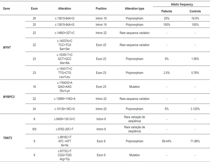
Table 2 - Alterations found in patients with HC and controls
Gene Exon Alteration Position Alteration type
Allelic frequency
Patients Controls
MYH7
20 c.13615-64A>G Intron 19 Polymorphism 25% 19.5%
20 c.13615-64A>G Intron 19 Polymorphism 100% 100%
22 c.14663+32T>C Intron 22 Rare sequence variation -
-22
c.14537A>C TCC>TCA
Ser>Ser
Exon 22 Rare sequence variation -
-23
c.15355 T>C GCT>GCC
Ala>Ala
Exon 23 Polymorphism 5% 1.56%
23
c.15431T>C TTG>CTG
Leu>Leu
Exon 23 Polymorphism 2.5% 0.78%
MYBPC3
16
c.11642G>A GAG>AAG
Glu>Lys
Exon 23 Mutation -
-22 c.13999+118G>A Intron 22 Rare sequence variation -
-24 c.15130+19C>G Intron 22 Polymorphism 5% 3.125%
TNNT2
8 c.8459+130 G>C Intron 8 Rara variação de
seqüência -
-8/9 c.8762-20C>T Intron 8 Rara variação de
seqüência -
-9
c.8816C>T ATC >ATT Ile>Ile
Exon 9 Polymorphism 69.44% 71.88%
9
c.8772C>T CGG>TGG Arg>Trp
Exon 9 Mutation -
-involved with the dilated form. There are two substitutions in each the TNNT2 and the α-tropomyosin genes, and there is one substitution in each the α-actin, the essential myosin light chain and the myosin regulatory light chain genes.
Moreover, when comparing the amino acid sequence of the MYBPC3 protein available in the NCBI database11 in several animals, it appears that the region where the substitution occurred is quite conserved in men (Homo sapiens), mice (Mus musculus), rats (Rattus norvegicus), dogs (Canis lupus familiaris), Oxen (Bos taurus) and chimpanzees (Pan troglodytes), which suggests that this is an important region in the protein.
To undoubtedly affirm that this mutation in the isolated form is able to cause the disease is only possible if the mutations in other exons of this gene and other sarcomeric genes are discarded through family studies. Together, however, the clinical and molecular data suggest that the p.E441K mutation is a pathogenic one.
TNNT2 gene, exon 9
The C → T substitution found in exon 9 of patient P5 generates a change of codon from CGG to TGG, changing
the amino acid from arginine to tryptophan. This mutation, p.Arg92Trp or p.R92W, was first described by Moolman et al16, with a high frequency among the patients studied, suggesting a founder effect. The clinical data of their patients showed a small hypertrophy, often undetectable, the mean maximum thickness of the wall of the patients was within the normal range and this was reflected in the low penetrance of the disease. However the prognosis associated with the disease was extremely poor, particularly for male adolescents and young adults. Furthermore, data from this study suggested that mutations in the TNNT2 gene lead to congestive heart failure at a lower frequency than the mutations in the MYH7 gene.
In a later study, Moolman-Smook et al17 investigated 40 unrelated patients and again the p.Arg92Trp mutation appeared with a high frequency and clinical data were similar to those seen in the previous study. According to the authors, the reduced life expectancy associated with this mutation and the well-preserved haplotype in many families, suggested a recent mutational event.
and reduced heart weight in comparison to patients with mutations in other genes. The study therefore suggests that the risk of sudden death in patients with mutations in this gene should be related to increased myocardial disarrangement, regardless of the hypertrophy degree.
However, according to Ackerman et al19, there are exceptions to all genotype-phenotype associations and this is the largest impediment to use genotyping alone as a clinical and prognostic tool for individual patients. In their study, one patient who had the p.Arg92Trp mutation presented extreme hypertrophy and his mother, who also had the mutation, did not present hypertrophy and there were no records of sudden deaths in the family. The authors mentioned that there are still many factors that may confound the phenotype, such as phenotypic expression and variability within families, the small size of the studied families, modifier genes, the role of polymorphisms and other non-genetic factors. These combined factors can weaken the premise that the risk of sudden death is associated to a particular mutation. They believe that the necessary knowledge to determine which HC mutation, combination of mutations and environmental factors may influence the clinical outcome is yet to be acquired. In 2003, Van Driest et al20 presented a study with TNNT2 gene mutations in which the patients did not present significantly lower hypertrophy than that in patients with mutations in other genes and none of them had family history of sudden death.
In our patient (P5), the p.Arg92Trp mutation led to a phenotype that can be considered severe, as the patient is very symptomatic, presenting moderate to severe hypertrophy and recurrence of syncope. Furthermore, we had access to an echocardiogram of his 18-year-old son, who presents massive hypertrophy (33 mm), corroborating the reports by Ackerman et al19.
Allele variants with frequencies lower than % Each of the alterations classified as allelic variants with a frequency lower than 1% appeared in only one individual (patient or control) among the 84 individuals analyzed. We found 5 alterations, 4 in intron regions and one in the exon, a silent-type polymorphism, which does not change the synthesized amino acid.
Changes in introns may or may not affect the protein. To correctly identify and bind RNA sequences that encode proteins, the exons must be differentiated from the introns. There are several preserved motifs in nucleotides sequences near the intron-exon boundaries that act as essential signals for splicing. These signals are involved with pre-messenger RNA intron excision and exon junction. Additional elements known as promoters and silencers are required to allow a normal splicing of the exonic sequences. These regulatory elements may be close to (to 50-100 bp) or far from (thousands of bp) splicing sites21.
Among the changes found, two were in regions close to the exon boundary (+32 to -20) and two were in more distant regions (+118 and +130). With the techniques used in this study only, there is no evidence that these changes affect the protein. Genomic variants that affect the splicing (or splicing-affecting genomic variants - SpaGVs) may occur in exonic or
intronic regulatory elements and are very difficult to be defined only by the sequence of nucleotides. However, these changes can lead to catastrophic splicing abnormalities. In particular, SpaGVs can induce the exclusion of exons, the activation of alternative splicing sites, or they may alter the balance of alternative splicing isoforms produced and thus cause disease phenotypes. The importance of functional genomic variants (GV) which are located far away from the splicing sites is even more difficult to be identified than those that are adjacent. Although they are not near any obvious regulatory sequences, such variants can cause, for instance, the activation of a new splicing site, which defines the boundaries of the exon21.
The other rare sequence variation found was of the silent type. Such changes are usually disregarded because they do not affect the synthesized protein, but studies show they may be important.
Silent mutations are normally classified as allelic polymorphisms and are considered neutral. These definitions may be correct in some cases, but when they are not based on the characterization of the mRNA level they can lead to errors, because mutations affecting the sequence are important for splicing modulation and may lead to a profound effect on the translated product. Many studies have correlated specific point mutations in coding regions to the exclusion of the exon containing the mutation. The fact that silent mutations theoretically cause exon exclusion is particularly significant, as these mutations must act at the RNA level. These mutations probably alter regulatory elements that are important for correct splicing. Still, it is likely that these mutations are infrequently reported, because they may be incorrectly perceived as neutral polymorphisms that do not deserve further characterization22.
It is not possible to affirm that the variations found here are pathogenic, as RNA studies would be needed in order to do that. However, even with sensitive techniques such as direct sequencing and the complete study of all 13 exons of the genes involved in the HC, the detection of mutations in patients is only successful in approximately 70% of the cases2,15. Thus, it is possible that some splicing mutations in introns or exons are being underestimated and that is one of the reasons why it is important to study and publish these alterations.
Variants with allele frequencies greater than % (polymorphisms)
A total of 6 polymorphisms, 3 in the intron region and 3 within the exon, were found in this study. Recently, it has been suggested that the genetic causes for susceptibility to complex diseases may be beyond the missense and nonsense mutations, which are commonly studied in simple genetic disorders. Within this spectrum, polymorphisms that alter the gene expression are suspected to play an important role23.
thickness and increased mass in the hypertrophied population. However, only the presence of the allele does not seem enough to cause significant cardiac hypertrophy. The results suggest therefore that the genotype predisposes individuals to cardiac hypertrophy.
Several studies have shown that polymorphisms in regions distant from the splicing site may be related to diseases, affecting regulatory elements. Hoogendoorn et al23 studied in
vitro the effect of polymorphisms on splicing promoters and found that a surprisingly high proportion of polymorphisms, approximately one third in their study, altered the expression of the gene in 50% or more of the cases.
The allelic frequency of the polymorphisms identified in this study was not statistically significant between patients and controls in this study. This suggests that the polymorphisms do not affect the phenotype. Furthermore, clinical data of patients who had and who did not have the polymorphism were similar.
Our knowledge of regulatory elements of the human genome is still limited and functional polymorphisms of these elements cannot be clearly distinguished from those with no effect, if considering only its sequence24. Therefore we have considered the study and the publication of the polymorphisms as being relevant, even though they are in intronic regions distant from the splicing sites.
Sensitivity of the method
In this study, mutations were identified in 2 of 20 patients, 10% of the sample. A higher proportion would be expected, as the studied exons account for approximately 31% of the mutations already described. Several possibilities can be suggested to explain the sensitivity below the expected and among them, three deserve attention.
Firstly, the method itself does not have 100% sensitivity and it markedly decreases with the increase in fragment size25. Studies suggest that, for fragments smaller than 200 base pairs, more than 90% of the sequence variations will be detected. When the fragment increases to 300 to 350 nucleotides, more than 80% of mutations will be detected. In our study, only two fragments had less than 350 bp. The SSCP was able to detect several substitutions, including the in larger fragments, but the sensitivity of the test can be improved by using smaller fragments or by changing other conditions, such as varying bisacrylamide, and glycerol concentrations, etc. Secondly, the sample size must be taken into account. In the case of a pilot project, with the use of a laborious method of genetic screening, involving several laboratory steps, the sample was initially considered sufficient, given the logistics available, but it has an impact when the sensitivity of the method is evaluated.
A third possibility is the local population profile. Most genotyping studies in patients with HC are from the northern hemisphere and the frequencies of mutations that cause the disease in South American countries such as Brazil
are yet to be known; additionally, little is known about their frequency in developing countries. Considering the miscegenation of the Brazilian population, some similarity with other populations worldwide is expected, but this is speculative at present and it is possible that the prevalence of affected genes in Brazil is different than that described for other populations. In 1999, a study in South Africa showed that the mutations described as predominant worldwide were apparently rare in the country17. A study of Spanish patients showed that, compared to other populations, mutations in the MYH7 and TNNT2 genes were rare among the studied patients26 and a very low rate of mutations in the MYH7 gene was found in a study with patients in Finland27. These data suggest that each region may have a single mutation profile and that may have important effects on the implementation of population screening.
Conclusions
Given the objectives of the study, we concluded that the genotyping of patients with HC is feasible in our country. With respect to the mutations, we concluded that it is possible that the p.E441K (p.Glu441Lys) mutation in exon 16 of the MYBPC3 gene is pathogenic in isolation, resulting in a less severe phenotype than when associated to another mutation, and the p.R92W (Arg92Trp) mutation in exon 9 of the TNNT2 gene has a phenotype that is not as homogeneous as previously described and may lead to severe hypertrophy. To the best of our knowledge, this is the first time that a HC-causing mutation has been described in Brazil. Depending on further analysis, one of the patients in this sample population may be carrying a novel mutation, when compared to the available databases.
Acknowledgments
To Fundação de Apoio à Pesquisa do Espirito Santo (FAPES) and the State Health Secretariat of the state of Espirito Santo, Brazil.
Potential Conflict of Interest
No potential conflict of interest relevant to this article was reported.
Sources of Funding
This study was partially funded by Secretaria de Saúde do Espírito Santo e Fundação de Apoio à Pesquisa do Espírito Santo (FAPES).
Study Association
References
1. Maron BJ. Hypertrophic cardiomyopathy. The Lancet. 1997; 350: 127-33. 2. Pascale R, Villard E, Charron P, Isnard R. The genetic bases of cardiomyopathies.
J Am Coll Cardiol. 2006; 48 (9): A79-A89.
3. Maron BJ. Hypertrophic cardiomyopathy: a systematic review. JAMA. 2002; 287: 1308-20.
4. Jarcho JA, McKenna W, Pare JA, Solomon SD, Holcombe RF, Dickie S, et al. Mapping a gene for familial hypertrophic cardiomyopathy to chromosome 14q1. N Engl J Med. 1989; 321: 372-8.
5. Geisterfer-Lowrance A, Kass S, Tanigawa G, Vosberg HP, McKenna W, Siedman CE, et al. A molecular basis of familial hypertrophic cardiomyopathy: a β-cardiac myosin heavy chain gene missense mutation. Cell. 1990; 62: 999-1006.
6. Cardiogenomics. Sarcomere protein gene mutation database. [Acesso em 2007 abr. 7]. Disponível em: <http://genetics.med.harvard.edu/~seidman/ cg3/index.html.
7. Tirone AP, Arteaga E, Pereira AC, Krieger JE, Buck PC, Ianni BM, et al. Research of markers for the genes of the heavy chain of cardiac â-myosin and myosin binding protein c in relatives of patients with hypertrophic cardiomyopathy. Arq Bras Cardiol. 2005; 84 (6): 467-72.
8. Instituto Brasileiro de Geografia e Estatística (IBGE). Estimativas populacionais para os municípios brasileiros em 01/07/2008. [Acesso em 2008 dez 28]. Disponível em: http://www.ibge.gov.br/home/estatistica/populacao/ estimativa2008
9. Miller SA, Dykes DD, Polesky HF. A simple salting out procedure for extracting DNA from human nucleated cells. Nucleic Acids Research. 1988; 16 (3): 1215.
10. Bassam BJ, Caetano-Anolles G, Gresshoff PM. Fast and sensitive silver staining of DNA in polyacrilamide gels. Anal Biochem. 1991; 196: 80-3.
11. National Center For Biotechnology Information. [Acesso em 2007 Feb. 10]. Disponível em: <http://www.ncbi.nlm.nih.gov>.
12. Cardiogenomics. Sarcomere protein gene mutation database. [Acesso em 2008 Feb. 20]. Disponível em: <http://genetics.med.harvard.edu/~seidman/ cg3/muts/MYBPC3_ Glu441Lys.html>.
13. Olivotto I, Girolami F, Ackerman MJ, Stefano N, Martijn BJ; Elisabetta Z, et al. Myofilament protein gene mutation screening and outcome of patients with hypertrophic cardiomyopathy. Mayo Clin Proc. 2008; 83 (6): 630-8. 14. Niimura H, Bachinski LL, Sangwatanaroj S, Watkins H, Chudley AE, McKenna
W, et al. Mutations in the gene for cardiac myosin-binding protein C and late-onset familial hypertrophic cardiomyopathy. N Engl J Med. 1998; 338 (18): 1248-57.
15. Richard P, Charron P, Carrier L, Ledeuil C, Cheav T, Pichereau C, et al. Hypertrophic cardiomyopathy: distribution of disease genes, spectrum of
mutations, and implications for a molecular diagnosis strategy. Circulation. 2003; 107 (17): 2227-32.
16. Moolman JC, Corfield VA, Posen B, Ngumbela K, Seidman C, Brink PA, et al. Sudden death due to troponin T mutations. J Am Coll Cardiol. 1997; 29 (3): 549-55.
17. Moolman-Smook JC, De Lange WJ, Bruwer EC, Brink PA, Corfield VA. The origins of hypertrophic cardiomyopathy-causing mutations in two South African subpopulations: a unique profile of both independent and founder events. Am J Hum Genet. 1999; 65 (5): 1308-20.
18. Varnava AM, Elliott PM, Baboonian C, Davison F, Davies MJ, Mckenna WJ. Hypertrophic cardiomyopathy: histopathological features of sudden death in cardiac troponin T disease. Circulation. 2001; 104 (12): 1380-4. 19. Ackerman MJ, Van Driest SL, Ommen SR, Will ML, Nishimura RA, Tajik AJ,
et al. Prevalence and age-dependence of malignant mutations in the beta-myosin heavy chain and troponin T genes in hypertrophic cardiomyopathy: a comprehensive outpatient perspective. J Am Coll Cardiol. 2002; 39 (12): 2042-8.
20. Van Driest SL, Ellsworth EG, Ommen SR, Tajik AJ, Gersh BJ, Ackerman MJ. Prevalence and spectrum of thin filament mutations in an outpatient referral population with hypertrophic cardiomyopathy. Circulation. 2003; 108 (4): 445-51.
21. Wang GS, Cooper TA. Splicing in disease: disruption of the splicing code and the decoding machinery. Nat Rev Genet. 2007; 8: 749-61.
22. Cartegni L, Chew SL, Krainer AR. Listening to silence and understanding nonsense: exonic mutations that affect splicing. Nat Rev Genet. 2002; 3: 285-98.
23. Hoogendoorn B, Coleman SL, Guy CA, Smith K, Bowen T, Buckland PR, et al. Functional analysis of human promoter polymorphisms. Hum Mol Genet. 2003; 12 (18): 2249-54.
24. Komamura K, Iwai N, Kokame K, Kim J, Yamagishi M, Morisaki T, et al. The role of a common TNNT2 polymorphism in cardiac hypertrophy. J Hum Genet. 2004; 49: 129-33.
25. Sheffield VC, Beck JS, Kwitek AE, Sandstrom DW, Stone EM. The sensitivity of single-strand conformation polymorphism analysis for the detection of single base substitutions. Genomics. 1993; 16: 325-32.
26. García-Castro M, Reguero JR, Batalla A, Diaz-Molina B, Gonzalez P, Alvarez V, et al. Hypertrophic cardiomyopathy: low frequency of mutations in the
β-myosin heavy chain (MYH7) and cardiac troponin t (TNNT2) genes among spanish patients. Clin Chem. 2003; 49 (8): 1279-85.