Abst ract
Submitted: April 30, 2016 0RGL¿FDWLRQ-XO\ Accepted: August 22, 2016
Oral m ucosa: an alt ernat ive epiderm ic
cell source t o develop aut ologous
derm al- epiderm al subst it ut es from
diabet ic subj ect s
Oral m ucosa has been highlight ed as a suit able sour ce of epider m al cells
due t o it s int r insic charact er ist ics such as it s higher pr oliferat ion rat e and it s
obt ainabilit y. Diabet ic ulcer s have a w or ldw ide pr evalence t hat is var iable
( 1% - 11% ) , m eanwhile t reat m ent of t his has been proven ineffect ive.
Tissue-engineered skin plays an im port ant role in w ound care focusing on st rat egies
such aut ologous der m al- epider m al subst it ut es. Obj ect ive: The aim of t his
st udy was t o obt ain aut ologous der m al- epider m al skin subst it ut es fr om oral
PXFRVD IURP GLDEHWLF VXEMHFWV DV D ¿UVW VWHS WRZDUGV D SRVVLEOH FOLQLFDO
applicat ion for cases of diabet ic foot . Mat erial and Met hods: Oral m ucosa was
obt ained fr om diabet ic and healt hy subj ect s ( n= 20 per gr oup) . Epider m al
FHOOVZHUHLVRODWHGDQGFXOWXUHGXVLQJDXWRORJRXV¿EULQWRGHYHORSGHUPDO
epider m al in vit r o subst it ut es by t he air- liquid t echnique w it h aut ologous
hum an serum as a supplem ent m edia. Subst it ut es were im m unocharact erized
ZLWKFROODJHQ,9DQGF\WRNHUDWLQDVVSHFL¿FPDUNHUV$6WXGHQWVWWHVW
was per for m ed t o assess t he differ ences bet w een bot h gr oups. Result s: I t
was possible t o isolat e epider m al cells fr om t he oral m ucosa of diabet ic and
healt hy subj ect s and develop aut ologous der m al- epider m al skin subst it ut es
using aut ologous ser um as a supplem ent . Differ ences in t he ex pr ession
RI VSHFL¿F PDUNHUV ZHUH REVHUYHG DQG WKH F\WRNHUDWLQ H[SUHVVLRQ
was low er in t he diabet ic subst it ut es, and t he collagen I V expr ession was
higher in t he diabet ic subst it ut es w hen com par ed w it h t he healt hy gr oup,
VKRZLQJDVLJQL¿FDQWGLIIHUHQFH&RQFOXVLRQ&HOOVIURPRUDOPXFRVDFRXOG
be an alt er nat ive and less invasive sour ce for skin subst it ut es and w ound
healing. A difference in collagen product ion of diabet ic cells suggest s diabet ic
subst it ut es could im pr ove diabet ic w ound healing. Mor e r esear ch is needed
t o det er m ine t he cr osst alk bet w een com ponent s of t hese skin subst it ut es
and dam aged t issues.
Ke y w or ds: Oral m ucosa. Skin subst it ut es. Biological dr essings.
Daniela GUZMÁN-URIBE1,2
Keila Neri ALVARADO-ESTRADA1
Mauricio PIERDANT-PÉREZ2
Bertha TORRES-ÁLVAREZ3
Jesus Martin SÁNCHEZ-AGUILAR2
Raúl ROSALES-IBÁÑEZ1,4
http://dx.doi.org/10.1590/1678-77572016-0217
1Universidad Autónoma de San Luis Potosí, Facultad de Estomatología, Grupo de Investigación en Ingeniería Tisular, San Luis Potosí, México.
2Universidad Autónoma de San Luis Potosí, Facultad de Medicina, Maestría en Ciencias en Investigación Clínica, San Luis Potosí, México.
3Hospital Central Dr Ignacio Morones Prieto, Departamento de Dermatología, San Luis Potosí, México.
4Universidad Nacional Autónoma de México, Facultad de Estudios Superiores Iztacala, Laboratorio en Ingeniería Tisular y Medicina Traslacional, Tlanepantla, México.
Corresponding address: Mauricio Pierdant Pérez Departamento de Epidemiología Clínica - Facultad de Medicina - Universidad Autónoma de San Luis Potosí
Avenida Venustiano Carranza #2405 - Colonia los Filtros - San Luis Potosí - SLP - CP.78210
I nt r oduct ion
$GYDQFHPHQWV LQ WKH ¿HOG RI WLVVXH HQJLQHHULQJ have allow ed t he w idespr ead use of sk in subst it ut es
t o cov er w ou n ds an d t o pr om ot e h ealin g1 9. Gr eat
p r o g r ess h as b een ach i ev ed i n t h e cu l t u r i n g o f NHUDWLQRF\WHVDQG¿EUREODVWVWKLVKDVEHHQHVSHFLDOO\ t r u e f o r k er at i n o cy t es si n ce t h e f i r st su ccessf u l
cult ur e by Rheinw ald and Gr een in 197525, t o t he
p r esen t d ay, w i t h t h e cr eat i o n o f sp eci al i zed i n
v it r o t ech n iq u es1 4 , 2 2 , 2 3 , 2 4. Alt h ou g h cu r r en t t issu e-engineer ed sk in subst it ut es have show n pr om ising
r esu lt s f or w ou n d h ealin g6 , 8 , 1 1 , 1 3 , 2 6, t h er e ar e st ill
several challenges t o over com e, t her efor e t her e is a
cont inuing sear ch for novel appr oaches t o w ound car e
and t r eat m ent s t hat could be m or e effect ive, w her e
all epider m al cells and m at r ices ar e obt ained fr om t he
sam e pat ient , w it h less invasive sour ces t o obt ain t he
biological com ponent s, and w it h new alt er nat ives t o
harvest t issue in pat ient s wit h physiological and et hical
lim it at ions due t o t heir condit ions.
Or al m u cosa h as been h igh ligh t ed as a v iable
alt er nat ive sour ce of epider m al cells, due t o it s easy
pr eparat ion and suit able feat ur es, such as higher cell
pr oliferat ion rat es, low er t er m inal cell differ ent iat ion
degrees and an increased biological act ivit y com pared
w it h epider m al kerat inocy t es22. This t issue also has
t h e ad v an t ag e t h at it s h ar v est in g p r od u ces less
disabilit y, and pr ov ides bet t er aest het ic out com es.
Wit h t hese special feat ur es, it is assum ed t hat t he
sk in subst it ut e obt ained fr om oral m ucosa can be
produced fast er. A possible disadvant age for t he use of
oral m ucosa as an epider m al cell sour ce could be t he
different iat ion of t he epit helial pat t ern of kerat inocyt es
from oral m ucosa, which differs from t hat of epiderm al
kerat inocy t es and sk in. The sk in is an ex am ple of an
or t h ok er at in ized epit h eliu m . I t display s a st r at u m
basale, st rat um spinosum , st rat um granulosum and
st rat um cor neum . Gingiva t issue also consist s of an
epit helium on a connect ive t issue m at r ix populat ed PDLQO\ZLWK¿EUREODVWVDQGHQGRWKHOLDOFHOOVKRZHYHU in cont rast t o skin, t he epit helium is parakerat inized27.
I n t his r egar d, Ueda, et al.26 ( 1995) r epor t ed in t heir
st u dy t h at a gr af t ed sh eet of k er at in ocy t es f r om
oral m ucosa changed t he epit helial pat t er n over a
per iod of 4 w eek s, ex plaining t his phenom enon due
t o epit helial- m esenchy m al int eract ions, suggest ing WKDWWKHVSHFL¿FLW\RIWKHHSLWKHOLXPLVGHSHQGHQWRQ WKHLQÀXHQFHRIWKHXQGHUO\LQJPHVHQFK\PDOWLVVXH
Wo r l d w i d e, d i ab et i c f o o t i s a m aj o r m ed i cal ,
social and econom ic pr oblem . I t is est im at ed t hat
appr ox im at ely 1 5 % of t h e m or e t h an 1 5 0 m illion
people w it h diabet es w or ld- w ide w ill at som e st age
develop diabet ic foot ulcerat ion7. Foot pr oblem s ar e indeed a global pr oblem and t her e is no ar ea in t he
w or ld t hat does not r epor t t he developm ent of foot
lesions as a consequence, m ainly of neur opat hy and
per ipheral vascular disease. The pr evalence of act ive
foot ulcerat ion varies from approxim at ely 1% in cert ain
Eur opean and Nor t h Am er ican st udies t o m or e t han
11% in report s from som e African count ries7. Alt hough t her e have been m any developm ent s in r ecent year s,
w hich encourages opt im ism for fut ur e im pr ovem ent
in diabet ic foot car e, t her e is st ill m uch t o be done.
The aim of t he pr esent st udy was t o obt ain an
aut ologous der m al- epider m al sk in subst it ut e using
or al m u cosa t issu e f r om diabet ic su bj ect s w it h a PHDVXUDEOHLPPXQRÀXRUHVFHQFHFKDUDFWHUL]DWLRQDVD ¿UVWVWHSWRSHUIHFWLQJWKHGHYHORSPHQWWHFKQLTXHZLWK t he m ain focus of fut ur e possible clinical applicat ions
on t h e t r eat m en t of d iab et ic f oot u lcer s. Also it SURSRVHVIRUWKH¿UVWWLPHPDLQWDLQLQJWKLVVXEVWLWXWH using aut ologous ser um , t hus av oiding t he use of
anim al pr oduct s and pr om ot ing t he developm ent of
t he const r uct .
Mat er ial and m et hods
This was an ex per im ent al in vit r o t est car r ied out
at Hospit al Cent ral “ Dr. I gnacio Mor ones Pr iet o”, t he
Basic Sciences Laborat or y, Facult y of St om at ology,
Univer sidad Aut ónom a de San Luis Pot osí. The st udy
w a s p er f o r m ed i n a cco r d a n ce w i t h t h e Hel si n k i
Declarat ion, and it w as appr ov ed by t he Facult y ’s
Et hical Boar d ( CEI FE- 033- 012) .
Subj ect s
Tw en t y ad u l t su b j ect s w i t h d i ab et es m el l i t u s
t y pe I I ( DM2) , diagnosed accor ding t o t he Am er ican
Diabet ics Associat ion gu idelin es1 6, an d 2 0 h ealt hy
cont r ols donat ed 2 0 m l of per ipheral blood and a
sam ple of or al m u cosa fr om t h e r et r om olar ar ea.
Blood was collect ed in labeled Vacut ainer t ubes, 10 m l
w it hout addit ives, and 10 m l w it h sodium cit rat e ( BD
Vacut ainer®, Frank lin Lakes, NJ, USA) and fr ozen at 20°C, unt il used. Oral m ucosa sam ples w er e obt ained
w it h a 3 m m biopsy punch ( Milt ex , Dav ies Dr ive Yor k ,
infer ior alveolar ner ve block age ( I AN) . Hem or r hage
w as con t r olled b y d ir ect m ean s f or 1 0 m in u t es.
Pat ient s r eceived inst r uct ions for w ound car e, and a
follow- up aft er 7 days. None of t he pat ient s pr esent ed
com plicat ions.
I solat ion and cell cult ur e
Each oral m ucosa sam ple was collect ed in 1.5 m l
t ubes cont aining phosphat e buffer ed saline ( PBS) , NjJPO VWUHSWRP\FLQ ,8PO SHQLFLOOLQ DQG NjJPODPSKRWHULFLQ%DOOIURP6LJPD$OGULFKSt . Louis, MO, USA) and pr eser ved at 4°C for 12 h. Oral
m ucosa was m acroscopically separat ed by region, wit h
a scalpel, int o epit helium and connect ive t issue. By
using t he ex plant t echnique, each r egion was gr ow n
separat ely in 25 cm2 cult ur e dishes ( Nunc, Roskilde,
Denm ark) . DMEM low glucose m edium ( Sigm a-Aldrich,
St . Lou is, MO,USA) su pplem en t ed w it h 1 0 % fet al
calf ser um ( Sigm a- Aldr ich, St . Louis, MO, USA) was XVHGDVFXOWXUHPHGLXPIRU¿EUREODVWV.HUDWLQRF\WHV ZHUH FXOWXUHG XVLQJ 41 PHGLXP D VSHFL¿F FXOWXUH m edium for kerat inocy t es) . Aft er 3 day s t he fet al calf
ser um concent rat ion was r educed t o 5% , and aft er a
fur t her 3 day s t o 1% . Blood sam ples w er e defr ost ed
and cent r ifuged at 1200 r pm for 10 m inut es. Ser um
and plasm a w er e isolat ed and kept at - 20°C unt il us e.
Aut olougus ser um was used for cell cult ur e inst ead of
t he fet al calf serum aft er 7 days, w hile t he plasm a was
used t o develop t he st r om a for t he subst it ut es. Bot h
cell lines w er e incubat ed at 37°C in an at m ospher e
of 5% CO2XQWLOWKHFXOWXUHVUHDFKHGDFRQÀXHQFHRI
80% . The cult ur e m edia was changed ever y t hir d day.
Cell cult ur es charact er izat ion
2QFHWKHFXOWXUHVUHDFKHGDFRQÀXHQFHRI par t of t he cell cult ur es w er e used for an indir ect LPPXQRÀXRUHVFHQFHDVVHVVPHQW,QRUGHUWRDVVXUH WKDWWKHFHOOVZHUH¿EUREODVWVDQGNHUDWLQRF\WHVWKH charact er izat ion of t hese cell cult ur es was per for m ed. 7KHVSHFL¿FPRQRFORQDODQWLERG\XVHGIRU¿EUREODVWV was ant i- collagen I ( Sant a Cr uz Biot echnology, Paso
Robles, CA, USA) m ainly because due t o it s abundance
in t he sk in, r ecognized as t he m ost abundant collagen LQWKHHSLGHUPLVDQGLWVVSHFL¿FLW\IRUWKLVFHOOW\SH For kerat inocy t e was ant i- cy t okerat ins 5- 14 ( Sant a
Cr u z Bi o t ech n o l o g y, Paso Ro b l es, CA, USA) . As VHFRQGDU\ DQWLERGLHV $OH[D ÀXRU $QWL5DEELW I gG polyclonal, I nv it r ogen, Walt ham , MA, USA) and
Alex a 594 ( Ant i- m ouse m onoclonal I gG, I nv it r ogen,
:DOWKDP0$86$ZHUHDOVRXVHG%ULHÀ\FHOOVZHUH gr ow n on r ound glass cover slips ( 12 m m ) . Aft er 12 h RILQFXEDWLRQWKHFXOWXUHVZHUH¿[HGIRUPLQXWHV w it h n eu t r al f or m alin , p er m eab ilized w it h Tr it on
X100 0.025% for 20 m in at r oom t em perat ur e t hen
blocked w it h bov ine ser um album ina ( BSA) 1% for
45 m in. The pr im ar y ant ibodies w er e incubat ed for 2
h at r oom t em perat ur e and t he secondar y ant ibodies
w er e incubat ed for 2 h at r oom t em perat ur e and w er e
pr ot ect ed fr om light . Bet w een each st ep, phosphat e
b u f f er ed sal i n e r i n ses w er e m ad e. Th e sam p l es
w er e obser ved by confocal m icr oscopy ( Leica, Model
DMI 4000B, Wet zlar,Ger m any ) and LASAF® soft war e
( Leica, Wet zlar, Ger m any ) .
Developm ent of t he derm o- epiderm al subst it ut e
St r om al phase
Fibr oblast s w er e det ach ed f r om cu lt u r e dish es
using TrypLE Express ( I nvit rogen, Walt ham , MA, USA) ,
cent r ifuged and suspended once m or e in t he m edium DQGTXDQWL¿HGE\2QH6FHSWHU0LOOLSRUH%LOOHULFD0$ USA) . Cells were again suspended in DMEM low glucose
m edium ( Sigm a- Aldr ich, St . Louis, MO, USA) w it h 1%
of hum an serum obt ained from each subj ect , reaching
a cell concent rat ion of 500,000 per 1 m L. A solut ion
was m ade by m ixing t he resuspended cells wit h plasm a DQGDJDURVHDWWRFUHDWH¿EULQDJDURVHJHOV5. 300 m icrolit ers of t he solut ion w ere placed in t he Transw ell
sy st em ( Cor ning, Midland, NC, USA) follow ing t he
air-liquid t echnique3. Cult ure m edium was placed covering
t he ent ir e subst it ut e. Each sy st em was incubat ed at
37°C in a 5% CO2 at m ospher e and w it h a r elat ive
hum idit y of 95% for 10 day s ( Figur e 1a, b) .
Epit helial phase
Cult ur ed kerat inocy t es w er e det ached fr om t he
cu lt u r e d ish es u sin g Tr y p LE Ex p r ess ( I n v it r og en ,
Walt ham , MA, USA) , cent r ifuged and r esuspended in
a QN m edium w it h 1% hum an ser um obt ained fr om , DQG TXDQWL¿HG E\ 2QH 6FHSWHU 0LOOLSRUH %LOOHULFD MA, USA) t o r each a concent rat ion of appr ox im at ely
500,000 cells per 1 m L. One hundr ed m icr olit er s of
t he cell solut ion w er e placed in t he Transw ell sy st em
( Cor ning, Midland, NC, USA) on t he t op of t he alr eady
set st r om a, and incubat ed for 10 day s using t he
air-liquid t echnique. Cult ur e m edium was placed cover ing ERWKFRPSDUWPHQWVRQWKH¿UVWGD\RIVHWWOHPHQWRQ t he four t h day, t he cult ur e m edium was placed only in
t he low er com par t m ent . Each sy st em was incubat ed
hum idit y for 10 fur t her day s.
Der m al- epider m al subst it ut e evaluat ion and
charact er izat ion
Subst it ut es were m echanically separat ed from each
sy st em and placed in dishes w it h 1X PBS t o r em ove H[FHVVRIWKHPHGLXP6XEVHTXHQWO\WKH\ZHUH¿[HG w it h 4% parafor m aldehyde and placed in a solut ion
of 3% sucr ose as a cr yopr ot ect or. Cr yosect ions of 6
m icr ons t hick ness w er e m ade at - 29°C in a cr yost at
( Leica Model CM1510S- 3, Wet zlar, Ger m any ) . Fr om
each sam ple, 100 cut s w er e m ade and placed on 50
slides ( t w o cut s per slide) . Each slide was t r eat ed for LWVLQGLUHFWLPPXQRÀXRUHVFHQFHDVVHVVPHQW
For t he charact erizat ion of t hese derm al- epiderm al VXEVWLWXWHVWKHVSHFL¿FPRQRFORQDODQWLERGLHVXVHG were ant i- collagen I V and ant icyt okerat ins 5- 14 ( Sant a
Cr uz Biot echnology, Paso Robles, CA, USA) . Collagen
I V was assessed in t his charact er izat ion due t o it s
r ole as t he pr im ar y collagen found in t he ex t racellular
basal m em branes separat ing a var iet y of epit helial
and endot helial cells. I t is a m aj or com ponent of t he
derm al- epiderm al j unct ion, where it is m ost ly found in
t he lam ina densa of t he basal m em brane9. The basal m em brane zone m ediat es t issue com part m ent alizat ion
and sends signals t o epit helial cells about t he ext er nal
m icr oenv ir onm ent , and also has im por t ant st r uct ural
and funct ional effect s on blood vessels, const it ut ing an
ex t racellular m icr oenv ir onm ent sensor for endot helial
cells and per icy t es9. Meanw hile, Cy t okerat ins 5- 14 ( CK5- 14) ar e ex pr essed on basal kerat inocy t es and
WKHH[SUHVVLRQRIWKHVH¿ODPHQWVDUHFKDUDFWHULVWLFRI FRPSOH[VWUDWL¿HGHSLWKHOLD1. Kerat in 14 ( CK14) is a pr ot ot y pic m ar ker of div iding basal kerat inocy t es and
helps in t he m aint enance of epider m al cell shape;
it also p r ov id es r esist an ce t o m ech an ical st r ess.
I nt er est ingly, t he CK5/ CK14 pair is ex pr essed in t he
basal layer of t he epiderm is, which cont ains epiderm al
st em cel l s an d t r an si en t am p l i f y i n g ( TA) cel l s2. 0RQRFORQDOVHFRQGDU\DQWLERGLHV$OH[DÀXRUDQG
594 ( I nv it r ogen, Walt ham , MA, USA) w er e also used. 7HQGLIIHUHQW¿HOGVZHUHFKRVHQUDQGRPO\IURPHDFK slide; each slide of ever y subst it ut e cr eat ed t o be
evaluat ed, was chosen by t he sam ple funct ion of t he
R pr ogr am , ver sion 3.0.1. The evaluat ion of t he slide
was per for m ed by an obser ver blinded t o t he nat ur e
of t he st udy. A t ot al of 20 im ages w er e obt ained;
obser vat ions w er e per for m ed by confocal m icr oscopy
( Leica, Model DMI 4000B, Wet zlar, Ger m any ) and t he
LASAF® soft war e ( Leica, Wet zlar, Ger m any ) . I m ages
obt ained were evaluat ed by I m age J program m anager
(1.46a ver sion, NI H) w it h t he ROI funct ion get t ing t he PHDQRIWKHDUELWUDU\ÀXRUHVFHQFHXQLWVper slide, per
ant ibody for fur t her st at ist ical analy sis.
Cell gr ow t h cur ve
Cell growt h curves were perform ed ( N= 8) . Cult ures
of 4 healt hy subj ect s and 4 age- m at ched diabet ic
subj ect s w er e select ed. Fibr oblast s w er e subcult ur e in
duplicat e in 6 w ell boxes by placing 100,000 cells per
w ell. The evaluat ion t im es w er e 0, 3, 6 and 9 day s.
St at ist ics
St at ist ical an aly sis at 9 5 % of con f id en ce w as
per for m ed using JMP 8 soft war e ( SAS I nst it ut e I nc.,
Cary, NC, USA) and R 3.1.320. An analysis of descript ive
st at ist ics, obt aining t he m easures of a cent ral t endency
and dispersion of all t he variables was t hen perform ed.
For t he bivar iat e com parat ive analy sis w e used t he
St udent ’s t - t est for cont inuous var iables based on
a nor m al dist r ibut ion calculat ed w it h plot of Fox10.
For cat egor ical var iables t he Chi- squar ed t est w as
applied. A pair ed St udent ´ s t t est for t im e 0 ver sus
3, 6 and 9 day s for t he gr ow ing cellular it y bet w een
WKHJURXSVZDVSHUIRUPHG6WDWLVWLFDOVLJQL¿FDQFHZDV
consider ed w it h a p value < 0.05. For t he cell gr ow t h
FXUYHVWDWLVWLFDOVLJQL¿FDQFHZDVFRQVLGHUHGZLWKD
Bonfer r oni cor r ect ion of alpha for 3 com par isons w it h
a p value of < 0.016.
Figure 1- $$LUOLTXLGWHFKQLTXHSKDVH'HYHORSPHQWRIVWURPDZLWKWKH¿EULQDJDURVHJHOV%6SHFL¿FFXOWXUHPHGLXPSODFHGLQD
Result s
St udy dem ographics
A t ot al of 40 oral m ucosa sam ples w er e collect ed
( n= 2 0 per gr oup) accor ding t o t he pr ev iously set
crit eria. Of t he t ot al of sam ples t aken, 19 corresponded
t o m en an d 2 1 t o w om en . Th e av er age age w as
52.15± 13.5 years. Variables st udied in each group are
show ed in Table 1. Charact er ist ics including age and
sex were relat ively hom ogenous bet ween bot h groups.
Obt aining and charact er izat ion of cell cult ur es
Fi b r o b l a s t a n d k e r a t i n o c y t e c u l t u r e s w e r e
est ab lish ed f r om t h e or al m u cosa f r om d iab et ic
and healt hy subj ect s ( Figur e 2 a, b) . Cell cult ur es
ach iev ed 8 0 % p er cen t of con f lu en ce at d if f er en t
t im es; diabet ic cult ur es had a cult ur e day average of
25.3± 3.09, m eanw hile t he healt hy subj ect s’ cult ur e
day average was 26.85± 1.78. Cell m orphology showed
by cult ur es of t he t w o cell lines cor r esponded t o t he W\SLFDOPRUSKRORJ\RI¿EUREODVWVDQGNHUDWLQRF\WHV Fibr oblast ´ cult ur es w er e posit ive for ant i- collagen
I , and kerat inocy t e cult ur es w er e posit ive for ant
i-cy t okerat ins 5 and 14 ( Figur e 2c, d) .
Cell gr ow t h cur ve
Cell p r olif er at ion r esu lt s sh ow ed a sig n if ican t
incr eased gr ow t h in t he diabet ic gr oup at t im e 0 v s
3 day s ( p= 0.009) , and t im e 0 v s 9 day s ( p= 0.004) ; KRZHYHU VWDWLVWLFDOO\ ZLGH FRQ¿GHQFH LQWHUYDOV DUH
DIABETIC SUBJECTS GROUP (N=20)
HEALTHY SUBJECTS GROUP (N=20)
P VALUE
Age 57.55*±11.59† 47*±15.92† 0.75‡
Sex (M/F) 9/11 10/10 0.02§
Culture days 25.3*±3.09† 26.85*±1.78† 0.05‡
AFU Col-IV 8.05*±1.59† 6.15*±3.16† 0.02‡
AFU Ck 5-14 9.56*±3.741† 10.47*±5.86† 0.05‡
AFU Total 17.61*±5.22† 16.62*±8.94† 0.67‡
*Mean
†Standard deviation ‡ Student´s t-test §Chi-Square
Match paired Student´s T-test
Table 1- Studied variables per group
show ed, indicat ing gr eat er var iabilit y in t he behav ior
of t he diabet ic group com pared wit h t he healt hy group
( Figur e 3, Table 2) .
Developm ent of t he derm o- epiderm al subst it ut e
Subst it ut es w er e developed using t he air- liquid
t echnique, as pr ev iously descr ibed. The cult ur e t im e
for t he st r om al and epit helial phase was 10 day s for
each. Once bot h phases w er e com plet ed, diabet ic and
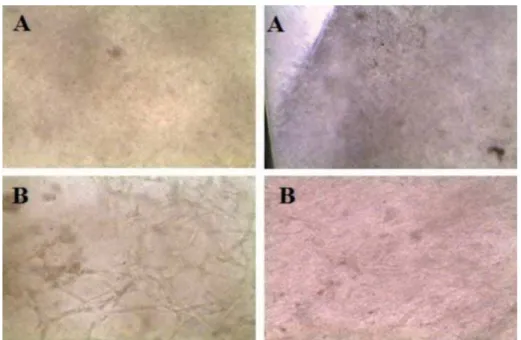
healt hy subst it ut es w er e obser ved w it h an inver t ed
m icr oscope at 4 0 X, sh ow in g in t er est in g f eat u r es. $Q H[WHQGHG FHOOXODU SDWWHUQ ZLWK PRUH ¿EHUV ZDV obser ved in t he diabet ic subst it ut es, w her eas healt hy
su bst it u t es sh ow ed a globu lar pat t er n w it h f ew er ¿EHUV)LJXUHDE7KHFOLQLFDODSSHDUDQFHRIWKH
subst it ut es was clear and hum id w it h an adher ence
t o sur faces ( Figur e 5) .
Der m al- epider m al subst it ut e evaluat ion and
charact er izat ion
Each subst it ut e was charact erized and assessed as
previously described. Ant i- cyt okerat in 5- 14 expression
show ed 1.14 DUELWUDU\ÀXRUHVFHQFHXQLWVAFU) less
in t he diabet ic subst it ut es com par ed w it h t he healt hy
cont r ol gr oup. Ant i- collagen I V ex pr ession was 1.9
AFU higher in t he diabet ic subst it ut es w hen com par ed
w it h t he healt hy gr oup. I n addit ion, der m o- epider m al
sk in subst it ut es w er e im m unost ained w it h Sy t ox Red
( I nv it r ogen, Walt ham , MA, USA) show ing t he nucleus
an d t h e cy t osk elet on w as st ain ed w it h Ph alloidin
DIABETIC SUBJECTS GROUP (N=4)
HEALTHY SUBJECTS GROUP (N=4)
P VALUE
Basal 100.000 100.000 1‡
Day 3 409,999*±258,686† 257,500*±20615† 0.009||
Day 6 1’089,999*±991,407† 585,000*±123693† 0.02||
Day 9 1’305,000*±1’075,648† 1’005,000*±185022† 0.004||
*Mean
†Standard deviation ‡ Student´s t-test § Chi-Square
||Match paired Student´s T-test
Table 2- Cell proliferation at the growth cell curve
Figure 3-&HOOJURZWKFXUYH&HOOSUROLIHUDWLRQRIWKH¿EUREODVWVRIGLDEHWLFVVXEMHFWVYVKHDOWK\VXEMHFWVDIWHUGD\VRILQFXEDWLRQ'LDEHWLF
( I nv it r ogen, Walt ham , MA, USA) , show ing t he cell
dist r ibut ion t hr oughout t he t hickness of t he subst it ut e
( Figur e 6) .
St at ist ical analysis
A St udent ’s T- t est was per for m ed as planned. All
var iables show ed a nor m al dist r ibut ion. Ther e was a VLJQL¿FDQWGLIIHUHQFHEHWZHHQWKHGLDEHWLFJURXSDQG t he healt hy gr oup for DUELWUDU\ÀXRUHVFHQFHXQLWVRI
collagen I V (AFU col- I V) p value= 0.02 and ar bit rar y
ÀXRUHVFHQFHXQLWVRIF\WRNHUDWLQAFU ck 5- 14)
p value= 0.05 ( Table 1) .
Discussion
Sk in t issu e en g in eer in g is a t h er ap eu t ic f ield
d ev elop ed t o aid in t h e h ealin g of in j u r ed sk in , HVSHFLDOO\IRUZRXQGVWKDWKDYHGLI¿FXOW\KHDOLQJLQ t he prim ary st age. This st udy showed t he developm ent
of t he der m o- epider m al subst it ut es of oral m ucosa
ob t ain ed f r om t h e r et r om olar ar ea w it h a 3 m m
pu n ch biopsy fr om diabet ic an d h ealt hy su bj ect s,
using t he air- liquid t echnique and it s assessm ent by LPPXQRÀXRUHVFHQFH FKDUDFWHUL]LQJ DQG TXDQWLI\LQJ LWVLQWHQVLW\LQ$)8DVD¿UVWVWHSWRZDUGVDSRVVLEOH f u t u r e cl i n i ca l a p p l i ca t i o n . Recen t st u d i es sh o w
oral m ucosa t o be a feasible, alt er nat ive sour ce of
epider m al cells and m at r ices, m ainly for it s int r insic
ch ar act er ist ics st u d ied an d d escr ib ed in p r ev iou s
st udies. I ida, et al.13 ( 2005) developed an epider m al
equivalent fr om oral m ucosa based on an acellular
allogeneic der m al m at r ix show ing good r esult s aft er
it s clinical applicat ion13,26. Ot her st udies r epor t ed t he
developm ent of sk in subst it ut es using cells obt ained
fr om biopsies of hum an sk in, m ainly t he for esk ins of
childr en or fr om est ablished epider m al cell lines. The
m ain disadvant age of t his appr oach is t he allogeneic
nat ur e of t he cells and t he lat ent possibilit y of shor t
-t er m r ej ec-t ion by -t he hos-t . S-t ill, w ound healing was
accom plished w it h it s clinical applicat ion6,8,11.
Figure 4- A. Dermal-epidermal substitutes of healthy subjects observed under the inverted microscope (40X). A globular pattern is REVHUYHG%'HUPDOHSLGHUPDOVXEVWLWXWHVRIGLDEHWLFVXEMHFWVREVHUYHGXQGHUWKHLQYHUWHGPLFURVFRSH;$¿EULOODUSDWWHUQLVREVHUYHG
3ULPDU\FXOWXUHVRI¿EUREODVWVDQGNHUDWLQRF\WHV w er e achieved fr om bot h populat ions ( diabet ic and
healt hy subj ect s) by t he ex plant t echnique. Eight y SHUFHQW RI FRQÀXHQFH ZDV DFKLHYHG LQ GD\V f or d iab et ic su b j ect s an d f or h ealt h y su b j ect s in
2 6 . 8 5 day s. I n sim ilar st u dies, t h e pr odu ct ion of SULPDU\¿EUREODVWDQGNHUDWLQRF\WHFXOWXUHVKDVEHHQ r epor t ed by t he ex plant t echnique alone8, and also
using enzy m at ic t echniques such as cell disr upt ion
w it hout m ent ioning t he t im e at w hich t hey achieved
con f lu en ce, p oin t in g ou t on ly t h e u se of cells in
different cult ure passages11,13,24. I n prim ary cell cult ure
t echniques, cell disint egrat ion is a st rat egy t hat can
r educe cult ur e t im es up t o 50% , yet has t he m ain
disadvant age of t he possibilit y of dam aging t issues and
cells12. I t is im por t ant t o point out t hat for t he clinical
applicat ion of t hese skin derm al- epiderm al subst it ut es,
cell cult ur e t im es should be r educed, and t her eby t he
enzy m at ic digest ion t echnique is a good st rat egy t o
be used for fut ur e st udies.
Regar ding t he developm ent of der m o- epider m al VXEVWLWXWHVWKHDLUOLTXLGWHFKQLTXHZDVDQHI¿FLHQWFHOO cult ure t echnique for obt aining t hese skin equivalent s.
I t pr ov ides t he condit ions and charact er ist ics needed
for t he obt ent ion of t his k ind of t issue, m ainly by t he
gas exchange ( CO2 and O2) of t he epit helial layer in WKH¿QDOVWDJH,WDOORZVIRUDSODFHVSHFL¿FFXOWXUH PHGLXPIRUHDFKFHOOOLQH¿EUREODVWVDQGPDWULFHVLQ t he lower com part m ent and kerat inocyt es in t he upper
com part m ent ) t hroughout t he ent ire cult ure process20.
I n our st udy, t he const ruct rem ained int act t hroughout
t he in vit r o pr ocess, allow ing for t he m anipulat ion of
bot h com par t m en t s in depen den t ly com par ed w it h
ot her cult ure t echniques and m at erials11,21,22,24. Cult ure
t im es r epor t ed t hr ough t he air- liquid t echnique range
fr om day 1 of t he st r om al st age, up t o 5 day s, and
t he epit helial phase for a per iod of 5 t o 10 day s21. I n
t his st udy w e used 10 day s in t he cult ur es for bot h
phases; st r om al and epit helial, obt aining good r esult s
in r elat ion t o t he est ablishm ent of t he st r om al cells DVHYLGHQFHGE\WKHLPPXQRÀXRUHVFHQFHHYDOXDWLRQ
I n t h e a s s e s s m e n t o f t h e s u b s t i t u t e s b y LPPXQRÀXRUHVFHQFHWKHUHZDVDVLJQL¿FDQWGLIIHUHQFH observed bet ween bot h groups ( p= 0.002 for AFU
COL-I V and p= 0.05 for AFU CK5- 14) . The r esult s show ed OHVVDUELWUDU\ÀXRUHVFHQFHXQLWVIRUNHUDWLQRF\WHV&. 14 ant ibody) in t he subst it ut es generat ed from t he oral
m ucosa of diabet ic subj ect s ( 9.56) com par ed t o t hose
generat ed from t he oral m ucosa of t he healt hy subj ect s
( 10.47) . I n r elat ion t o t he above, r ecent st udies show
an associat ion of Neurot ensin ( NT) , a neurot ransm it t er
involved in t he pr ocess of w ound healing. Moura, et
al.1 5 ( 2 0 1 4 ) st u died t h e effect s of n eu r ot en sin on
hum an kerat inocy t es under hy per glycem ic condit ions
and nor m al condit ions at differ ent funct ional levels,
m ainly NT r ecept or s, cy t ok ines and gr ow t h fact or
ex pr ession and pr oliferat ion and also m igrat ion of t he
epider m al cells. They obser ved t hat t he neur ot ensin
did not affect t he v iabilit y of kerat inocy t es, how ever,
neur ot ensin and t he ex pr ession levels of all r ecipient s ZHUH VLJQL¿FDQWO\ UHGXFHG 1HXURWHQVLQ WUHDWPHQW st im ulat ed t he ex pr ession of neur ot ensin r ecept or
2 ( NTR2) , w her eas ex pr ession levels of neur ot ensin
r ecept or s 1 an d 3 ( NTR1 , NTR3 ) did n ot ch an ge.
Kerat inocy t e pr oliferat ion was not affect ed, but t he
m igrat ion of kerat inocy t es was r educed. Their r esult s
show t hat hy per glycem ic condit ions adver sely affect
t he endogenous ex pr ession of neur ot ensin and it s
r ecep t or s, esp ecially in k er at in ocy t es NTR2 w it h
im por t ant consequences for it s funct ion. This effect
of neur ot ensin in hy per glycem ic condit ions has also
been w ell st udied in ot her cells such as m acr ophages,
w her e it was obser ved t hat t his neur ot ransm it t er r uns FHOOPLJUDWLRQSDWWHUQVDQGWKHLQÀDPPDWRU\UHVSRQVH
in w ound healing18*LYHQWKHVH¿QGLQJV0RXUDHW
al.16,17 ( 2014) conduct ed t w o addit ional st udies w her e
neur ot ensin was added as a com ponent t o t he der m o-HSLGHUPDOVXEVWLWXWHVVSHFL¿FDOO\IRUWKHVFDIIROGRU biom at er ial in w hich cells ar e suspended. They t est ed
in m ediu m s of collagen an d ch it osan obser v in g a UHGXFWLRQLQLQÀDPPDWRU\LQ¿OWUDWHVXSWRGD\DIWHU im plant at ion in rat s w it h induced diabet es, and also
an im pr ov em en t in w ou n d h ealin g pr om ot in g cell
m igrat ion and deposit ion of collagens I and I I I16,17.
Fut ure st udies concerning t he effect of NT are required
in or der t o assess if in t hese neur ot ensin subst it ut es
have an inhibit or y pr oliferat ion r ole.
Fibr osis is a com plicat ion of chr onic hyper glycem ia
involv ing t he excessive pr oliferat ion of ex t racellular
m at r ix ( ECM) and it s accum ulat ion in var ious t issues
and organs, t he m ost prom inent being m icrocirculat ion,
k idn ey, h ear t , r et in a an d w ou n d h ealin g. Ch r on ic
hy per glycem ia affect s t he cells r esponsible for t he
pr oduct ion of collagen I V causing t his accum ulat ion4.
Ou r r esu lt s su ppor t t h is st at em en t , as it sh ow ed LQFUHDVHG LPPXQRÀXRUHVFHQFH IRU DQWLFROODJHQ,9 DQG ¿EUREODVWV RI WKH GHUPDOHSLGHUPDO VXEVWLWXWHV generat ed fr om diabet ic subj ect s ( 8. 05) com par ed
w it h h ealt hy su bj ect s ( 6 . 1 5 ) . Ber lan ga- Acost a, et
al.5 ( 2010) r epor t ed t hat under t he high glucose load
LPSRVHGE\GLDEHWHVVNLQDQGVNLQ¿EUREODVWVZHUH dist ur bed in r ecr eat ed in vit r o clinical m odels show ing DOWHUDWLRQV WR WKH QRUPDO SK\VLRORJ\ RI ¿EUREODVWV as w ell as ex t racellular m at r ix secr et ion, t her efor e,
it has been suggest ed t hat high concent rat ions of
glucose is t he m aj or t r igger of a cascade of m olecular FKDQJHVWRVNLQ¿EUREODVWV,QRXUVWXG\FHOOFXOWXUHV RIGLDEHWLFVXEMHFWVUHDFKHGDQFRQÀXHQFHLQD shor t er t im e ( 25.3 days) t han healt hy subj ect s ( 26.85
days) suggest ing a perm anent effect of hyperglycem ic
condit ions in cell phy siology ; how ever fur t her st udy
is needed.
Differ ences in t he collagen pr oduct ion of diabet ic
subj ect s’ cells could suggest t hat diabet ic subst it ut es
m ight im pr ov e t he healing of diabet ic foot ulcer s,
how ever m or e r esear ch is needed t o det er m ine t he
clinical im pact of t he differ ences found. Addit ionally
t hese subst it ut es m ay be used t o t est new dr ugs in
an env ir onm ent t hat is closer t o t hat of r eal t issues.
Re su l t s o f o u r st u d y w e r e m e a su r a b l e a n d TXDQWL¿DEOHLQWHQVLW\RIFROODJHQ,9DQGDQWLF\WRNHUDWLQ 5 - 1 4 i n a r b i t r a r y f l u o r e sce n ce u n i t s) sh o w i n g
n u m er ical var iat ion s in t h e am ou n t of ex pr ession
of bot h ant ibodies in bot h populat ions; diabet ic and
healt hy subj ect s, suggest ing differ ences in cellular
funct ion under t his clinical condit ion and, wit h possible
im plicat ions in t he clinical applicat ion of t he subst it ut e.
Also it is necessar y t o em phasize t hat t he cells not
only sur v ived t hr ough t he developm ent pr ocess of
t he subst it ut e, but also t hey w er e int egrat ed in t o
t h e der m al m at r ix an d secr et ed basal m em br an e VXSSRUWHGE\WKHSRVLWLYHH[SUHVVLRQRIWKHVSHFL¿F m ar ker s CK5- 14 and COL I V.
'HVSLWHWKHDERYHWKHVLJQL¿FDQFHRIWKHVH¿QGLQJV is n ot y et k n ow n , f u r t h er st u d ies ar e n eed ed t o
elucidat e t hem . However t his dat a m ay provide a basis
t o per fect / im pr ove t he developm ent t echnique of t he
der m al- epider m al subst it ut es fr om t he oral m ucosa of
diabet ic pat ient s and t o answer t he biological quest ions
raised by it .
Wit hin t he const raint s of our st udy, it is w or t h
m ent ioning t hat no in vit r o t est s w er e per for m ed t o
evaluat e t he funct ionalit y of t he developed subst it ut e,
such as m igrat ion essays. I n t hat m at t er, t he next st ep
w ould be in vivo and in vit r o essay s of funct ionalit y of
t hese subst it ut es developed by t his t echnique and t he
charact er izat ion pr oposed in t his st udy. This w ill lead
us t o t he fut ur e clinical applicat ion of t hese diabet ic
sk in subst it ut es.
Conclusions
I t i s p o ssi b l e t o d e v e l o p d e r m a l - e p i d e r m a l
subst it ut es fr om t he isolat ion of t he epider m al cells
of oral m ucosa fr om diabet ic and healt hy subj ect s
using t he air- liquid t echnique and it s assessm ent by LPPXQRÀXRUHVFHQFHFKDUDFWHUL]LQJDQGTXDQWLI\LQJLWV LQWHQVLW\LQDUELWUDU\ÀXRUHVFHQFHXQLWV$)8
D e r m a l - e p i d e r m a l s k i n s u b s t i t u t e s o f
LPPXQRÀXRUHVFHQFH LQWHQVLW\ ZKHQ FRPSDUHG WR t h e su b st it u t es f r om h ealt h y su b j ect s; w h ile f or ¿EUREODVWVDQG&2/,9VXEVWLWXWHVRIGLDEHWLFVXEMHFWV show ed a gr eat er int ensit y, w it h a higher AFU value. 7KHVH¿QGLQJVVXJJHVWGLIIHUHQFHVLQFHOOXODUIXQFWLRQ under t his clinical condit ion; how ever m or e r esear ch
is needed t o det er m ine t he cr osst alk w hich occur s
bet w een t he com ponent s of t hese sk in subst it ut es
and t he dam aged t issues. The follow ing long- t er m
goal in t his r esear ch is t he clinical applicat ion of t hese
subst it ut es; fur t her in vivo anim al st udies t o evaluat e
funct ionalit y w ill be per for m ed t o cont inue w it h t his
m at t er.
Refer ences
1 - Abr eu - Velez A, How ar d M. Collagen I V in n or m al sk in an d in
pat hological pr ocesses. N Am J Med Sci. 2012; 4( 1) : 1- 8.
2- Alam H, Sehgal L, Kundu ST, Dalal S, Vaidya M. Novel funct ion
RINHUDWLQVDQGLQSUROLIHUDWLRQDQGGLIIHUHQWLDWLRQRIVWUDWL¿HG
epit helial cells. Mol Biol Cell. 2011; 22: 4068- 78.
$PHULFDQ 'LDEHWHV $VVRFLDWLRQ 'LDJQRVLV DQG FODVVL¿FDWLRQ RI
diabet es m ellit us. Diabet es Car e. 2014; 37( Suppl.1) : S81- 90.
4- Ban CR, Tw igg SM. Fibr osis in diabet es com plicat ions: pat hogenic
m echanism s and cir culat ing and ur inar y m ar ker s. Vasc Healt h Risk
Manag. 2008; 4( 3) : 575- 96.
5- Ber langa- Acost a J, Valdéz- Pér ez C, Sav igne- Gut iér r ez W,
Mendoza-Marí Y, Franco- Pérez N, Vargas- Machiran E, et al. Cellular and m olecular
insight s int o t he w ound healing m echanism in diabet es. Biot ecnol Apl.
2010; 27( 4) : 255- 61.
6 - Boelsm a E, Gibbs S, Faller C, Pon ec M. Ch ar act er izat ion an d
co m p a r i so n o f r e co n st r u ct e d sk i n m o d e l s: m o r p h o l o g i ca l a n d
im m unohist ochem ical evaluat ion. Act a Derm Venereol. 2000; 80( 2) :
82-8.
7- Boult on AJ. The diabet ic foot : a global v iew. Diabet es Met ab Res
Rev. 2000; 16( Suppl 1) : S2- 5.
8- Chang DW, Sanchez LA, Veit h FJ, Wain RA, Ok hi T, Suggs WD. Can
a t issue- engineer ed sk in graft im pr ove healing of low er ex t r em it y foot
w ounds aft er r evascular izat ion? Ann Vasc Sur g. 2000; 14( 1) : 44- 9.
9 - Ch u PG, Weiss LM. Ker at in ex pr ession in h u m an t issu es an d
neoplasm s Hist opat hology. 2002; 40( 5) : 403- 39.
10- The Com prehensive R Archive Net work. RMS: Regression m odeling
st rat egies [ hom epage] . Vienna: R Foundat ion for St at ist ical Com put ing;
2016 [ cit ed 2016 Dec 13] . Available fr om : ht t ps: / / cran.r- pr oj ect .or g/
w eb/ pack ages/ r m s/ .
11- Falanga V, Mar golis D, Alvar ez O, Aulet t a M, Maggiacom o F, Alt m an
M, et al. Rapid healing of venous ulcers and lack of clinical rej ect ion wit h
an allogeneic cult ur ed hum an sk in equivalent . Hum an Sk in Equivalent
I nvest igat or s Gr oup. Ar ch Der m at ol. 1998; 134( 3) : 293- 300.
12- Fr eshney RI . Cult ur e of anim al cells: a m anual of basic t echnique.
5t h ed. Wiley- Black w ell: Hoboken; 2005.
13- I ida T, Tak am i Y, Yam aguchi R, Shim azak i S, Har ii K. Developm ent
of a t issue- engineer ed hum an oral m ucosa equivalent based on an
acellular allogeneic der m al m at r ix : a pr elim inar y r epor t of clinical
applicat ion t o bur n w ounds. Scand J Plast Reconst r Sur g Hand Sur g.
2005; 39( 3) : 138- 46.
14- Llam es SG, Del Rio M, Lar cher F, Gar cía E, Gar cía M, Escam ez
MJ, et al. Hum an plasm a as a der m al scaffold for t he generat ion
of a com p let ely au t olog ou s b ioen g in eer ed sk in . Tr an sp lan t at ion .
2004; 77( 3) : 350- 5.
15- Moura LI , Cr uz MT, Car valho E. The effect of neur ot ensin in hum an
kerat inocyt es - im plicat ion on im pair ed w ound healing in diabet es. Exp
Biol Med ( May w ood) . 2014; 239( 1) : 6- 12.
16- Moura LI , Dias AM, Leal EC, Car valho L, Sousa HC, Car valho
( &KLWRVDQEDVHG GUHVVLQJV ORDGHG ZLWK QHXURWHQVLQ DQ HI¿FLHQW
st rat egy t o im pr ov e ear ly diabet ic w ound healing. Act a Biom at er.
2014; 10( 2) : 843- 57.
17- Moura LI , Dias AM, Suesca E, Casadiegos S, Leal EC, Font anilla
05HWDO1HXURWHQVLQORDGHGFROODJHQGUHVVLQJVUHGXFHLQÀDPPDWLRQ
and im pr ove w ound healing in diabet ic m ice. Biochim Biophy s Act a.
2014; 1842( 1) : 32- 43.
18- Moura LI , Silva L, Leal EC, Tellechea A, Cr uz MT, Car valho E.
1HXURWHQVLQ PRGXODWHV WKH PLJUDWRU\ DQG LQÀDPPDWRU\ UHVSRQVH
of m acr ophages under hy per glycem ic condit ions. Biom ed Res I nt .
2013; 2013: 941764.
1 9 - Pom ah ac B, Sv en sj ö T, Yao F, Br ow n H, Er ik sson E. Tissu e
engineer ing of sk in. Cr it Rev Oral Biol Med. 1998; 9( 3) : 333- 44.
2 0 - R Cor e Team . R: A lan gu age an d env ir on m en t for st at ist ical
co m p u t i n g [ h o m e p a g e ] . Vi e n n a : R Fo u n d a t i o n f o r St a t i st i ca l
Com put ing; 2012 [ cit ed 2016 July 20] . Available fr om : ht t p: / / w w
w.R-pr oj ect .or g/ .
5HLFKO60OOHU*R\PDQQ&&7KHXVHRIDSRUFLQHRUJDQRW\SLF
cor nea const r uct for per m eat ion st udies fr om for m ulat ions cont aining
befunolol hydr ochlor ide. I nt J Phar m . 2003; 250: 191- 201.
22- Rolin G, Placet V, Jacquet E, Tauzin H, Robin S, Pazar t L, et al.
Developm ent and charact er izat ion of a hum an der m al equivalent w it h
physiological m echanical propert ies. Skin Res Technol. 2012; 18( 2) :
251-8.
23- Sanchez- Quevedo MC, Alam inos M. Capit an LC. Moreu G, Garzon I ,
Crespo PV, et al. Hist ological and hist ochem ical evaluat ion of hum an oral
m ucosa const ruct s developed by t issue engineering. Hist ol Hist opat hol.
2007; 22: 631- 40.
24- Seet WT, Manira M, Khair ul Anuar K, Chua KH, Ahm ad I r fan AW,
Ng MH, et al. Shelf- life evaluat ion of bilayer ed hum an sk in equivalent ,
0\'HUP3/R62QHH
25- Tat sioni A, Balk E, O’Donnell T, Lau J. Usual care in t he m anagem ent
of chr onic w ounds: a r ev iew of t he r ecent lit erat ur e. J Am Coll Sur g.
2007; 205( 4) : 617- 24.
26- Ueda M, Hat a K, Hor ie K, Tor ii S. The pot ent ial of oral m ucosal
cells for cult ur ed epit helium : a pr elim inar y r epor t . Ann Plast Sur g.
1995; 35( 5) : 498- 504.
27- Vr iens AP, Waaij m an T, van den Hoogenband HM, de Boer EM,
Scheper RJ, Gibbs S. Com par ison of aut ologous full- t hick ness gingiva
and skin subst it ut es for wound healing. Cell Transplant . 2008; 17: