The effect of esophageal acidification on bronchial
obstruction in asthmatics with gastroesophageal reflux
ANA CARLA SOUSA DE ARAUJO, LÍLIAN ROSE OTOBONI APRILE, JOÃO TERRA FILHO, ROBERTO OLIVEIRA DANTAS, MILTON ARRUDA MARTINS, ELCIO OLIVEIRA VIANNA(TE SBPT)
*Study carried out in the Pulmonology and Gastroenterology Sectors of the Clinical Medicine Department of the Faculdade de Medicina de Ribeirão Preto (FMRP) - Universidade de São Paulo. Sponsored by CAPES and FAPESP
Correspondence to: Elcio O. Vianna - Seção de Pneumologia, Hospital das Clínicas de Ribeirão Preto - Campus da USP - Av. Bandeirantes, 3900 CEP 14048-800, Ribeirão Preto, SP. Phone: 55-16 602 2631 - E-mail: [email protected]
Submitted: 20 October 2003. Accepted, after review: 15 September 2004.
J Bras Pneumol 2005; 31 (1): 13-9.
Key words: Asthma. Gastroesophageal reflux. Respiratory function tests. Endoscopy/methods.
Background: The relationship between asthma and gastroesophageal reflux is, as yet, not completely understood. Among the mechanisms thought to be responsible for gastroesophageal reflux-related worsening of asthma symptoms are the vagovagal reflex and microaspiration.
Objective: To assess forced expiratory volume in one second after acid infusion.
Method: This study investigated the effect of acid infusion in 13 volunteers with moderate asthma and gastroesophageal reflux. Spirometry was performed before and after insertion of an 8F nasogastric tube and a pH meter. After 15 minutes of saline solution infusion into the midpoint between the upper esophageal sphincter and lower esophageal sphincter, and again after 15 minutes of esophageal acidification (with hydrochloric acid) of the same area, forced expiratory volume in one second was reassessed. Acidification was repeated every 5 minutes until forced expiratory volume in one second values stabilized (variation: < 5%).
Results: Mean forced expiratory volume in one second values remained stable during the tube insertion, saline infusion, initial hydrochloric acid infusion and subsequent hydrochloric acid infusion procedures (p = 0.72). Lower forced expiratory volume in one second values were seen resulting from the tube insertion in two patients (drops of 11% and 22%, respectively), after saline infusion in another two (drops of 13% and 14%) and after acid infusion in 1 (a drop of 22%).
INTRODUCTION
A s t h m a c a n b e t r i g g e r e d b y m u l t i p l e determinants, such as environmental factors, respiratory infections, occupational exposure, c l i m a t i c c h a n g e s , p h y s i c a l e x e r c i s e a n d gastroesophageal reflux (GER).
Three main mechanisms point to GER as an asthma trigger: the vagovagal reflex, in which afferent nerves submitted to a stimulus (acid or irritating substances in the reflux) generate action potentials that result in peripheral liberation of proinflammatory neuropeptides, which (present in the pulmonary and esophageal mucosa) can promote inflammation of the airway mucosa and peribronchial smooth muscle contraction; reflux causing bronchial hyperreactivity, due to the bronchial inflammation caused by the neural mechanism described; and microaspiration of the acid into the larynx and upper airways(1,2).
The elevated prevalence of GER disease (GERD) in asthmatics has led various researchers to study the relationship between these two diseases in an attempt to clarify the possible mechanisms that make GER an asthma-inducing agent. Nevertheless, there is no consensus regarding the physiopathology of asthma in relation to GER, as well as regarding the effect of reflux on pulmonary function.
In this context, the objective of the present article was to study the spirometry of asthmatics with GER during esophageal acidification and assess the roles played by reflux and microaspiration in the accompanying bronchospasm.
METHODS
A total of thirteen patients with clinically controlled asthma, diagnosed based on the presence of (partially or totally reversible) airway o b s t r u c t i o n , o b s t r u c t i o n - r e l a t e d e p i s o d i c symptoms and absence of other diagnoses, were selected from the Hospital das Clínicas of the Faculdade de Medicina of Ribeirão Preto. The patients also met the diagnostic criteria for asthma in terms of the concentration of methacholine provoking forced expiratory volume in one second (FEV1) to drop by 20% (PC20) < 8 mg/mL or a response to 200 mcg of inhaled salbutamol presenting an improvement in FEV1 equal to or greater than 15%. In order to classify severity, asthma was considered moderate when the patient presented FEV1 or peak expiratory flow (PEF) values
between 60%-80% of the expected values, daily symptoms, daily use of a bronchodilator, nocturnal symptoms more often than once a week or PEF variation higher than 30%, according to the standards of IIConsenso Brasileiro no Manejo da A s m a ( I I B r a z i l i a n C o n s e n s u s o n A s t h m a Management)(3). Selection criteria also included
GER symptoms, upper digestive endoscopy showing esophagitis (or esophageal pH monitoring confirming GER with a DeMeester score > 14.72), age 18-70, stable clinical profile and capacity to understand and execute the procedures involved in the study. Smokers and pregnant women, as well as patients with pulmonary diseases other than asthma, serious extrapulmonary diseases, or diseases that might interfere in the execution or interpretation of the study, were excluded from the selection, as were patients with a history of esophageal, gastric or pulmonary surgery and patients on antibiotics or systemic corticosteroids. Participation of patients who had presented respiratory infection during the last six weeks prior to the study was postponed.
Patients who met the selection criteria were informed of the objectives of the study, the procedures involved and the risks. The volunteers gave written informed consent. The study and the consent form were approved by the Ethics in Research Committee of the Hospital das Clínicas of the Faculdade de Medicina de Ribeirão Preto.
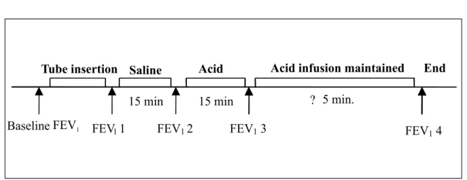
The study consisted of spirometry performed before and after the following procedures: insertion of the esophageal tubes (manometry, pH m onitoring and an 8F nasogastric tube ) ; esophageal infusion of normal saline solution (pH 5.5 to 6.0) at 10 to 20 microdrops/min for 15 minutes; and infusion with hydrochloric acid (HCl) 0.1N (pH 1.0 to 1.5), using the same drip rate and duration. These solutions were infused through the 8F intranasal tube into the midpoint between the upper esophageal sphincter and lower esophageal sphincter, previously defined through esophageal manometry. After the initial 15 minutes of e s o p h a g e a l a c i d i f i c a t i o n , n e w s p i r o m e t r y measurements were taken every 5 minutes until FEV1 values stabilized (variation: < 5%), thus
Function Testing Laboratory of the Pulmonology Department of the Faculdade de Medicina de Ribeirão Preto of the University of São Paulo and were all performed in the morning, using the same equipment, and by the same certified professional technician. The schematic summary of the protocol is illustrated in Figure 1.
The equipment used in the spirometry was a Koko spirometer, calibrated on a daily basis, and the corresponding software (PDS Instrumentation, Inc., Louisville, CO, USA). The measurements were analyzed according to the values of normality described by Polgar & Promahaldt(4). Exams were
carried out in accordance with the guidelines established by the I Consenso Brasileiro de Espirometria (I Brazilian Consensus on Spirometry)(5)
and always by the same technician. Patients were instructed to suspend the use of long-acting bronchodilators 24 hours prior to the exam and to suspend the use of short-acting bronchodilators 12 hours prior to the exam.
For all patients, esophageal pH was monitored during the procedures and for 24 hours thereafter through the use of a catheter with an antimony (channel 2) electrode at the end (Synectics Multi-use Ph-Catheter-Synectics Medical, Stockholm, Sweden) and another electrode positioned 15 cm above (channel 1), calibrated with pH 7.0 and 1.0 buffer solutions, respectively. The pH-meter was inserted nasally and placed 5 cm (channel 2) above the upper esophageal sphincter, previously located through esophageal manometry, and 20 cm above the lower esophageal sphincter (channel 1) and c o n n e c t e d t o a p o r t a b l e d i g i t a l r e c o r d e r
Figure 1 – Sequence of FEV1 procedures and measurements
(Digitrapper MK III- Synectics Medical, Stockholm, Sweden). Routine pre-pH monitoring procedures were adopted, such as a minimum 8-hour fast and suspension of antacids and prokinetics (72 hours prior to the exam), as well as suspension of proton pump inhibitors or drugs that could interfere with digestive motility (seven days prior to the exam). The period of continuous esophageal acid infusion was excluded from the analysis of the total amount of abnormal GER.
The statistical approach utilized in the analysis of this data set was parametric, using the analysis of variance (ANOVA) method for repeated measurements and formation of contrasts through difference, comparing the average of each situation, except for baseline values, with the average of the previous situation, also known as the Helmert method of reversed contrasts. The comparisons of FEV1 variation between the patients with positive DeMeester scores(6) and those with
negative DeMeester scores were calculated through the non-paired two-tailed Student’s t-test. The same test was utilized to compare patients with Bernstein test positivity to those with Bernstein test negativity. The level of significance adopted was 5% (pd” 0.05). Data are expressed as average
± standard deviation.
RESULTS
All thirteen individuals submitted to the study protocol performed the tests without difficulties or incidents, except for case number 7. This patient presented serious bronchospasm after 15 minutes of esophageal HCl infusion and was therefore
Baseline FEV
1FEV
11
FEV1
2
FEV1
3
Tube insertion
Saline
Acid
Acid infusion maintained
End
15 min
15 min
?
5 min.
unable to complete the pulmonary function tests designated, in this study, HCl and HCl stability. Partial data related to this case were used in the analysis and demonstration of the results. The general characteristics of the patients, pulmonary f u n c t i o n a n d G E R D c h a r a c t e r i s t i c s a r e demonstrated in Table 1. All of the patients presented moderate asthma (clinical criteria, FEV1
or PEF) during the selection. Nevertheless, at the time of the exam, two cases that had met the criteria for moderate asthma presented FEV1 values
below 60%, although without signs of acute exacerbation in the clinical profile. Due to the stability of the clinical profile, together with the willingness and the consent of these patients, we decided to proceed with the protocol.
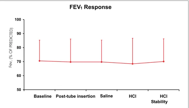
Pulmonary function remained stable (p = 0.72) during the tube insertion, saline solution infusion, initial hydrochloric acid infusion and subsequent hydrochloric acid infusion procedures (Table 2 and Figure 2). In the initial stage, we observed an FEV1
response after tube insertion in two cases, with a drop of over 10% (11% and 22%). In the following stage, two cases other than the ones who had experienced drop related to the tube insertion, presented FEV1 drops (13% and 14%) related to
saline solution infusion. After the acid infusion, one case presented lower FEV1 (22% variation),
and this was one of the cases that had already presented a reaction to the tube insertion. Case 7 also reacted to acid, presenting bronchospasm (unmeasured). However, no individual or group changes were found during the HCl stability stage. Seven patients (54%) presented GER symptoms during esophageal acidification (Bernstein test
TABLE 1
General characteristics of the patients studied (n=13)
Age (years) 41.9 (± 10.8)
Gender (% females) 69.2
FEV1 (% of predicted) 70.5 (± 14.7)
FVC (% of predicted) 89.1 (± 16.0)
FEF25-75% (% of predicted) 42.9 (± 22.5)
PEF (% of predicted) 68.4 (± 20.4)
FEV1/FVC % 78.5 (± 14.9)
Daily GER symptoms (% patients) 38.4
Esophagitis seen in UDE (% patients) 84.6
Positive pH monitoring (% patients) 77
Data presented as average (± standard deviation) FEV1: forced expiratory volume in one second; FVC: forced
vital capacity; FEF25-75%: forced expiratory flow between 25
and 75% of forced vital capacity; PEF: peak expiratory flow; GER: gastroesophageal reflux; UDE: upper digestive endoscopy positive pH monitoring = positive DeMeester score
TABLE 2
FEV1 response (L) to tube insertion and esophageal infusions
Patient No FEV1 (L) FEV1 (L) FEV1 (L) FEV1 (L) HCl FEV1 (L)
Baseline Post-insertion Saline HCl Stability
1 1.18 1.05 1.05 0.82 1.05
2 1.67 1.30 1.55 1.44 1.52
3 2.79 2.77 2.79 2.85 2.79
4 1.56 1.62 1.71 1.62 1.69
5 2.25 2.44 2.58 2.58 2.66
6 1.94 2.31 2.42 2.33 2.28
7 2.28 2.14 1.83 * *
8 2.46 2.37 2.06 2.17 2.26
9 3.20 3.30 3.36 3.41 3.32
10 1.68 1.63 1.67 1.61 1.60
11 2.54 2.70 2.57 2.53 2.73
12 2.96 2.89 2.92 2.91 2.94
13 1.23 1.19 1.18 1.13 1.15
Average 2.13 ± 0.62 2.13 ± 0.68 2.13 ± 0.67 2.11 ± 0.76 2.16 ± 0.71 *Patient number 7 could not be given the last two tests due to a bronchospasm
positivity). All of the patients maintained proximal pH above 6 during HCl infusion. The cases presenting Bernstein test positivity or a positive DeMeester score did not present behavior different from that of the cases presenting Bernstein test negativity (p = 0.24) or a negative DeMeester score (p = 0.45).
DISCUSSION
Pulmonary function remained stable during the procedures of tube insertion, saline solution infusion, initial hydrochloric acid infusion and subsequent hydrochloric acid infusion. However, four of the thirteen patients presented bronchial reactions to these procedures: two reacted to the tube insertion and two reacted to the saline solution infusion. Of these four patients, two patients presented additional worsening during the acid infusion. The effect of esophageal acidification has been described in previous studies(7), although the effect of the tube
insertion had not yet been investigated.
Various indices are used in esophageal acidification studies, some, such as flow at 50 percent of vital capacity (V50), the flow at 25% of vital capacity
(V25), PEF or airway resistance, presenting good
sensitivity but less specificity. In a review carried out by Field(7), a significant (8%) drop in the FEV
1 values
of asthmatics with GER during acidification was
reported in only one study. Nevertheless, although this FEV1 variation was high in relation to those seen in other studies, the authors concluded that there was little reaction and therefore no clinical relevance. However, there was no FEV1 variation in the other nine studies of asthmatics with GER mentioned in that review(7). Harding et al.(8) obtained a 6% PEF
drop during esophageal acidification in a group of asthmatic patients and reported that, despite pH normalization, recovery was not immediate after discontinuation of the infusion. The authors observed a 7% increase in airway resistance in this group of patients and, during the recovery phase, when the patients remained in supine position for 40 minutes, airway resistance increased by 27% over pre-HCl infusion values. Fiss(9), in a study of the
esophageal-bronchial reflex in patients with asthma, concluded that 60 minutes of HCl instillation in the proximal third of the esophagus provokes a significant reduction in FEV1 and triggers the reflex, thereby increasing airway resistance, mainly in patients with GER. However, in order to rule out microaspiration, proximal esophageal pH was not measured during HCl infusion.
Comparing our results with those of other authors who found variations in pulmonary function only detected by more sensitive tests such
Figure 2 – FEV1 response (% of predicted) to tube insertion and esophageal infusions
FEV Response
150 60 70 80 90 100
Baseline Post-tube insertion Saline HCl HCl
Stability
Fe
v
(%
O
FP REDICTED)
as determination of airway resistance and PC20(2,10,11), we verified that the HCl concentration
used, the amount of acid instilled and the time of infusion were similar to those employed in the present study(12,8). Wilson et al.(10) stated that the
esophageal bronchial reflex response is directly proportional to acid concentration and time of exposure and emphasized the importance of the choice of liquid instilled or swallowed. The authors stimulated the esophagus with HCl swallowed at a concentration of 0.001 N, and they obtained no reduction in airway flow, although they detected significant drops in PC20. The same authors also mentioned examples of other substances with pH values of approximately 3, such as citric juices, which, when ingested, which might trigger bronchospasm crises(10). Case number 7 in our study
was a patient who reported ingesting lemon juice and suffering subsequent dyspnea that diminished after the use of a bronchodilator. During the protocol, after the saline solution infusion, this patient presented a 20% drop in FEV1 in
comparison to the baseline value and a 14% drop in comparison to the pre-infusion value. After 15 minutes of HCl infusion, the patient presented severe bronchospasm. We concluded that the acidification of the ingested solution seems to have had an influence on some patients, and the anamnesis may identify the phenomenon.
Seven (54%) of the patients in our study presented GER symptoms during HCl esophageal infusion (Bernstein positivity). However, only two patients presented worsening of obstruction during esophageal acidification: one presenting Bernstein test positivity and the other Bernstein test negativity. When we compared the Bernstein-positive group with the Bernstein-negative group, no difference was observed in the spirometric behavior during the procedures. Tan et al.(13) used the Bernstein test to
establish GER or chronic esophagitis, based on the good acceptance of this clinical test for GER diagnosis, and concluded that the presence or absence of esophagitis (defined by the authors as Bernstein test positivity) does not alter the pulmonary response to esophageal acidification. Harding et al.(8)
found no correlation between Bernstein test positivity and the bronchoconstriction induced by esophageal acidification. Kjellen et al.(14), however, concluded that
Bernstein test positivity is necessary to demonstrate an airway response to esophageal acidification in the
asthmatic population they studied. In our study, of the two patients who presented relevant responses to HCl infusion, only one had endoscopically diagnosed esophagitis. However, the identification of esophagitis by endoscopy is subject to variations since it depends on macroscopic observation and individual interpretation.
The pH monitoring showed positive DeMeester scores for 77% of the patients in our study. One of the patients who had a negative DeMeester score presented an FEV1 drop upon HCl esophageal
infusion. Case 7 of our study, who presented severe bronchospasm, had a negative DeMeester score. In his study of esophageal-bronchial reflex in asthmatics, Fiss(9) related that 30% of the patients
who presented significant FEV1 drops also
presented normal 24-hour esophageal pH monitoring. Wilson et al.(10) referred to a silent
reflex, in which esophagitis can occur without the corresponding GER. Another hypothesis is that, although the pH measurement did not demonstrate GER on the day of the exam, we cannot rule out the possibility that it was present(15). Therefore, just
as spirometry is an exam done at a specific point in time and may not reflect the clinical and functional worsening of the patients over a period of time, so may pH monitoring be similarly limited. In conclusion, esophageal acidification does not lead to spirometric alterations in asthmatics as a group. However, in isolated cases, acidification as well as esophageal tube insertion may result in bronchospasm. The size of the sample studied does not allow inferences as to the frequency of reactions to esophageal procedures. However, in this sample of thirteen individuals, approximately 40% presented bronchospastic responses to tube insertion or infusions. Defining the characteristics of these individuals that make them prone to bronchospastic responses is an important step for the future of knowledge regarding asthma physiopathology, just as in studies involving larger patient samples it is relevant to know the exact frequency of patients that present immediate responses to esophageal infusion or tube insertion.
ACKNOWLEDGEMENTS
REFERENCES
1. Tuchman DN, Boyle JT, Pack AL, Scwartz J, Kokonos M, Spitzer AR, et al. Comparison of airway responses following tracheal or esophageal acidification in the cat. Gastroenterology. 1984;87:872-81.
2. Mansfield LE, Stein MR. Gastroesophageal reflux and asthma. A possible reflex mechanism. Ann. Allergy. 1978;41:224.
3. Sociedade Brasileira de Pneumologia e Tisiologia. II Consenso Brasileiro no Manejo da ASMA. J Pneumol. 1998;173:24.
4. Polgar G, Promadhat V. Pulmonary function testing in children: techniques and standards. Philadelphia: WB Saunders; 1991. p.87-208.
5. Sociedade Brasileira de Pneumologia e Tisiologia. I Consenso de Espirometria. J Pneumol. 2002;22:122-49. 6. Johnson LF, Demeester TR. Development of the 24-hour intraesophageal pH monitoring composite scoring system. J Clin Gastroenterol. 1986;8(Suppl. 1):52-8.
7. Field SK. A critical review of the studies of the effects of simulated or real gastroesophageal reflux on pulmonary function in asthmatic adults. Chest. 1999;115:848-56.
8. Harding SM, Guzzo MR, Maples RV. Gastroesophageal reflux induced bronchoconstriction: vagolytic doses of atropine diminish airway responses to esophageal acid infusion. Am J Respir Crit Care Med. 1995;151:A589. 9. Fiss E. Estudo do reflexo esôfago-brônquico em
pacientes portadores de asma. [tese]. São Paulo: Faculdade de Medicina da Universidade de São Paulo; 1993. 60p.
10. Wilson NM, Charette L, Thomson AH, Silverman M. Gastroesophageal reflux and chidhood asthma: the acid test. Thorax. 1985;40:592-7.
11. Ekstrom T, Tibbling L. Esophageal acid perfusion, airway function and symptoms in asthmatic patients with marked bronchial hyperreativity. Chest. 1989;96:995-8. 12. Schan CA, Harding SM, Haile JM, Bradley LA, Richter JE. Gastroesophageal reflux induced bronchoconstriction: an intraesophageal acid perfusion study using state-of-the-art technology. Chest. 1994;106:731.
13. Tan WC, Martin RJ, Pandey R, Ballard RD. Effects of spontaneous and simulated gastroesophageal reflux on sleeping asthmatics. Am Rev Respir Dis. 1990;141:1394-9. 14. Kjellen G, Tibbling L, Wranne B. Bronchial obstruction after esophageal acid perfusion in asthmatics. Clin Physiol. 1981;92:285-92.