Effects of fatty acids on liver regeneration in rats
Effects of fatty acids on liver regeneration in rats
Effects of fatty acids on liver regeneration in rats
Effects of fatty acids on liver regeneration in rats
Effects of fatty acids on liver regeneration in rats
Efeitos dos ácidos graxos sobre a regeneração hepática em ratos
Efeitos dos ácidos graxos sobre a regeneração hepática em ratos
Efeitos dos ácidos graxos sobre a regeneração hepática em ratos
Efeitos dos ácidos graxos sobre a regeneração hepática em ratos
Efeitos dos ácidos graxos sobre a regeneração hepática em ratos
J
OSÉU
LISSESDES
OUZAM
ELO, TCBC-CE
1; J
EFFERSONM
ENEZESV
IANAS
ANTOS2,; O
SAMUDES
ANDESK
IMURA2; M
ANOELM
ESSIASC
AMPOSJ
ÚNIOR3; R
ADAMÉSB
EZERRAM
ELO4; P
AULOR
OBERTOL
EITÃODEV
ASCONCELOS5A B S T R A C T
A B S T R A C T
A B S T R A C T
A B S T R A C T
A B S T R A C T
Objective ObjectiveObjective
ObjectiveObjective: To study the effects of polyunsaturated fatty acids (PUFA) omega-3 and omega-6 in the oxidative stress and in liver regeneration in rats subjected to 70% partial hepatectomy (PH, 70% hepatectomy, Higgins- Anderson partial hepatectomy). Methods
MethodsMethods
MethodsMethods: 72 young male Wistar rats were randomly divided into four equal-sized groups: control (G1), partially hepatectomized (G2), partially hepatectomized with two weeks daily intraperitoneal infusion of omega-3 (G3) and partially hepatectomized with two weeks daily intraperitoneal infuison of omega-6 (G4). In moments 36h (T1), 168h (T2) and 336h (T3) post-PH, thiobarbituric acid reactive substances (TBARS) and reduced glutathione (GSH) were measured in plasma and liver tissue, while glucose and total bilirubin were measured in blood. The mass of the residual liver in the same moments was the parameter used to estimate the evolution of liver regeneration. ResultsResultsResultsResultsResults: omega-3 PUFA inhibited liver regeneration and induced reduction of hepatic GSH concentration seven days post-PH. Omega-6 PUFA, in contrast, showed no inhibitory effect on regeneration. There was an increase of lipid peroxidation both in blood and liver with administration of omega-6. Conclusion:Conclusion:Conclusion:Conclusion:Conclusion: Omega-3 PUFA retarded post-PH liver regeneration, probably through inhibition of oxidative stress. Omega-6 PUFA increased TBARS concentrations in blood and liver but did not alter the evolution of the liver regenerative process.
Key words: Key words: Key words:
Key words: Key words: Liver regeneration. Oxidative stress. Fatty acids. Lipid peroxidation. Rats.
Work done at the Laboratory of Experimental Surgery (LABCEX) of the UFC Medical School, Fortaleza – CE – BR.
1. PhD in Surgery, Federal University of Ceará (UFC) - Fortaleza – BR; 2. M.D., UFC - Fortaleza – BR; 3. Resident, General Surgery, Fortaleza General Hospital, CE - Fortaleza – BR; 4. Dentistry School Graduate, UFC- Fortaleza – BR; 5. PhD, Oxford University – England.
INTRODUCTION
INTRODUCTION
INTRODUCTION
INTRODUCTION
INTRODUCTION
T
he mammalian liver has a remarkable capacity for
regeneration after tissue damage, including partial
hepatectomy
1. After resection of the two largest lobes of
the rat liver, left lateral and median (PH, 70% partial
hepatectomy, Higgins-Anderson partial hepatectomy,
2/3 partial hepatectomy), the residual right lateral and
caudate lobes trigger an essentially hyperplastic
response with cell and tissue regeneration
2, culminating,
in 3 to 14 days, in restoration of the original volume of
the gland
2-4. Reactive oxygen species (ROS), antioxidants
and lipid peroxidation (LPO) have been implicated as
influencing the mechanisms controlling cell growth and
proliferation
5-7. The administration of exogenous
antioxidants such as alpha-tocopherol (vitamin E) and
reduced glutathione (GSH) slows the progression of
hepatic regeneration
7,8. Moreover, many studies have
reported that the genesis and/or the formation of free
radicals is an important factor in the phenomenon of
liver regeneration and necessary for their natural
course
6,9. Omega-3 and omega-6 PUFA were studied in
a classical experimental model of partial hepatectomy
by Higgins and Anderson
1to evaluate their influences
on oxidative stress and liver regeneration.
METHODS
METHODS
METHODS
METHODS
METHODS
The study followed the standards of the Brazilian
College of Animal Experimentation (COBEA) and was
approved by the Ethics Committee on Animal Research
(CEPA) under the number of CFU protocol 14/06 of August
11
th,
2006.
temperature was maintained at 23 ± 4° C, relative humidity
between 40 and 70% and free access to water and
balanced chow proper for the species composed of 4%
lipids, 21% protein, 52% carbohydrate and the remaining
of non-digestible residue (Guabi Nutrilabor®, Mogiana
Alimentos, São Paulo, SP)
The animals were randomly distributed to four
groups of 18 rats. Group 1 (G1) was the control group: the
animals underwent only laparotomy (without PH) at time
T0. All other groups underwent the classical
Higgins-Anderson 70% hepatectomy (PH)
1at time T0 and received,
for 14 days, intraperitoneal (ip) injections of: NaCl 0, 9%
(saline), 0.1 ml/kg (Group 2 – G2); omega-3 PUFA, 0.1 g/
kg (Group 3 – G3); and omega-6 PUFA, 0.1 g/kg (Group 4
– G4).
Six randomly picked anesthetized rats of each
group underwent complementary hepatectomy (HC)
-except for G1, who underwent total hepatectomy (TH) - in
each of the following moments: 36h (T1), 168h (T2) and
336h (T3) after the initial procedure (T0). In T1, T2 and T3
samples were collected from blood and residual liver
tissue. Blood collection was performed under direct vision
by puncturing the abdominal vena cava just before the
residual or total hepatectomy. All surgeries were performed
under inhalation anesthesia of diethyl ether and PH consisted
in the removal of the two anterior lobes of the rat liver, as
originally described by Higgins and Anderson
1, via bilateral
subcostal oblique transverse laparotomy (bilateral Kocher
incision) with approximately 4cm extension. Blood glucose
and bilirubin were used as parameters for evaluation of
liver metabolism and regeneration under the action of the
GSH antioxidant, and Thiobarbituric Acid Reactive
Substances (TBARS), both in liver and blood, to measure
oxidative stress. Blood samples were heparinized and, after
10 minutes of centrifugation (4,000 rpm), placed in sealed
test tubes and frozen in liquid nitrogen at 70° C for further
preparation and analysis. The hepatic tissue samples were
in the same way frozen and stored in test
tubes. Thiobarbituric acid (TBA) was purchased from Sigma
Chemical Co., St. Louis, USA. TAP kit (TA-01) was
purchased from Oxford Biomedical Research (Oxford, MI,
USA). Saline (NaCl 0.9%) was obtained from
Pharmaceutical Chemistry (Gaspar Viana, Brazil) and
Omegaven® (omega-3) and Lipovenos® (omega-6) were
purchased from Fresenius Kab (GmbH, Austria).
Lipid peroxidation (LPO) was determined by
measuring malondialdehyde (MDA) as the reactive
substance to TBA
10. For determination of GSH, we
calculated the content of non-protein sulfhydryl groups
by the technique of Sedlak & Lindsay
11. D glucose was
estimated according to the method of Slein as described
by Vasconcelos
12and total bilirubin levels were
determined via the Meiteis modification of the Mallory
and Evelyn procedure
13. Liver regeneration was
evaluated by measurements of residual masses in the
livers of mice.
GraphPad Prism 4.0 (GraphPad Software, San
Diego, California, USA) was used for computer analysis
and statistical comparisons with the Dunnett test. The
regeneration of liver tissue after PH is represented by linear
regression lines obtained by the method of the least squares;
after the distribution of the full set of groups was guaranteed
to be approximately normal (not shown), the lines were
compared in pairs by Student t test for angular coefficients
(â). Statistical significance was set at 95% (p<0.05).
RESULTS
RESULTS
RESULTS
RESULTS
RESULTS
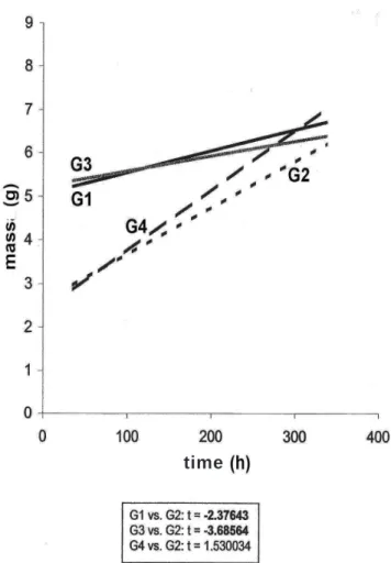
The comparison between G1 and G2 (Figure 1)
represents “non-hepatectomy vs. hepatectomy”.
Figure 2 shows a statistically significant increase
in hepatic GSH concentration of G2 at moment T2. Similar
significant increases occurred in T1 and T3 (not shown). In
addition, hepatic GSH decreased in a statistically significantly
way in G3 at moment T2 (Figure 2), which also occurred at
moments T1 and T3 (not shown).
Figure 1 Figure 1 Figure 1 Figure 1
Figure 1 - Evolution of the Post-PH residual livers. G1 = Iaparotomy group (without PH) = control group; G2 = PH + saline, G3 = PH + Omega 3, G4 = PH + Omega 6; Student t test of angular coefficients of the regression lines shows statistic significance G1xG2 and G3xG2, p<0.05 (t values in bold).
The concentrations of liver TBARS was
significantly increased (p<0.01) in G4 (Figure 3) at moment
T2, but this growth also occurred at T1 (not shown).
Plasma GSH after PH was significantly higher
(p<0.01) in G2 in T1 and T2 and at all times in G3. Figure 4
presents these facts at moment T2.
Both G2 and G3 did not have significant changes
in plasma TBARS after PH, but the MDA was shown with
significantly increased concentrations in G4 at moments T1
and T2. Figure 5 shows this at T2.
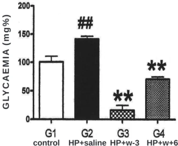
Statistically significant hyperglycaemia (p<0.01)
occurred with G2 rats at T2 and T3, while significant
hypoglycaemia (p<0.01) was evident in G3 and G4 at all
times. Figure 6 displays these facts at T2.
Total bilirubin levels did not change significantly
in either group at any time (not shown).
DISCUSSION
DISCUSSION
DISCUSSION
DISCUSSION
DISCUSSION
Figure 1 shows the evolution of residual livers in
each group by means of regression lines (interpolation). As
expected, the G1 did not show regenerative behavior once
their line of evolution of liver mass is practically horizontal. In
fact, the animals in this group did not undergo any surgery
of the liver or were subjected to any injection of drug. The
slope of G1 is not null - it’s small, but positive - probably
because it expresses the small natural growth of the liver in
young rats. G2, in contrast, displays aggressive behavior of
regeneration, with inclination (â) significantly greater than
G1 (Figure 1). It is the natural liver regeneration, without
stimulating or inhibiting drugs.
G1 and G3 showed very similar slopes (Figure 1),
indicating that the regenerative growth in these two groups
(non-PH and PH omega 3) are not different. In other words,
the administration of omega-3 PUFA resulted in inhibition
of liver regeneration, since in G1 there is not, strictly
speaking, any ongoing regeneration. The slopes of G4 and
G2 lines, on the other hand, are not significantly different,
which in essence confirms that the intake of PUFA
omega-6 (G4) does not interfere significantly with the natural
development of liver regeneration (G2).
Partial hepatectomy per se is related to the
formation of free radicals
14,15, and the animals only partially
hepatectomized (G2) showed increased GSH, both in
plasma and liver (Figures 4 and 2, respectively), which can
be understood as an attempt to mitigate the oxidative stress
caused by PH. In general, cells respond to oxidative stress
with higher concentration of GSH in order to inhibit potential
oxidative injury. In fact, many studies have shown increased
production of free radicals after partial hepatectomies
9,16,17.
The present study demonstrates that the
phenomenon of liver regeneration was significantly reduced
after infusion of omega-3 (Figure 1, G3 compared to G2);
at the same time, there was a decrease of hepatic GSH
(Fig. 2, G3 compared to G2). Omega-3 PUFA thus functions
Figure 2 Figure 2 Figure 2 Figure 2
-Figure 2 - Post-PH Hepatic GSH, T2. Statistically significant (p<0.01) increase of GSH in G2 compared to G1; significant decrease (p<0,01) in G3 and no significant changes in G4, both compared to G2.
Figure 3 Figure 3 Figure 3 Figure 3
-Figure 3 - Post-PH Hepatic TBARS, T2. Statistically significant increase (p<0,01) of TBARS in G4 compared to G2. Absence of significant changes in G3 compared to G2 and in G2 compared to G1.
Figure 4 Figure 4 Figure 4 Figure 4
Figure 4 - Post-PH Plasmatic GSH, T2. Statistically significant increases (p<0,01) in G2 compared to G1 and in G3 compared to G2. Absence of significant changes in G4 compared to G2.
control HP+saline HP+w-3 HP+w+6
control HP+saline HP+w-3 HP+w+6
as an antioxidant agent that inhibits rat liver regeneration
after PH. Corroborating these findings, several researchers
have shown that omega-3 PUFA inhibits the generation of
free radicals
18,19and, with respect to the regenerative process,
many studies attest to the importance of lipid peroxidation
(LPO)
7,16,20. Such papers are unanimous in pointing out the
genesis and formation of free radicals among the prominent
factors for natural, physiological liver regeneration
6,9.
Furthermore, administration of exogenous antioxidant
blocks hepatic regeneration
5,6. For example, the exogenous
supply of vitamin E affects hepatic regeneration in partially
hepatectomized rats
7and the administration of GSH blocks
liver regeneration in rats
8. Thus, omega-3 PUFA exerts an
inhibitory factor on liver regeneration after PH in rats
probably through its antioxidative properties.
Unlike omega-3, the results obtained here show
that the administration of omega 6 PUFA did not inhibit
the regeneration process of rat liver and since both plasma
Figure 5 Figure 5 Figure 5 Figure 5
-Figure 5 - Post-PH Plasmatic TBARS, T2. Absence of statistically significant changes in G2 compared to G1 and in G3 compared to G2. Significant increase of TBARS in G4 compared to G2.
Figure 6 Figure 6 Figure 6 Figure 6
-Figure 6 - Post-PH GLYCAEMIA, T2. Statistically significant (p<0,01) hiperglycaemia in G2 compared to G1 and hipoglycaemia in G3 and G4, both compared to G2.
and liver TABRS increased significantly with the infusion of
omega-6 at T1 and T2 (Figures 5 and 3, respectively), this
study confirms that omega-6, though inducing increased
lipid peroxidation, does not interfere with the evolution of
liver regeneration. Some studies have obtained even more
conclusive results and showed that omega-6, besides
increasing oxidative stress
18,20, increases liver regeneration
in rats
20.
The divergent behavior of 3 and
omega-6 in relation to oxidative stress and liver regeneration can
be summarized as follows: while omega-3 inhibits liver
regeneration and acts as an antioxidant, omega-6 does
not interfere with changes in the liver regenerative process
and has pro-oxidant properties.
Epidemiological studies with the Greenland
Eskimo population
21as well as investigations in clinical
nutrition
22,23and laboratory research with cytokines and
eicosanoids
24,25have shown that omega-6 has
proinflammatory properties, while omega-3 exhibits
anti-inflammatory activities
26. Similar properties were seen here.
They explain the differences between these PUFA on liver
regeneration.
Malignancies are living examples of major cell
proliferation as it occurs in the liver regeneration
phenomenon. Several investigators have suggested that
omega-3 works to suppress cancer cells while omega-6
stimulates carcinogenesis
27-29. There is evidence suggesting
that the incidence of aggressive carcinoma correlates in a
straightforward way with the intake of omega-6
30and, on
the other hand, there is a study reporting decreased
proliferation of tumor cells in the colon of rats fed with fish
oil 3) when compared to corn oil
(omega-6)
31. Several surveys are therefore conclusive in pointing
out that diets rich in omega-6 PUFA induce tumor growth
(cell proliferation), whereas diets high in omega-3 PUFA
show inhibitory effects
32. Such studies are in line with this
research in relation to cell proliferation – the compensatory
hyperplasia of the liver regenerative process: omega-3
blocks, omega-6 does not.
A large number of studies have reported that
the residual liver lobes after hepatectomy maintain all the
liver functions necessary to keep homeostasis in normal
organic levels despite the ongoing regeneration
process
3,33.34. In partially hepatectomized rats, plasma insulin
concentrations have a significant decrease
35,36, while the
levels of glucagon increase
37,38, which probably constitutes
part of the homeostatic response resulting, among other
things, in proper blood glucose levels
36. In the group with
omega-3 intake, as well as in the one with omega-6, plasma
glucose values were significantly decreased (Figure 6). These
are probably consequences of the hypoglycemic action of
lipids on the mechanism of insulin secretion. In fact, research
has revealed that the secretion of insulin, previously deemed
to be dependent on a unique and essential glucose stimulus,
is now being more and more related to the interaction of
glucose and lipid metabolism
39. When high levels of
control HP+saline HP+w-3 HP+w+6
control HP+saline HP+w-3 HP+w+6
GL
YCAEMIA
circulating free fatty acids in rats fasted for one day are
suddenly reduced via infusion of a lipolytic agent (i.e.,
nicotinic acid), a subsequent stimulation of glucose
secretion by insulin is totally inhibited, but it becomes
normal if the concentration of free fatty acids is kept high
by co infusion of lipid emulsion with heparin
40,41. Clinical
trials show similar results with two day fasting humans
42. In
fed rats, since the level of circulating free fatty acids is
low, the response of insulin secretion after a glucose
stimulus is normal
40, but an artificial increase in the
concentration of circulating free fatty acids results in
dramatic increase in insulin secretion after a glucose
stimulus
43. Moreover, the insulinotropic power of fatty
acids is directly correlated to the length of their chains
and their degree of saturation
41.Presumably, all animals
of groups G3 and G4, nourished daily with proper chow
plus the artificial intraperitoneal infusion of omega-3 or
omega-6 PUFA, consequently presented with higher insulin
secretion and therefore hypoglycaemia (Figure 6 ). In
addition, antioxidants induce greater insulin action
44due
in part to the protection of beta cells from free radicals’
injuries
45. Omega-3 PUFA, possessing the two properties,
should, in theory, have higher hypoglycemic potential: in
fact, the G3 mice displayed a steeper glycemic fall.
Total plasma bilirubin showed no significant
change at all moments. Recent researches show that the
administration of fish oil or vitamin E or eicosapentaenoic
acid (EPA) did not induce any significant change in hepatic
function tests
34, which agrees with this study on oxidative
stress
46,47.
Thus, the present experimental results corroborate
the hypothesis that oxidative stress plays a significant role
in the phenomenon of liver regeneration in rats after partial
hepatectomy, and that the influence of omega-3 and
omega-6 are distinct, both in oxidative stress as in liver
regeneration itself.
R E S U M O
R E S U M O
R E S U M O
R E S U M O
R E S U M O
Objetivo: Objetivo: Objetivo:
Objetivo: Objetivo: Estudar os efeitos dos ácidos graxos poli-insaturados (PUFA) ômega-3 e ômega-6 no estresse oxidativo e na regeneração hepática em ratos submetidos à hepatectomia parcial à 70% (HP, hepatectomia a 70%, hepatectomia parcial à Higgins-Anderson). Métodos:Métodos:Métodos:Métodos:Métodos: 72 ratos Wistar machos jovens foram aleatoriamente distribuídos em quatro grupos de mesmo tamanho: controle, parcialmente hepatectomizados, e parcialmente hepatectomizados com aporte diário intraperitoneal, por duas semanas, de ou ômega-3 ou ômega-6. Nos tempos 36h (T1), 168h (T2) e 336h (T3) pós-HP, substâncias reativas ao ácido tiobarbitúrico (TBARS) e glutationa reduzida (GSH) foram medidos no plasma e no tecido hepático, enquanto glicose e bilirrubina total foram aquilatados no sangue. A massa do fígado residual, nos mesmos tempos, foi o parâmetro utilizado para estimar a evolução da regeneração hepática. Resultados:Resultados:Resultados:Resultados: PUFA ômega-3 inibiu a regeneraçãoResultados: hepática e induziu redução na concentração de GSH hepático sete dias pós-HP. PUFA ômega-6, ao contrário, não mostrou efeito inibitório sobre a regeneração. Houve aumento da peroxidação lipídica tanto no sangue como no fígado com a administração de ômega-6. Conclusão:Conclusão:Conclusão:Conclusão:Conclusão: PUFA ômega-3 retardou a regeneração hepática pós-HP provavelmente por inibição do estresse oxidativo. PUFA ômega-6 aumentou as concentrações de TBARS no sangue e no fígado mas não alterou a evolução do processo regenerativo hepático.
Descritores: Descritores: Descritores:
Descritores: Descritores: Regeneração hepática. Estresse oxidativo. Ácidos graxos. Peroxidação de lipídeos. Ratos.
REFERENCES
REFERENCES
REFERENCES
REFERENCES
REFERENCES
1. Higgins GM, Anderson RM. Experimental pathology of the liver: I. Restoration of the liver of the white rat following partial surgical removal. Arch Path. 1931;12:186-202.
2. Steer CJ. Liver regeneration. FASEB J. 1995;9(14):1396-400. 3. Michalopoulos GK. Liver regeneration: molecular mechanisms of
growth control. FASEB J. 1990;4(2):176-87.
4. Hockings PD, Roberts T, Campbell SP, Reid DG, Greenhill RW, Polley SR, et al. Longitudinal magnetic resonance imaging quantification of rat liver regeneration after partial hepatectomy. Toxicol Path. 2002;30(5):606-10.
5. Van Noorden CJ. Effects of n-3 and n-6 polyunsaturated fatty acid-enriched diets on lipid metabolism in periportal and pericentral compartments of female rat liver lobules and the consequences for cell proliferation after partial hepatectomy. J Lipid Res. 1995;36(8):1708-20.
6. Nakatani T, Inouye M, Mirochnitchenko O. Overexpression of antioxidant enzymes in transgenic mice decreases cellular ploidy during liver regeneration. Exp Cell Res. 1997;236(1):137-46.
7. Trejo-Solís C, Chagoya De Sánchez V, Aranda-Fraustro A, Sánchez-Sevilla L, Gómez-Ruíz C, Hernàndez-Muñoz R. Inhibitory effect of vitamin E administration on the progression of liver regeneration induced by partial hepatectomy in rats. Lab Invest. 2003;83(11):1669-79.
8. Holocek M, Skopec F, Sprongl L. Influence of buthionine sulfoximine, S-adenosylmethionine and glutathione on liver regeneration following partial hepatectomy. Arzneimittelforschung. 2000;50(12):1093-8.
9. Kurir TT, Markotiæ A, Kataliniæ V, Bozaniæ D, Cikes V, Zemunik T, et al. Effect of hyperbaric oxygenation on the regeneration of the liver after partial hepatectomy in rats. Braz J Med Biol Res. 2004;37(8):1231-7. Epub 2004 Jul 20.
10. Mihara M, Uchiyama M. Determination of malonaldehyde in tissues by thiobarbituric acid test. Anal Biochem. 1978;86(1):271-8. 11. Sedlak J, Lindsay RH. Estimation of total protein-bound, non-protein
sulphydril group in tissue with Ellman´s reagent. Anal Biochem. 1968;25:192-205.
13. Malloy HT, Evelyn KA. The determination of bilirubin with the photoelectric colorimeter. J Biol Chem. 1937;119(2):481-90. 14. Aguilar-Delfín I, López-Barrera F, Hernández-Muñoz R.
Selective enhancement of lipid peroxidation in plasma membrane in two experimental models of liver regeneration: partial hepatectomy and acute CCl4 ad min is tratio n . Hepatology. 1996;24(3):657-62.
15. Carnovale CE, Scapini C, Alvarez ML, Favre C, Monti J, Carrillo MC. Nitric Oxide release and enhancement of lipid peroxidation in regenerating rat liver. J Hepatol. 2000;32(5):798-804.
16. Guerrieri F, Vendemiale G, Grattagliano I, Cocco T, Pellecchia G, Altomare E. Mitochondrial oxidative alterations following partial hepatectomy. Free Rad Biol Med. 1999;26(1-2):34-41.
17. Hernández-Muñoz R, Sánchez-Sevilla L, Martínez-Gómez A, Dent MA. Changes in mitochondrial adenine nucleotides and in permeability transition in two models of rat liver regeneration. Hepatology. 2003;37(4):842-51.
18. Wu G, Meininger CJ. Regulation of nitric oxide synthesis by dietary factors. Annu Rev Nutr. 2002;22:61-86. Epub 2002 Jan 4. 19. Takahashi M, Tsuboyama-Kasaoko N, Nakatani T, Ishii M,
Tsutsumi S, Aburatani H, Ezaki O. Fish oil feeding alters liver gene expression to defend against PPARalpha activation and ROS production. Am J Physiol Gastrointest Liver Physiol. 2002;282(2):G338-48.
20. Ok E, Yilmaz Z, Karakücük I, Akgün H, Sahin H. Use of olive oil based emulsions as an alternative to soybean oil based emulsions in total parenteral nutrition and their effects on liver regeneration following hepatic resection in rats. Ann Nutr Metab. 2003;47(5):221-7.
21. Kromann N, Green A. Epidemiological studies in the Upernavik district, Greenland. Incidence of some chronic diseases 1950-1974. Acta Med Scand. 1980;208(5):401-6.
22. Wu D, Meydani SN. n-3 polyunsaturated fatty acids and immune function. Proc Nutr Soc. 1998;57(4):503-9.
23. Harbige LS. Dietary n-6 and n-3 fatty acids in immunity and autoimmune disease. Proc Nutr Soc. 1998;57(4):555-62. 24. Endres S, Ghorbani R, Kelley VE, Georgilis K, Lonnemann G, Van
der Meer JWM, et al. The effect of dietary supplementation with n-3 polyunsaturated fatty acids on the synthesis of interleukin-1 and tumor necrosis factor by mononuclear cells. N Eng J Med. 1989;320(5):265-71.
25. Engström K, Luostarinen R, Saldeen T. Whole blood production of thromboxane, prostacyclin, and leukotriene B4 after dietary fish oil
supplementation in man: effect of vitamin E. Prostaglandins Leukot Essent Fatty Acids. 1996;54(6):419-25.
26. Azevedo RB, Silva LP, Lemos APC, Miyasaka CK, Lacava ZGM. Controle da resposta inflamatória por ácidos graxos. In: Curi R, Pompéia C, Miyasaka CK, Procópio J. Entendendo a gordura – Os ácidos graxos. 1ª ed. São Paulo: Manole; 2002. p. 379-91.
27. Fritsche KL, Johnston PV. Effect of dietary alpha-linolenic acid on growth, metastasis, fatty acid profile and prostaglandin production of two murine mammary adenocarcinomas. J Nutr. 1990;120(12):1601-9.
28. Rao CV, Simi B, Wynn TT, Garr K, Reddy B. Modulating effect of amount and types of dietary fats on colonic mucosal phospholipase A2 phosphatidylinositol-specific phosphalipase C activities and cyclooxigenase metabolite formation during different stages of colon promotion in male F344 rats. Cancer Res. 1996;56(3):532-7. 29. Rose DP. Effect of dietary fatty acids on breast and prostate
cancers: evidence from in vitro experiments and animal studies. Am J Clin Nutr. 1997;66(6 Suppl):1513S-22S.
30. Falconer JS, Ross JA, Fearon KC, Hawkins RA, O´Riordain MG, Carter DC. Effect of eicosapentaenoic acid and other fatty acids on the growth in vitro of human pancreatic cancer cell lines. Br J Cancer. 1994;69(5):826-32.
31. Woutersen RA, Appel MJ, van garderen-Hoetmer A, Wijnands MV. Dietary fat and carcinogenesis. Mutat Res. 1999;443(1-2):111-27.
32. Lima MMR, Moreira NX, Santos BMA, Mancini Filho J, Fernandes LC. Ácidos graxos e câncer. In: Curi R, Pompéia C, Miyasaka CK, Procópio J. Entendendo a gordura – Os ácidos graxos. 1ª ed. São
Paulo: Manole; 2002. p. 523-36.
33. Anderson WR, Zieve L, Lindblad S. Ultrastructural study of hepatic regeneration following one-lobe, two-lobe, and subtotal hepatectomy in the rat. Exp Pathol. 1990; 38(1):61-72.
34. Kirimlioglu V, Kirimlioglu H, Yilmaz S, Ozgor D, Coban S, Karadag N, Yologlu S. Effect of fish oil, olive oil, and vitamin E on liver pathology, cell proliferation, and antioxidant defense system in rats subject to partial hepatectomy. Transplant Proc. 2006;38(2):564-7.
35. Bucher NRL, Swaffield MN. Regulation of hepatic regeneration in rats by synergistic action of insulin and glucagon. Proc Natl Acad Sci USA. 1975;72(3):1157-60.
36. Michalopoulos G, DeFrances MC. Liver regeneration. Science.1997;276(5309):60-6.
37. Bucher NRL, Patel U, Cohen S. Hormonal factors concerned with liver regeneration. In: Porter R, Whelan J. Hepatotrophic factors – CIBA Foundation Symposium. Amsterdam: Elsevier; 1978.
38. Petenusci SO, Freitas TC, Roselino ES, Migliorini RH. Glucose homeostasis during the early stages of liver regeneration in fasted rats. Can J Physiol Pharmacol. 1983; 61(3):222-8.
39. Martins EF, Carpinelli AR. Ácidos graxos e secreção de insulina. In: Curi R, Pompéia C, Miyasaka CK, Procópio J. Entendendo a gordura – Os ácidos graxos. 1ªt ed. São Paulo: Manole; 2002. p.
271-86.
40. Stein DT, Esser V, Stevenson BE, Lane KE, Whiteside JH, Daniels MB, et al. Essentiality of circulating fatty acids for glucose-stimulated insulin secretion in fasted rats. J Clin Invest. 1996;97(12):2728-35.
41. Stein DT, Stevenson BE, Chester MW, Basit M, Daniels MB, Turley SD, McGarry JD. The insulinotrophic potency of fatty acids is influenced profoundly by their chain length and degree of saturation. J Clin Invest. 1997;100(2):398-403.
42. Dobbins RL, Chester MW, Stevenson BE, Daniels MB, Stein DT, McGarry JD. A fatty acid-dependent step is critically important for both glucose- and non-glucose-stimulated insulin secretion. J Clin Invest 1998;101(11):2370-6.
43. Dobbins RL, Szczepaniak LS, Myhill J, Tamura Y, Uchino H, Giacca A, McGarry JD. The composition of dietary fat directly influences glucose-stimulated insulin secretion in rats. Diabetes. 2002;51(6):1825-33.
44. Koksal C, Bozkurt AK, Cangel U, Ustundag N, Konukolu D, Musellim B, Sayin AG. Attenuation of ischemia/reperfusion injury by N-acetylcysteine in a rat hind limb model. J Surg Res. 2003;111(2):236-9.
45. Facchini FS, Humphreys MH, DoNascimento CA, Abbasi F, Reaven GM. Relation between insulin resistance and plasma concentrations of lipid hydroperoxides, carotenoids, and tocopherols. Am J Clin Nutr. 2000;72(3):776-9.
46. Greca FH, Biondo-Simões MLP, Martins VDM, Araújo FH, Milano JB. Os ácidos graxos de cadeia curta na cicatrização de anastomoses colônicas: estudo experimental em ratos. Rev Col Bras Cir. 2003;30(4):268-74.
47. Branco AL, Campos ACL, Ferreira M, Ferreira M, Andriguetto PC, Sakamoto DG, Trubian PS, Matias JEF. Regeneração hepática em ratos submetidos à hepatectomia parcial: efeito da desnutrição protéica e da renutrição pré-operatória. ABCD Arq Bras Cir Dig. 2006;19(4):159-66.
Received in 04/08/2009
Accepted for publication in 08/10/2009 Conflict of interest: none
How to cite this article: How to cite this article: How to cite this article: How to cite this article: How to cite this article:
Melo JUS, Santos JMV, Kimura OS, Campos Júnior MM, Melo RB, Vasconcelos PRL. Effects of fatty acids on liver regeneration in rats . Rev Col Bras Cir. [periódico na Internet] 2010; 37(5). Disponível em URL: http://www.scielo.br/rcbc
Correspondence address: Correspondence address: Correspondence address: Correspondence address: Correspondence address: Dr. Ulisses S. Melo