Comparison of the electric activity of the suprahyoid
muscles during different lingual exercises
Comparação da atividade elétrica dos músculos supra-hióideos
durante a realização de diferentes exercícios linguais
Renata Maria Moreira Moraes Furlan1, Bárbara Antunes Rezende2, Andréa Rodrigues Motta3
ABSTRACT
Purpose: To analyze the electric activity of suprahyoid muscles in eight different isometric exercises and to suggest the most appropriate exercise for data normalization. Methods: Twenty two female volunteers, ages between 19 and 38 years (Avg=24,1 and SD=3,8) participated of the study. At first, the participants underwent a clinical evaluation of the tongue. Electric activity of submental region was recorded, by means of the electromyographic assessment, with the participant keeping the tongue in rest position and accomplishing the following exercises: tongue suction on the palate; tongue pressure on the palate; tongue apex pressure against the palate; tongue apex pressure against incisive papilla; exaggerated tongue retraction; tongue lateralization; and tongue protrusion. The exercises were randomly accomplished by the participants. Collected data were analyzed using Kruskal Wallis and Mann-Whitney tests. It was adopted as the significance level for all analyzes value of p≤0.05. Results: Electromyographic activity of all the exercises was different from the one recorded in rest position. There was no statistical significant difference between different exercises.
Conclusion: There was no difference in submental electrical activation in different exercises. Therefore none was more appropriate then the other for data normalization.
Ke y wo r d s : E l e c t r o m y o g r a p hy ; To n g u e ; M u s c l e s t r e n g t h ; Stomatognathic system; Evaluation
RESUMO
Objetivo: Pesquisar a atividade elétrica dos músculos supra-hióideos em oito diferentes exercícios isométricos e sugerir o exercício mais apropria-do para normalização apropria-dos daapropria-dos. Métodos: Participaram do estudo 22 indivíduos do gênero feminino, com idades entre 19 e 38 anos (M=24,1 anos e DP=3,8 anos).Primeiramente, os participantes foram submetidos à avaliação clínica da língua. Em seguida, por meio de eletromiografia, foi feito o registro da atividade elétrica da região submentual durante o repouso e realização dos exercícios isométricos: sucção de língua no palato; pressão de língua no palato; pressão de ápice de língua contra o palato; pressão de ápice de língua contra a papila palatina; retração exa-gerada de língua; lateralização de língua e protrusão de língua. A ordem de realização dos exercícios foi randomizada entre os participantes. Os dados coletados foram analisados, utilizando-se os testes Kruskal Wallis e Mann-Whitney. Adotou-se como nível de significância em todas as análises, valor de p≤0,05. Resultados: A atividade elétrica encontrada para todos os exercícios diferiu apenas daquela medida durante o re-pouso lingual, não apresentando diferença com significância estatística quando os exercícios foram comparados entre si. Conclusão: Não houve diferença na ativação elétrica da musculatura supra-hióidea nos diversos tipos de exercícios realizados. Portanto, nenhum destes exercícios foi mais apropriado, em relação aos demais, para normalização dos dados.
Descritores: Eletromiografia; Língua; Força muscular; Sistema estoma-tognático; Avaliação
Work done at Universidade Federal de Minas Gerais – UFMG – Belo Horizonte (MG), Brazil.
(1) Post-Graduate Program in Structural Engineering, Universidade Federal de Minas Gerais – UFMG – Belo Horizonte (MG), Brazil. (2) Post-Graduate Program in Speech Therapy Sciences, Universidade Federal de Minas Gerais – UFMG – Belo Horizonte (MG), Brazil. (3) Departament of Speech Therapy, Universidade Federal de Minas Gerais – UFMG – Belo Horizonte (MG), Brazil.
Conflict of interests: No
Authors’ contribution: RMMMF participated in the elaboration of the research, collection, analysis and interpretation of the data and writing of the article; BAR
participated in the elaboration of the research, collection, analysis and interpretation of the data and writing of the article; ARM participated in the conception and outline of the study, in the analysis and interpretation of the data, writing, critical review of the article and approval of the final version.
Correspondence address: Andréa Rodrigues Motta. Departamento de Fonoaudiologia, Faculdade de Medicina, Universidade Federal de Minas Gerais. Av. Alfredo Balena, 190, sala 249, Belo Horizonte (MG), Brazil, CEP: 30130-100. E-mail: [email protected]
INTRODUCTION
During swallowing, the tongue plays a fundamental role in the propulsion of the food from the oral cavity to the pharynx and esophagus(1). Research shows that dysphagic individuals
may have weakness in the musculature of the tongue(2,3) and
that the strength training through isometric exercises improves performance in the swallowing(4,5).
The suprahyoid muscles also participate in the swallowing. There is a relationship between the activity of suprahyoid muscles and the strength exerted by the tongue against the palate during the swallowing(6). In addition to helping in the
food propulsion pressure, the suprahyoid musculature plays an important role in the hyolaryngeal excursion and consequen-tly the opening of the pharyngoesophageal segment(7-9). The
reduction in the hyolaryngeal excursion is a frequent cause of aspiration in dysphagic patients(10).
Because it represents the activity of suprahyoid muscles, the electromyographic assessment of the submental region is commonly performed in the investigation of swallowing disor-ders(11,12). In the submental electromyographic assessment, it is
obtained the electrical activity of mylohyoid, anterior belly of the digastric and geniohyoid muscles (13).
Authors compared the electrical activity of the suprahyoid musculature in the tongue pressure exercises against the pa-late and head elevation from the supine position, and greater submental electrical activity was obtained during the pressure exercise of the tongue on the palate(11). According to the authors,
exercises which focus on increasing the contact effort tongue--palate may form effective strategies to exercise suprahyoid musculature.
Given the great variability inter and intra subject in the electromyographic signal, for the comparison of the electrical activity among individuals (or between exercises) to be pos-sible, it is necessary to use a standard reference of muscular contraction. It is a procedure called normalization of signal(14).
It is recommended that the normalization be performed based on the maximum voluntary isometric contraction(14). A survey
investigated among six muscular maneuvers, the most suitable for the normalization of the electromyographic signal in the case of the suprahyoid muscles. The maneuvers were: incom-plete swallowing with effort; tongue pressure on the palate, with open mouth and closed mouth; tongue retraction with open mouth and closed mouth and push a wall. The authors cited the incomplete swallowing maneuver with effort as the most suitable for the normalization of the signal, due to its lower coefficient of variation and greater statistical significance(15).
In clinical practice, there are several other exercises to the strength of the tongue, such as: pressing the tongue against the palate(16); protrude the tongue and press it against a
woo-den spatula positioned near the patient’s mouth(17); suck the
tongue on the palate(18); press the tip of your tongue against
the incisive papilla(19); stretch and retract the tongue(19); push
the tongue against the cheeks(19). In the literature, there are few
researches that compare the submental electrical activity during the performing of different exercises.
Therefore, this study aimed to investigate the electromyo-graphic activity of submental region in eight different isome-tric exercises of tongue (tongue suction on the palate, tongue pressure on the palate, tongue apex pressure against the incisive papilla, exaggerated retraction of tongue, tongue lateralization and protrusion and tongue apex pressure against the palate) and verify which one would be the most suitable for use as a reference for the normalization of the electromyographic signal.
The hypothesis of the authors is that the exercises involving the press of the palate or the cheek with the tongue have greater muscular recruitment than the others.
METHODS
Observational cross-sectional study conducted at Hospital das Clínicas, Universidade Federal de Minas Gerais (UFMG), after approval by the Research Ethics Committee under number 0415.0.203.000-10.
Participants
The sample consisted of 22 female subjects, aged between 19 and 38 years (average of 24.1 years), students and employees of UFMG.
Inclusion criteria were: (a) aged between 18 and 40 years; (b) female; (c) not undergoing orofacial myofunctional treat-ment; (d) absence of neuromuscular or hormonal disorders; (e) the absence of temporomandibular dysfunction; (f) absence of head and neck cancer history; (g) agree to participate in the survey (Signature of Informed Consent Form); (h) absence of cognitive problems that could affect the understanding of the language. Items (a) to (f) were collected through anamnesis. Exclusion criterion consisted in not completing all the requi-red demanded activities or in presenting claims related to oral motor.
Clinical evaluation
First, participants underwent clinical evaluation of tongue in order to characterize the sample. The evaluation was carried out by two speech therapists experienced in the treatment of orofacial myofunctional alterations The following aspects were observed: strength, mobility, habitual position, morphology, presence of tremor and the lingual frenulum aspect. The result was obtained by consensus between the two evaluators.
motion was kept without tremor or deformation of the tongue; “reduced only in the anterior third,” when only the anterior re-gion showed a deformation; “slightly reduced” when presented mild tremor and tongue tip folding and “reduced” when the tongue performed only a slight strength of counter resistance, presenting tremor and deformation. The evaluator also recorded when only the tongue apex had reduced strength(20).
To assess tongue mobility, participants were asked to per-form movements of protrusion and retraction, touch the right and left corner of the mouth and the central region of the upper and lower lips. When the participant was unable to perform some of these movements precisely, the mobility of the tongue was considered abnormal.
In assessing the habitual position, the evaluator asked the participant to point to the location where her tongue generally remained during rest. The tongue position could be classified as: between the teeth, on the lower teeth, on the upper teeth, on the upper alveolar region or on the inferior alveolar region.
As to the morphological aspect, the tongue was classified as: enlarged (lingual edges supported on the surfaces of the lower teeth), fissured (presence of grooves on the lingual surface), geographical (varied colorations on the tongue mucosa), with presence of groove in the central region, or unchanged (tongue taking oral floor, touching only the lingual surfaces of the lower teeth and absence of color and/or shape).
The tremor was also investigated by visual inspection and recorded as absent or present. If present, it was verified in which task it occurred.
The frenulum was evaluated by requesting the participant to open her mouth, protrude the tongue, elevate towards the palate and touch the incisive papilla. The lingual frenulum was classified as normal when its fixation to the floor of the mouth was visible from the sublingual caruncles, had sublingual fixation in the middle region of the tongue and the tongue was able to perform the protrusion and touch the incisive papilla. It was considered altered when its fixation to the floor of the mouth was visible from the inferior alveolar crest, had sublingual fixation between the medium and the apex of the tongue, the difference between the opening of the mouth touching and without touching the incisive papilla was less than or equal to 50%, or when the tongue tip was oblong, square, or heart-shaped(21).
Electromyographic evaluation
The electromyographic evaluation was performed in an acoustically treated room, with participants sitting in a chair, feet flat on the floor on a rubber mat, hands relaxed and sup-ported on legs and back against the backrest of the chair and upright head. It was asked to the participant to keep this posture during the exercises.
The skin surfaces of the submental region and the left wrist of each participant were wiped with gauze soaked in
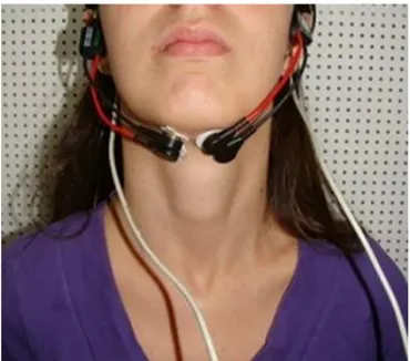
70% alcohol solution. It was expected an approximate time of 30 seconds, until the skin was dry, to position the electrodes. The electrical signals were obtained using disposable surface electrodes (Ag/AgCl), the Hal® brand, pre-jellified, circular, double and self-adhesive, with 10 mm diameter and 20 mm inter electrode distance, center to center, bilaterally positioned on the skin in the submental region, between the mandible and the hyoid bone(12), as shown in Figure 1.
The ground electrode was placed on the left wrist of the participants, as directed by the equipment manufacturer. The distance between the electrodes, center to center, was 20 mm. The electrodes were connected to an electromyography device of eight channels (EMG System do Brasil Ltda®), which re-corded the muscular electrical activity in microvolts (µV). The signal was filtered by high-pass filters of 20 Hz and low-pass of 500Hz, amplified with a gain of 1000x and rejection ratio of common mode >120 dB. Data were processed by a 16-bit analog to digital converter (EMG System do Brasil Ltda®), with a sampling frequency of 1 kHz. The active electrodes had an amplification gain of 20x. Data were processed through a specific software to data acquisition and processing (AqDados Software, version 5.5, Lynx Tecnologia Eletrônica Ltda®). It
was used only one channel. The other channels are disabled. Before starting the measurements, the participants were trained to perform the eight different exercises for the strength of the tongue: 1) tongue suction on the palate; 2) tongue pres-sure on the palate; 3) tongue apex prespres-sure against the palatal papilla; 4) exaggerated tongue retraction; 5) tongue laterali-zation to the right; 6) tongue lateralilaterali-zation to the left; and 7) tongue protrusion; 8) tongue apex pressure against the palate. The instructions for performing each exercise are in Chart 1.
After pasted the electrodes, the measurements were perfor-med. The first electromyographic signal was recorded during
the tongue rest and then while the participants were performing each exercise once. The exercises were performed randomly by the participants, with an interval of five minutes of rest between each one. The exercises were held for five seconds for the record. The recording was initiated at the time the participant started the exercise and ended after five seconds of recording by the computer program. The five seconds of the exercise were analyzed in full. For each exercise, the mean values, standard deviation, minimum, maximum, median and Pearson coefficient of variation were calculated.
The collected data were analyzed using STATA 12.0 pro-gram. For comparison of the RMS values among the exercises, it was used the Kruskal-Wallis test. To identify which variable had median value different from the other ones, it was used the Mann-Whitney test. It was adopted as the significance level, in all analyzes, the value of p≤0.05.
For choosing the activity to be suggested in the data stan-dardization process, it was considered that the exercise had the highest value of RMS, lower coefficient of variation and greater statistical significance when compared to the other ones(15).
RESULTS
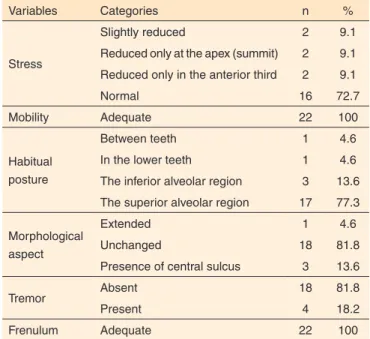
Regarding the distribution of the tongue evaluation varia-bles, most of the participants presented normotension, habitual position in superior alveolar region, unchanged morphology and lingual frenulum, absence of lingual tremor and adequate mobility (Table 1).
Table 2 shows the descriptive analysis of RMS measures for the electrodes positioned on the right and left sides for each performed exercise and for the rest. At rest it was found the lowest mean value of RMS and the lowest standard deviation.
The highest RMS mean value was observed in the activity of pushing the tongue against the right cheek for both electrodes, the right and the left ones. The lowest coefficient of variation was also found in this activity.
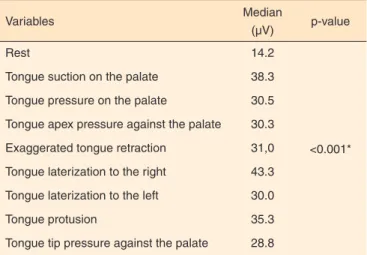
To compare the activities to each other, it was regarded as the mean value between right and left. The value of p indicates that at least one variable showed a different median value from the other ones (Table 3).
Regarding the difference of the median of the variable rest with each of the analyzed exercises, it was observed that the exercises showed no differences among the medians. Therefore, none of the exercises performed has reached the stipulated
Chart 1. Instructions for performing the exercises
Exercises Instructions
1) Tongue suction on the palate With the half-open mouth, suck the tongue on the palate and hold for 5 seconds.
2) Tongue pressure on the palate With the closed mouth, press the entire back of the tongue against the palate as strong as possible and hold for 5 seconds.
3) Tongue apex pressure against the palatal papilla With closed mouth, press only the tongue apex against the incisive papilla as strong as possible and hold for 5 seconds.
4) Exaggerated tongue retraction With the half-open mouth, retract the tongue as much as possible and hold for 5 seconds.
5) Tongue laterization to the right With the occluded lips, press the tongue in the jugal mucosa right and hold for 5 seconds.
6) Tongue laterization to the left With the occluded lips, press the tongue in the left jugal mucosa and hold for 5 seconds.
7) Tongue protusion Place a wooden spatula vertically next to the mouth and press the
tongue against the spatula as strong as possible and hold for 5 seconds. 8) Tongue apex pressure against the palate With the closed mouth, press only the lingual apex against the palate
as strong as possible and hold for 5 seconds.
Table 1. Distribution of tongue evaluation variables
Variables Categories n %
Stress
Slightly reduced 2 9.1
Reduced only at the apex (summit) 2 9.1 Reduced only in the anterior third 2 9.1
Normal 16 72.7
Mobility Adequate 22 100
Habitual posture
Between teeth 1 4.6
In the lower teeth 1 4.6
The inferior alveolar region 3 13.6 The superior alveolar region 17 77.3 Morphological
aspect
Extended 1 4.6
Unchanged 18 81.8
Presence of central sulcus 3 13.6
Tremor Absent 18 81.8
Present 4 18.2
It is noteworthy that the RMS was higher in absolute terms for the exercise of lateralization of the tongue to right and left, i.e., there was greater muscle recruitment in this activity. However, the electrical activity found for all exercises differed only from that measured during the tongue rest, with no statistically significant difference when the exercises were compared to each other.
A survey of 53 healthy subjects, aged between 21 and 60 ye-ars, compared the electrical activity of suprahyoid musculature in two types of exercises: tongue pressure against the palate and head elevation from the supine position(11). The authors found
more muscle activation in the exercise of tongue pressure on the palate than in the exercise of head elevation. Another survey, with a population of 20 healthy women between 19 and 33 years, compared the electrical activity between the exercises of pressing the chin to the chest and raise the head in the supine position and it was found that the first one produced higher values in electromyographic evaluation than the second one(9).
The authors believe that the counter resistance exercises are more effective for the musculature training.
Studies indicate that the tongue pressure on the palate exer-cises are effective in the rehabilitation of lingual strength(16,22)
and promote great activation of suprahyoid musculature(11).
The results of this study suggest that the other lingual exerci-ses performed in this research can be equally effective as they showed the same degree of muscle activation of the exercises of tongue pressure on the palate. However, additional studies
Table 2. Descriptive analysis of Root Mean Square measures, in µV, at rest and in each exercise
Electrode Exercises Mean Standard deviation Minimum Median Maximum PCV
Right
Rest 15.7 6.5 8.6 14.8 27.6 41.3
E1 39.2 18.2 17.1 35.6 84.5 46.9
E2 34.0 15.6 13.8 33.1 83.2 45.9
E3 36.9 24.2 12.5 28.1 94.7 65.5
E4 29.5 11.0 13.9 42.2 48.6 37.4
E5 42.7 17.2 18.7 42.0 72.5 40.3
E6 35.8 15.5 18.9 30.5 81.8 43.4
E7 41.7 20.9 13.1 38.9 88.3 50.2
E8 31.2 16.5 1.4 31.2 66.1 52.9
Left
Rest 14.6 8.6 5.7 14.0 41.0 59.0
E1 41.4 20.9 14.8 35.0 88.3 50.5
E2 33.0 16.8 7.1 29.0 77.3 51.0
E3 33.1 19.4 8.2 27.5 76.4 58.5
E4 35.4 23.2 10.8 30.8 114.0 65.5
E5 47.0 21.4 13.1 51.7 78.7 45.6
E6 38.4 24.5 11.8 26.2 91.9 63.6
E7 35.1 16.5 16.2 30.7 89,4 46.9
E8 34.8 27.7 7.5 26.1 111.0 70.7
Note: PCV = Pearson’s coefficient of variation; E1 = tongue suction on the palate; E2 = tongue pressure on the palate; E3 = tongue apex pressure against the palatal papilla; E4 = exaggerated tongue retraction; E5 = tongue laterization to the right; E6 = tongue laterization to the left; E7 = tongue protusion; E8 = tongue apex pressure against the palate
Table 3. Comparison of muscle electrical activity in Root Mean Square, during different isometric exercises
Variables Median
(µV) p-value
Rest 14.2
<0.001* Tongue suction on the palate 38.3
Tongue pressure on the palate 30.5 Tongue apex pressure against the palate 30.3 Exaggerated tongue retraction 31,0 Tongue laterization to the right 43.3 Tongue laterization to the left 30.0
Tongue protusion 35.3
Tongue tip pressure against the palate 28.8
*Significant values (p<0.05) – Kruskal Wallis test
criterion to be suggested as the basis for normalization of the data (Table 4).
DISCUSSION
Different exercises recruit different muscle groups and it is expected, therefore, different results on the electromyo-graphic evaluation(12). In this study, it was found that there
are needed to verify the real effectiveness of these exercises for the strength gain of the suprahyoid musculature and of the tongue as a whole.
The great variability among individuals, as evidenced by the high coefficients of variation obtained, is confirmed in the literature(23). This happens because the surface
electromyo-graphic signal can be influenced by several factors, distinct from subject to subject, such as thickness of adipose tissue, muscle resting length, speed of contraction, muscle mass, fiber type predominance, subtle changes in posture, inter electrode distance and skin impedance(24). Given this variability, when a
comparison between individuals is required, the amplitude of the EMG signal must be normalized(14). In this research, there
was no conclusion that one of the exercises performed were more intense in muscle activation than the others. Therefore, it was not obtained an exercise to use as a comparison. Some authors indicate incomplete swallowing maneuver for normali-zation of the submental electromyographic signal(15). For future
studies, it is suggested that such a maneuver is incorporated into the research.
The submandibular positioning of the electrodes, as perfor-med in this study, has been employed by several authors(9,11),
as a way of evaluating the hyolaryngeal elevation. The elec-tromyographic signal captured in the submental region reflects the activity of the mylohyoid, geniohyoid and anterior digastric belly muscles, with small contribution of the genioglossus one(13). There is a great intercorrelation among the suprahyoid
muscles and it is not possible to capture, with surface electro-des, the electrical activity of only one of them. Therefore, the obtained signal represents the entire complex(6).
Some exercises were performed with open mouth, others with closed mouth. The literature indicates that the increase of mouth opening results in the decrease of the electrical acti-vity of the suprahyoid muscles during swallowing, as the jaw rotation causes the shortening of these muscles fibers(25). The
individuals of this study were told to keep the mouth open, half-open or closed, in a specific way for each exercise. Some exercises, such as the tongue protrusion one, for example, must
be performed with open mouth. It is suggested that the degree of mouth opening is a controlled factor in future researches.
Most participants showed adequate tongue stress, habitual posture in superior alveolar region, unchanged morphological aspect and absence of lingual tremor, which was expected, since the participants were young adults who were not in orofacial myofunctional treatment. The reproduction of the study in individuals with dysphagia, or with severe alterations in the tongue strength could generate results different from the ones that were found.
All participants of the research managed to perform the requested exercises without difficulty, which probably would not be possible in a population with major alterations, or that has undergone surgery or radiotherapy. It is suggested that further researches should be conducted with a larger sample and with participants with serious alterations in the tongue strength. It is possible that, in individuals with lingual hypotension, the comparison among the exercises get a different result. However, this comparison could not be performed in this study due to the small number of participants with this feature in the sample.
In this study, the clinical evaluation of the tongue was per-formed to characterize the sample and the relationship between this assessment and the electromyographic activity was not researched. It is suggested that this relationship is investigated in studies with larger sample and include individuals with and without orofacial myofunctional alterations.
It was a limitation of this study to carry out only a repetition of each exercise(25). This happened to prevent fatigue of the
participants, as eight exercises were tested in maximal isometric contraction. It is suggested that future researches carry out at least three repetitions of the same task and make further surveys to prove the effectiveness of each exercise in increasing the strength of tongue and swallowing rehabilitation.
CONCLUSION
There was no difference in the electrical activation of suprahyoid musculature in the different exercises performed.
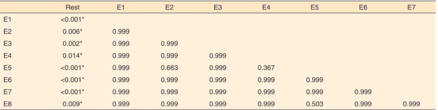
Table 4. Comparison of electrical activity in RMS, between pairs of isometric exercises
Rest E1 E2 E3 E4 E5 E6 E7
E1 <0.001*
E2 0.006* 0.999
E3 0.002* 0.999 0.999
E4 0.014* 0.999 0.999 0.999
E5 <0.001* 0.999 0.663 0.999 0.367
E6 <0.001* 0.999 0.999 0.999 0.999 0.999
E7 <0.001* 0.999 0.999 0.999 0.999 0.999 0.999
E8 0.009* 0.999 0.999 0.999 0.999 0.503 0.999 0.999
*Significant values (p<0.05) – Mann Whitney test
None of the exercises reached the stipulated criterion to be suggested as a basis for normalization of the data.
REFERENCES
1. Pouderoux P, Kahrilas PJ. Deglutitive tongue force modulation by volition, volume, and viscosity in humans. Gastroenterology. 1995;108(5):1418-26. doi:10.1016/0016-5085(95)90690-8 2. Stierwalt JA, Youmans SR. Tongue measures in individuals with
normal and impaired swallowing. Am J Speech Lang Pathol. 2007;16(2):148-56. doi:10.1044/1058-0360(2007/019)
3. Yoshida M, Kikutani T, Tsuga K, Utanohara Y, Hayashi R, Akagawa Y. Decreased tongue pressure reflects symptom of dysphagia. Dysphagia. 2006;21(1):61-5. doi:10.1007/s00455-005-9011-6 4. Yeates EM, Molfenter SM, Steele CM. Improvements in tongue
strength and pressure-generation precision following a tongue-pressure training protocol in older individuals with dysphagia: three case reports. Clin Interv Aging. 2008;3(4):735-47.
5. Steele CM, Bailey GL, Polacco RE, Hori SF, Molfenter SM, Oshalla M et al. Outcomes of tongue-pressure strength and accuracy training for dysphagia following acquired brain injury. Int J Speech-Language Pathol. 2013;15(5):492-502. doi:10.3109/17549507.2012.752864 6. Palmer PM, Jaffe DM, McCulloch TM, Finnegan EM, Van Daele
DJ, Luschei ES. Quantitative contributions of the muscles of the tongue, floor-of-mouth, jaw, and velum to tongue-to-palate pressure generation. J Speech Lang Hear Res. 2008;51(4):828-35. doi:10.1044/1092-4388(2008/060)
7. Kahrilas PJ, Dodds WJ, Dent J, Logemann JA, Shaker R. Upper esophageal sphincter function during deglutition. Gastroenterology. 1988;95(1):52-62.
8. Shaw DW, Cook IJ, Gabb M, Holloway RH, Simula ME, Panagopoulos V et al. Influence of normal aging on oral-pharyngeal and upper esophageal sphincter function during swallowing. Am J Physiol. 1995;268(3):G389-96.
9. Watts CR. Measurement of hyolaryngeal muscle activation using surface electromyography for comparison of two rehabilitative dysphagia exercises. Arch Phys Med Rehabil. 2013;94(12):2542-8. doi:10.1016/j.apmr.2013.04.013
10. Perlman AL, Booth BM, Grayhack JP. Videofluoroscopic predictors of aspiration in patients with oropharyngeal dysphagia. Dysphagia. 1994;9(2):90-5. doi:10.1007/BF00714593
11. Yoshida M, Groher ME, Crary MA, Mann GC, Akagawa Y. Comparison of surface electromyographic (sEMG) activity of submental muscles between the head lift and tongue press exercises as a therapeutic exercise for pharyngeal dysphagia. Gerodontology. 2007;24(2):111-6. doi:10.1111/j.1741-2358.2007.00164.x 12. Lenius K, Carnaby-Mann G, Crary M. The relationship between
lingual-palatal pressures and submental surface electromyographic signals. J Oral Rehabil. 2009;36(2):118-23. doi:10.1111/j.1365-2842.2008.01921.x
13. Palmer PM, Luschei ES, Jaffe D, McCulloch TM. Contributions of individual muscles to the submental surface electromyogram during swallowing. J Speech Lang Hear Res. 1999;42(6):1378-91. doi:10.1044/jslhr.4206.1378
14. De Luca CJ. The use of surface electromyography in biomechanics. J Appl Biomech. 1997;13:135-63.
15. Balata PMM, Silva HJ, Nascimento GKO, Moraes KLR, Pernambuco LA, Freitas MCR et al. Incomplete swallowing and retracted tongue maneuvers for electromyographic signal normalization of the extrinsic muscles of the larynx. J Voice. 2012,26(6):813e1-7. doi:10.1016/j.jvoice.2012.03.006
16. Robbins J, Gangnon RE, Theis SM, Kays SA, Hewitt AL, Hind JA. The effects of lingual exercise on swallowing in older adults. J Am Geriatr Soc. 2005;53(9):1483-9. doi:10.1111/j.1532-5415.2005.53467.x
17. Clark HM, O’Brien K, Calleja A, Corrie SN. Effects of directional exercise on lingual strength. J Speech Lang Hear Res. 2009;52(4):1034-47. doi:10.1044/1092-4388(2009/08-0062) 18. Guimarães KC, Drager LF, Genta PR, Marcondes BF, Lorenzi-Filho
G. Effects of oropharyngeal exercises on patients with moderate obstructive sleep apnea syndrome. Am J Respir Crit Care Med. 2009;179(10):962-6. doi:10.1164/rccm.200806-981OC
19. Bacha SMC; Camargo AFFP, Enne J, Ribeiro JML, Volpe MRFT. Exercícios de motricidade orofacial: anatomia e fisiologia [DVD]. São Paulo: Pró-Fono; 1998. 1 DVD: 32 min.
20. Almeida LD, Furlan RMMM, Las Casas EB, Motta AR. Influence of height, weight and body mass index in the axial tongue force. J Soc Bras Fonoaudiol. 2012;24(4):381-5. doi:10.1590/S2179-64912012000400015
21. Marchesan IQ. Lingual frenulum protocol. Int J Orofacial Myology. 2012;38:89-103.
22. Robbins J, Kays SA, Gangnon RE, Hind JA, Hewitt AL, Gentry LR et al. The effects of lingual exercise in stroke patients with dysphagia. Arch Phys Med Rehabil. 2007;88(2):150-8. doi:10.1016/j. apmr.2006.11.002
23. Vaiman M, Eviatar E, Segal S. Surface electromyographic studies of swallowing in normal subjects: a review of 440 adults. Report 2. Quantitative data: amplitude measures. Otolaryngol Head Neck Surg. 2004;131(5):773-80. doi:10.1016/j.otohns.2004.03.014
24. Belo LR, Coriolano MGWS, Menezes DC, Lins OG. Valores referenciais da eletromiografia de músculos envolvidos na deglutição: uma revisão sistemática. Rev CEFAC. 2012;14(1):156-63. doi:10.1590/S1516-18462011005000072