*Correspondence: D.J. Macrini. Instituto de Ciências da Saúde, Universidade Paulista – UNIP, Av. Dr. Bacelar, 1212, 04026-002 - São Paulo - SP, Brasil, Tel: 55 11 5586-4074; FAX: 55 11 276-0500. E-mail: [email protected].
A
rti
Pharmaceutical Sciences vol. 45, n. 2, abr./jun., 2009
Extracts from Amazonian plants have inhibitory activity against
tyrosinase: an
in vitro
evaluation
Daclé Juliani Macrini
1*, Ivana Barbosa Suffredini
2, Antonio Drauzio Varella
2,
Riad Naim Younes
2, Mitsuko Taba Ohara
31Health Sciences Institute, University Paulista, 2Laboratory of Extraction, University Paulista, 3Faculty of Pharmaceutical Sciences, University of São Paulo.
Dermatological disorders related to pigmentation result in tenuous hyper or hypopigmentation Cosmetic and pharmaceutical products containing depigmenting substances are used in the treatment of patients who have high pigmentation disorders, such as melasma or chloasma, post-inlammatory hyperpigmentation, senile lentigo and ephelides. Skin lightening agents are not yet totally effective or safe and therefore intensive research for the discovery of new agents is continuous. Enzyme inhibitors involved in melanogenesis, such as tyrosinase, have been discovered in Asian countries, including those isolated from plant extracts. The Brazilian lora has the highest species diversity in the world, and the chemical, pharmacological and cosmetic potential for the discovery of new skin whitening agents is in proportion with this biodiversity. For these reasons, 25 aqueous and 24 organic extracts obtained from 19 plants native to the Amazon rain forest and to the Atlantic forest, belonging to 11 different families, were evaluated as tyrosinase inhibitors. Nine out of 49 extracts showed inhibitory activity in the screening process. The 50% inhibitory activity (IA50) was calculated, revealing that the most active extracts were
the organic extracts from the leaves and stem of Ruprechtia sp. (IA50 33.76 mg.mL-1) and the organic
extract from the aerial organs of Rapanea parvilora (IA50 64.19 mg.mL-1).
Uniterms: Tyrosinase. Melanin. Plant extract/in vitro evaluation. Skin lightening agents. Amazonian plants/evaluation/dermatological use.
Problemas dermatológicos relacionados com a pigmentação resultam em hiperpigmentações ou hipopigmentação cutâneas. Produtos cosméticos e farmacêuticos com atividade despigmentante são utilizados para o tratamento de pacientes que apresentam distúrbios de hiperpigmentação, tais como melasma ou cloasma, hiperpigmentação pós-inlamatória, lentigem senil e efélides. Os despigmentantes atualmente utilizados não são totalmente eicazes ou seguros, razão pela qual há intensa pesquisa, principalmente em países asiáticos, com a inalidade de se obter novos agentes com esta ação, em especial inibidores de enzimas envolvidas na melanogênese, como a tirosinase. Considerando-se que algumas substâncias obtidas de plantas apresentam essa atividade, a lora brasileira constitui-se uma fonte potencial de obtenção de novos despigmentantes. Por essa razão, 25 extratos aquosos e 24 orgânicos obtidos de 19 plantas da Floresta Amazônica e Mata Atlântica, provenientes de 11 diferentes famílias, foram avaliados quanto à atividade de inibição da tirosinase. Do total de 49 extratos testados, 9 mostraram atividade. Os valores de concentração da atividade inibitória 50% (AI 50%), foram calculados e o mais ativo foi o extrato orgânico das folhas e caule de Ruprechtia sp. (AI50 33,76 mg.mL-1) seguido do extrato orgânico
dos órgãos aéreos de Rapanea parvilora (AI50 64,19 mg.mL-1).
Unitermos: Tirosinase. Melanina. Extrato de plantas/avaliação in vitro. Despigmentantes. Plantas amazônicas/avaliação/uso dermatológico.
INTRODUCTION
For centuries, humans have tried to artificially
et al., 2002). As the world population ages it becomes even more important to treat pigment disorders (Benech, 2002). Severe anomalies may also hamper an individual in social relationships (Grimes, 1999).
Both hyper and hypopigmentation are treated with cosmetic or pharmaceutical products containing depigmenting substances, which are synthetic or of na-tural origin. Pigmentation disorders can also be treated with mechanical peeling allied to chemical substances or treatments with laser rays (Grimes, 1999).
The skin produces a complex mixture of enzymes for protection from the constant exposition to
environ-mental oxidative stress (Podahaisky et al., 2002), but in
some situations this is insuficient to prevent disorders from appearing. However, the fact that drugs can be
absorbed by the skin (Thorsteinsson et al., 1999) aids
in the development of cosmetic preparations to treat skin pigmentation or depigmentation, particularly drugs whose active substance is a natural product.
One of the most effective depigmenting substan-ces is hydroquinone, which inhibits the synthesis of melanin. Enzyme inhibition is one of the mechanisms involved in melanogenesis (Lee, Choi, 1998).
Many screening studies are reported in the li-terature, with emphasis on those products made with
tyrosinase inhibitors from African plants (Baurin et
al., 2002; Kubo, Hori, 1999b, Momtaz et al., 2008),
Bolivia (Kubo et al., 1995), China (Iida et al., 1995;
Masamoto et al., 1980; Miao et al., 1997), Japan (No
et al., 1999), Bangladesh (Khanom et al., 2000) and others. Positive results in relation to activity were obtai-ned and some authors continued this work by isolating active substances.
Considering that current therapies have shown less than satisfactory results in the treatment of various derma-tological disorders such as melasma, post-inlammatory or senile lentigo and ephelides, and that the side effects of the therapy include high cytotoxicity and mutagenicity, poor skin penetration and low stability of formulations
(Grimes, 1999; Nerva et al., 2003), new drugs with
enzy-me inhibitory activity are needed(Su, 1999).
Satisfactory results obtained from screening
stu-dies previously conducted by other groups, reporting the identiication of inhibitory activity of melanoge-nesis by plant extracts, allied with the fact that Bra-zilian biodiversity corresponds to 20% of the world’s
biodiversity (Suffredini et al., 2004) justify the current
project of screening Brazilian plant extracts such as
tyrosinase inhibitors in vitro, so that agents may be
found and eventually used in cosmetic and pharma-ceutical products.
MATERIAL AND METHODS
Plant extracts
The plant material listed in Table I was collected
from igapó, terra irme and Atlantic forests, according
to a chemosystematic approach. Vouchers were depo-sited in the Herbarium UNIP, where they were identi-ied. After collection, the material was ground up and 24h-macerated with a mixture of dichloromethane and methanol (1:1), followed by 24h-maceration with Milli-Q water. The organic extracts were evaporated under
reducedpressure (Büchi) and the aqueous extracts were
lyophilized (Virtis). All the extracts were stored in a
freezer (-27 ºC, Revco) until use (Younes et al., 2000).
Tyrosinase enzymatic reaction assay
The assay was realized with modiications
(Kha-nom, 2000; Kobayashi et al., 1995), such that 10 mL of
the solution made with 120 U.mL-1 (1st screening) or
480 U.mL-1 (2nd screening) of tyrosinase obtained from
mushrooms (Sigma) were added to 96-well micropla-tes, as were 70mL of pH 6.8 buffer solution and 60 mL of the plant extract solutions (water or DMSO 50% in water, were used as vehicles). A negative control was used, as was a positive control made with a solution of
kojic acid. To this mixture, 70 µL of L-tyrosine (Sigma)
were added, completing the inal volume up to 210 µL.
The absorbance was taken at 490nm in a microplate spectrophotometer reader (Biotek) in the beginning of the reaction, as the time zero reading. Microplates
were incubated at (30±1) ºC for 120 minutes in the irst
screening, and for 60 minutes in the second screening. Optical densities were registered on a computer coupled to the spectrophotometer reader. Inhibitory activity of 50% was obtained according to the formula
IA (%) = [((C-T0)-(S- T0))/(C- T0)] x 100
where IA = inhibitory activity, C= control absorbance
at 490 nm, S= test absorbance at 490 nm and T0= time
zero, and each parameter was a mean of 8 measures. The percentage of inhibitory activity of tyrosi-nase obtained from the extracts was compared to that obtained from the kojic acid.
Tyrosinase inhibitory activity by plant extracts
The irst screening was performed as previously
concentration in the assay was 80 µg/mL (µg.ml-1)
for the extracts, and 5 µg.mL-1 for kojic acid, used as
a positive control. Readings were taken 120 minutes after the addition of the extract or kojic acid. The active extracts were submitted to a second screening, whose
enzyme concentration was 480 U.mL-1.
Determination of the inhibitory activity at 50%, of kojic acid using tyrosinase at concentrations
of 120 U.mL-1 and 480 U.mL-1
The test was performed as irst described.
Tyro-sinase was diluted to 120 U.mL-1 and 480 U.mL-1, in
the irst and second screening, respectively. Kojic acid concentrations used in the test were 5.0, 4.0, 3.0, 2.5,
1.25, 0.625 and 0.3125 µg.mL-1 in the irst screening,
and 10.0, 5.0, 2.5, 1.25 and 0.625 µg.mL-1 in the second
screening.
Determination of the inhibitory activity at 50%, of the active extracts
The test was executed as irst described, substitu-ting the kojic acid by plant extracts, whose
concentra-tions were 80.0, 40.0, 20.0 and 10.0 µg.mL-1.
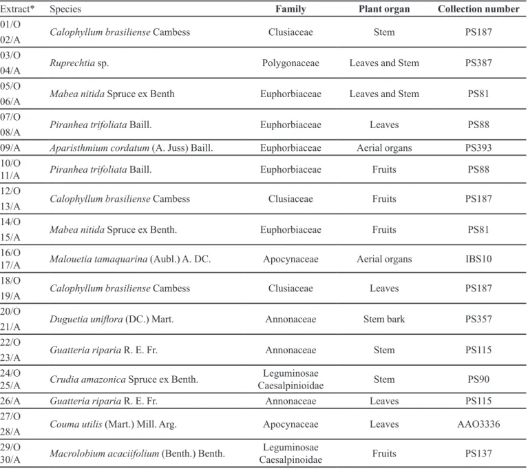
TABLE I - List of Amazonian Rain Forest plant extracts tested against tyrosinase
Extract* Species Family Plant organ Collection number
01/O
Calophyllum brasiliense Cambess Clusiaceae Stem PS187 02/A
03/O
Ruprechtia sp. Polygonaceae Leaves and Stem PS387 04/A
05/O
Mabea nitida Spruce ex Benth Euphorbiaceae Leaves and Stem PS81 06/A
07/O
Piranhea trifoliata Baill. Euphorbiaceae Leaves PS88 08/A
09/A Aparisthmium cordatum (A. Juss) Baill. Euphorbiaceae Aerial organs PS393 10/O
11/A Piranhea trifoliata Baill. Euphorbiaceae Fruits PS88 12/O
Calophyllum brasiliense Cambess Clusiaceae Fruits PS187 13/A
14/O
Mabea nitida Spruce ex Benth. Euphorbiaceae Fruits PS81 15/A
16/O
17/A Malouetia tamaquarina (Aubl.) A. DC. Apocynaceae Aerial organs IBS10 18/O
Calophyllum brasiliense Cambess Clusiaceae Leaves PS187 19/A
20/O
Duguetia unilora (DC.) Mart. Annonaceae Stem bark PS357 21/A
22/O
Guatteria riparia R. E. Fr. Annonaceae Stem PS115 23/A
24/O
25/A Crudia amazonica Spruce ex Benth. CaesalpinioidaeLeguminosae Stem PS90 26/A Guatteria riparia R. E. Fr. Annonaceae Leaves PS115 27/O
Couma utilis (Mart.) Mill. Arg. Apocynaceae Leaves AAO3336 28/A
29/O
RESULTS AND DISCUSSION
IA50 of the kojic acid obtained from the reaction
with tyrosinase at 120 U.mL-1, calculated from the
re-gression line formula generated after 120 minutes of
reaction, was 2.99 µg.mL-1. The IA50 of the kojic acid,
obtained from regression line formula generated from the
reaction with tyrosinase at 480 U.mL-1 after 60 minutes
was 6.85 µg.mL-1.
Results obtained in the first screening of plant extracts as inhibitors of tyrosinase are shown in Table II
(statistical analysis, T test). The IA50 concentrations are
shown in Table III.
Skin clarity is an important characteristic, as hyper and hypopigmentation affect the self-esteem of an indivi-dual, but fortunately can be treated, or at least minimized. In the last few years the number of whitening products on the market has increased dramatically, but therapies have not always shown satisfactory results (Su, 1999;
Tengamnuay et al., 2006), This is principally due to the
high toxicity of the whitening substances, such as that observed for hydroquinone (Grimes, 1999). The present observations highlight the need for intensive research on new depigmenting substances. The identiication of
enzyme inhibitors involved in melanogenesis, such as hydroquinone and kojic acid, is especially important.
Given that plants may contain enzyme inhibitory substances, screening methodologies show greater eficacy
in the identiication of active substances (Lee et al., 1999),
and there are calls to search for active compounds from Brazilian plants, the present screening was delineated in order to evaluate plant extracts obtained from plants che-mosystematically related to groups whose phenolic
com-pound production is signiicant (Iida et al., 1995; Kubo,
Hori, 1999a; Kubo, 1995; No, 1999). Plants belonging to the families: Annonaceae, Apocynaceae, Clusiaceae, Dil-leniaceae, Euphorbiaceae, Flacourtiaceae, Leguminosae, Myrsinaceae, Passiloraceae, Polygonaceae and Rubiaceae were assayed. Some plants of these families have already been evaluated and have been shown to contain phenolic tyrosine inhibitor compounds such as lavonoids and
tan-nins (Iida et al., 1995; Khanom et al., 2000).
The tyrosinase inhibitory effect of 49 aqueous and organic extracts obtained from 19 Brazilian plants belonging to 11 families was evaluated using a spectro-photometric method. Kojic acid was chosen as a positive control because the substance is an effective inhibitor of
tyrosinase in vitro (Sandoval, 1999).
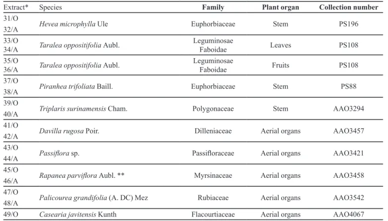
Extract* Species Family Plant organ Collection number
31/O
Hevea microphylla Ule Euphorbiaceae Stem PS196 32/A
33/O
34/A Taralea oppositifolia Aubl. Leguminosae Faboidae Leaves PS108 35/O
36/A Taralea oppositifolia Aubl. Leguminosae Faboidae Fruits PS108 37/O
Piranhea trifoliata Baill. Euphorbiaceae Stem PS88 38/A
39/O Triplaris surinamensis
Cham. Polygonaceae Stem AAO3294
40/A 41/O
Davilla rugosa Poir. Dilleniaceae Aerial organs AAO3457 42/A
43/O
Passilora sp. Passiloraceae Aerial organs AAO3421 44/A
45/O
Rapanea parvilora Aubl. ** Myrsinaceae Aerial organs AAO3458 46/A
47/O
Palicourea grandifolia (A. DC) Mez Rubiaceae Aerial organs AAO3542 48/A
49/O Casearia javitensis Kunth Flacourtiaceae Aerial organs AAO4067 *O = organic extract; A = aqueous extract; ** = Atlantic Forest
The concentration of tyrosinase used in the irst
scre-ening was 120 U.mL-1 in order to increase the sensitivity
and chances of inding active extracts. As the chemical composition of such extracts is not known, further studies were needed in order to verify the presence of tyrosinase inhibitor substances isolated by bioguided fractionation, and will be discussed below.
Calculations of the extracts’ inhibitory activity on melanogenesis were based on the comparison between data obtained from tests with extracts, and from negative controls. The extracts that showed signiicant inhibitory activity in the 120 minute screening obtained from the irst screening (03/0, 07/0, 45/0, 11/A, 50/A, 21/A, 32/A, 34/A and 45/A), and the kojic acid, were submitted to a second screening in order to determine the inhibitory
ac-tivity (IA%) and IA50, using a concentration of tyrosinase
of 480 U.mL-1.
As shown in Table III, all the extracts except 34/A, demonstrated positive inhibitory activities. Nonetheless,
the linearity of the curves obtained to calculate IA50 were
not always signiicant (R2> 0.90), as was the case with
extracts 45/O, 15/A and 21/A.
The relative comparison of test and control IA50
showed that IA50 obtained for kojic acid was 6.85µg.
mL-1 and that the extract 03/O (Ruprechtia sp. - stem and
leaves - Polygonaceae, IA50 33.76µg.mL-1) was the most
potent enzyme inhibitor. Nonetheless, the dose dependent
relationship was not as significant (R2=0.803) as that
obtained for extract 45/O (Rapanea parvilora - aerial
or-gans - Myrsinaceae, IA50 64.19 µg.mL-1, R2=0.9935). Both
extracts can be considered potential anti-tyrosinase agents
based on the IA50 and R2 values found in the present study
and on comparison with the values obtained by Khanom
et al. (2000), where kojic acid yielded IA50 = 7.5 µg.mL-1,
TABLE III - Tyrosinase inhibitory activity of plant extracts.
Extract IA %* Equation R2 IA
50*
10 20 40 80
03/O 28.99 52.61 68.22 82.73 y = 0.9282x + 18.664 0.8032 33.76
07/O 5.50 4.40 5.40 14.23 y = 0.1529x + 1.3198 0.8748 318.37
45/O 10.91 14.85 34.02 61.00 y = 0.7559x + 1.4798 0.9935 64.19
11/A 11.31 10.54 11.16 7.97 y = 0.0446x + 6.8585 0.0872 nd
15/A 6.70 5.80 8.70 16.21 y = 0.1764x + 2.1911 0.9077 271.03
21/A 2.74 8.95 20.23 33.67 y = 0.4354x + 0.0568 0.9845 114.70
32/A 0.71 2.87 3.55 2.31 y = 0.0027x + 1.0773 0.3298 nd
34/A -0.90 -1.32 -0.81 -1.61 y = - 0.0144x – 0.4975 0.5511 nd
44/A 1.93 3.31 2.06 -1.32 y = 0.0293x + 2.0743 0.2537 nd
IA (%) = percentage of inhibitory activity; R2 = coeficient of linear correlation;
IA50 = inhibitory activity at 50%; nd = not determined; *=µg.mL
TABLE II - Tyrosinase reaction in the presence of plant extracts at different concentrations after 60 minutes of exposure
Extract
Extract concentration µg.mL-1
Control** 10 20 40 80
Abs SD Abs SD p Abs SD p Abs SD p Abs SD p
and the extract obtained from Glycyrrhyza glabra had IA50 = 21.2 µg.mL-1.
CONCLUSIONS
The extracts obtained from the leaves and stem of
Ruprechtia sp. and from the aerial organs of Rapanea parvilora were considered the most active in the present
assay. Extracts obtained from the leaves of Piranhea
tri-foliata, fruits of P. trifoliata, fruits of Mabea nitida, stem
bark of Duguetia unilora, stem of Hevea microphylla,
leaves of Taralea oppositifolia and the aerial organs of
Passilora sp. were found to exhibit anti-tyrosinase acti-vity. These may be interesting candidates for evaluation in more complex biological system assays such as toxicity
in vitro, melanocyte cultures and eventually in human in vivo assays.
ACKNOWLEDGMENTS
We thank UNIP, especially Dr. Nicolau Tortamano (in memoriam) and Dr. José Jam de Melo; Fapesp (Grant 99/05904-6) and the technicians involved in the process.
REFERENCES
BAURIN, N.; ARNOULT, E.; SCIOR, T.; DO, Q. T.; BERNARD, P. Preliminary screening of some tropical plants for anti-tyrosinase activity. J. Ethnopharmacol., v.82, p.155-158, 2002.
BENECH, G. Novo ativo clareador extraído de cítricos.
Cosmetics & Toiletries, v.14, p.51-53, 2002.
GRIMES, P. E. The safety and eficacy of salicylic acid chemical peels in darker racial-ethnic groups. Dermatol. Surg., v.25, p.18-22, 1999.
IIDA, K.; HASE, K.; SHIMOMURA, K.; SUDO, S.; KADOTA, S.; NAMBA, T. Potent inhibitors of tyrosinase activity and melanin biosynthesis from Rheum oficinale. Planta Med.,
v.61, p.425-428, 1995.
KHANOM, F.; KAYAHARA, H.; TADASA, K. Tyrosinase inhibitory activity of bangladeshi indigenous medicinal plants. Biosci. Biotechnol. Biochem., v.64, p.1967-1969, 2000.
KOBAYASHI, Y.; KAYAHARA, H.; TADASA, K.; NAKAMURA, T.; TANAKA, H. Synthesis of amino acid derivates of kojic acid and their tyrosinase inhibitory activity. Biosci. Biotechnol. Biochem., v.59, p.1745-1746, 1995.
KUBO, I.; HORI, I. K. Flavonol from saffron lower: tirosinase inhibitory activity and inhibition mechanism. J. Agric. Food Chem., v.47, p.4121-4125, 1999a.
KUBO, I.; HORI, I. K. 2-Hidroxy-4-methoxybenzaldehyde: a potent tyrosinase inhibitor from African medicinal plants.
Planta Med., v.65, p.19-22, 1999b.
KUBO, I.; YOKOKAWA, Y.; HORI, I. K. Tyrosinase inhibitors from Bolivian medicinal plants. J. Nat. Prod., v.58, p.739-743, 1995.
LEE, K. K.; CHOI, J. D. Areca catechu L. extract. II. Effects on inlammation and melanogenesis. J. Cosmet. Sci., v.49, p.351-359, 1998.
LEE, K. K.; KIM, J. H.; CHO, J. J.; CHOI, J. D. Inhibitory effect of 150 plant extracts on elastase activity and their anti-inlammatory effects. Int. J. Cosmet. Sci.,v.21, p.71-82, 1999.
MASAMOTO, Y.; IIDA, S.; KUBO, M. Inhibitory effect of Chinese crude drugs on tyrosinase. Planta Med., v.40, p.361-365, 1980.
MIAO, Z.; KAYAHARA, H.; TADASA, K. Superoxide-scavenging and tyrosinase-inhibitory activities of the extracts of some Chinese medicines. Biosci. Biotechnol. Biochem., v.61, p.2106-2108, 1997.
MOMTAZ, S.; MAPUNYA, B. M.; HOUGHTON, P. J.; EDGERLY, C.; HUSSEIN, A.; NAIDOO, S.; LALL, N. Tyrosinase inhibition by extracts and constituents of Sideroxylon inerme L. stem bark, used in South Africa for skin lightening. J. Ethnopharmacol., v.119, n.3, p.507-512, 2008.
NERVA, O.; MUSA, R.; IZRAEL, S.; TAMIR, S. Glabrene and isoliquiritigenin as tyrosinase inhibitors from licorice roots.
J. Agric. Food Chem., v.51, p.1201-1207, 2003.
PODHAISKY, H. P.; RIEMSCHNEIDER, S.; WOHLBAB, W. UV light and oxidative damage of the skin. Pharmazie, v.57, p.30-33, 2002.
SANDOVAL, B. Tratamiento del melasma con ácido kójico.
Fol. Derm. Per., v.10, p.53-56, 1999.
SHEVLIN, E. J. Skin lighteners and bleach creams. In:
BALSAN, M. S.; SAGARIN, E., (Eds.). Cosmetics. New York: John Wiley, 1974. p.223-239.
SU, E. G. Formulando com branqueadores da pele. Cosmetics & Toiletries, v.11, p.57-63, 1999.
SUFFREDINI, I. B.; SADER, H. S.; GONÇALVES, A. G.; REIS, A. O.; GALES, A. C.; VARELLA, A. D.; YOUNES, R. N. Screening of antibacterial active extracts obtained from plants native to Brazilian Amazon rain forest and Atlantic forest. Braz. J. Med. Biol. Res., v.37, p.379-384, 2004.
TENGAMNUAY, P.; PONGRUNGRUANGGWANG, K.;
PHEANSRI, I. LIKHITWITAYAWUID, K. Artocarpus
eakoocha heartwood extract as a novel cosmetic ingredient: evaluation of the in vitro anti-tyrosinase and in vivo skin whitening activity. Int. J. Cosmet. Sci., v.28, p.269-276, 2006.
THORSTEINSSON, T.; MÁSSON, M.; LOFTSSON, T.; HARALDSSON, G. G.; STEFÁNSSON, E. Diacyl glyceryl ester prodrugs for slow release in the skin: synthesis and
in vitro degradation and absorption studies for naproxen derivatives. Pharmazie, v.54, p.831-836, 1999.
YOUNES, R. N.; SUFFREDINI, I. B.; VARELLA, A. D. Extração e rastreamento de novas drogas em plantas brasileiras. Acta Oncol. Bras., v.20, p.15-19, 2000.
Received for publication on 03rd September 2008.