www.bjorl.org
Brazilian Journal of
OTORHINOLARYNGOLOGY
1808-8694/$ - see front matter © 2014 Associação Brasileira de Otorrinolaringologia e Cirurgia Cérvico-Facial. Published by Elsevier Editora Ltda. All rights reserved.
DOI: 10.5935/1808-8694.20140026
ORIGINAL ARTICLE
Complications in the endoscopic and endoscopic-assisted
treatment of juvenile nasopharyngeal angiofibroma with
intracranial extension
,
Maria Dantas Costa Lima Godoy
a,b,*, Thiago Freire Pinto Bezerra
c,
Fabio de Rezende Pinna
c, Richard Louis Voegels
ca Universidade de São Paulo (USP), São Paulo, SP, Brazil
b Hospital do Servidor Público Estadual de São Paulo, São Paulo, SP, Brazil
c Hospital das Clínicas da Faculdade de Medicina, Universidade de São Paulo (USP), São Paulo, SP, Brazil
Received 8 April 2013; accepted 12 October 2013
KEYWORDS Angioibroma;
Endoscopy;
Video-assisted surgery; Recurrence;
Intraoperative
complications;
Postoperative
complications
Abstract
Introduction: Although it is a rare neoplasm, juvenile nasopharyngeal angioibroma (JNA) is associated with high rates of morbidity and mortality, with the potential for intracranial exten
-sion. Surgical excision is the main treatment. The external approach has largely been replaced by the endoscopic approach in small lesions, and it can be used as a complement in more ad
-vanced cases. However, there is no consensus in the literature regarding the complications of surgical treatment of JNAs with intracranial extension.
Aim: To assess the prevalence of complications in endoscopic or endoscopic-assisted surgical
treatment of JNA with minimal intracranial invasion.
Methods: This was a retrospective cohort study of all patients with JNA with intracranial exten
-sion (Radkowski grade IIIa) treated with endoscopic, endoscopic-assisted, and external surgery from January of 1996 to May of 2010.
Results: Thirteen patients underwent surgery. Endoscopic surgery was performed in three pa
-tients, without postoperative complications; endoscopic-assisted surgery in three others, with two instances of complications, and external surgery in seven.
Conclusions: Operative treatment of nasopharyngeal angioibroma with intracranial extension is one of the major challenges of ENT and neurosurgical practice. The success rates and low intra- and postoperative complication rates of endoscopic surgery suggest that this route has been gaining ground in the management of Radkowski grade IIIa JNAs.
© 2014 Associação Brasileira de Otorrinolaringologia e Cirurgia Cérvico-Facial. Published by Elsevier Editora Ltda. All rights reserved.
Please cite this article as: Godoy MDCL, Bezerra TFP, Pinna FR, Voegels RL. Complications in the endoscopic and endoscopic-assisted treatment of juvenile nasopharyngeal angioibroma with intracranial extension. Braz J Otorhinolaryngol. 2014;80:120-5.
Study conducted at Hospital das Clínicas da Faculdade de Medicina da Universidade de São Paulo, São Paulo, SP, Brazil.
* Corresponding author.
Introduction
Although it is a rare neoplasm, accounting for less than 0.5% of all head and neck tumors,1,2 juvenile nasopharyngeal an -gioibroma (JNA) is associated with high rates of morbidity and mortality, with the potential for intracranial extension, which occurs in 10% to 20% of cases.3
Surgical excision is considered the treatment of choice for JNA, regardless of the presence of intracranial invasion.4
A variety of external surgical approaches have been des
-cribed, such as midfacial degloving, transpalatal approach, lateral rhinotomy, and several craniofacial routes.5 All of these approaches are associated with increased morbidity, due to the need for oral or facial incisions and for removal
of bone to gain access to the target lesion.1
For treatment of small lesions, external surgical appro
-aches are gradually being replaced by endoscopic surgery, which can also be combined with conventional approaches in cases of more advanced disease.6 However, there is no consensus in the literature regarding to the complications of surgical treatment, whether endoscopic or open, of JNAs with intracranial extension.1,4,7-9 The objective of this study was to assess the prevalence of complications of endoscopic or endoscopic-assisted surgical treatment of JNAs with mi
-nimal intracranial invasion.
Materials and methods
This was a retrospective cohort study. The sample compri -sed patients with a clinical and radiographic diagnosis of
JNA with erosion of the skull base and minimal intracranial extension, meeting criteria for grade IIIa of the Radkowski classiication.6,10 Patients were treated between January of 1996 and May of 2010.
According to the adopted categorization scheme, the -se patients would correspond to grade IIIb of the Andrews
classiication.8
All patients underwent preoperative embolization at a
single interventional radiology service. For the purposes of
this study, the presence of intraoperative or postoperative complications were evaluated and the rates of recurrence and reoperation were measured. Patients requiring cranio
-tomy for neurosurgical access were excluded from the sam
-ple, since the indings associated with this route fell outside
the scope of the present study.
This study was approved by the local research ethics
committee under protocol No. 0459/10.
Results
Thirteen patients met the criteria for inclusion and unde
-rwent surgical treatment. The demographic proile, surgi
-cal techniques used, immediate and delayed complications, and recurrence rate of the sample are listed in Table 1.
The chosen approach was fully endoscopic in three patients, fully external in seven, and combined (endoscopic-assisted) in three. Craniotomy was not performed in any of the patients. Mean patient age was 15.8 ± 2.5 years (range: 11–19 years). En
-doscopic-assisted surgical approaches were used in 23% of pa
-tients (3/13), and reoperation was indicated in one case. All patients in the sample had Radkowski grade IIIa10 or
Andrews grade IIIb8 disease (Figs. 1-4). The Radkowski and
Andrews classiications are summarized in Tables 2 and 3
respectively.
Based on the Snyderman classiication,3 residual
vascu-larity following preoperative embolization was present in all cases included the sample. Therefore, all cases correspon
-ded to Snyderman grade IV (Table 4). PALAVRAS-CHAVE
Angioibroma;
Endoscopia;
Cirurgia videoassistida; Recidiva;
Complicações
intraoperatórias;
Complicações
pós-operatórias
Complicações no tratamento do nasoangioibroma juvenil com invasão intracraniana
Resumo
Introdução: Apesar de ser uma neoplasia rara, o nasoangioibroma juvenil (NAJ) está associado a el
-evadas taxas de morbimortalidade e potencial invasão intracraniana. Excisão cirúrgica é o tratamento de escolha. O acesso endoscópico transnasal tem substituído a abordagem cirúrgica externa nas lesões pequenas, podendo ser utilizados de forma conjunta nos casos mais avançados.
Objetivo: Deteminar a prevalência de complicações no tratamento cirúrgico endoscópico ou guiado por endoscopia nos NAJ com mínima invasão intracraniana.
Método: Trata-se de um estudo retrospectivo realizado nos pacientes com NAJ classe IIIA de Radkowski, com mínima invasão intracraniana, submetidos à cirurgia endoscópica guiada por endoscopia ou acesso cirúrgico externo, entre janeiro de 1996 e maio de 2010.
Resultados: No total, 13 pacientes foram submetidos a tratamento cirúrgico. O acesso endoscópico ex
-clusivo foi realizado em três pacientes, sem complicações pós-operatórias. Cirurgia guiada por endosco
-pia foi realizada em três pacientes, com duas complicações pós-operatórias. Acesso cirúrgico externo foi realizado em sete pacientes.
Conclusão: O tratamento cirúrgico do nasoangioibroma com invasão intracraniana constitui um grande desaio a otorrinolaringologistas e neurocirurgiões. Neste aspecto, os índices de sucesso associado à baixa taxa de complicação intra e pós-operatória parecem ser indicativos de que o acesso endoscópico vem ganhando espaço no manejo do NAJ IIIA da classiicação de Radkowski.
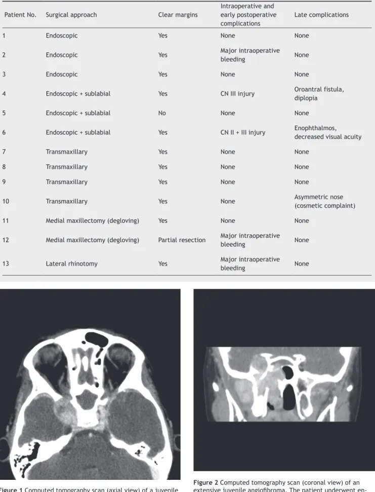
Figure 1 Computed tomography scan (axial view) of a juvenile angioibroma extending into the right cavernous sinus.
Figure 2 Computed tomography scan (coronal view) of an
extensive juvenile angioibroma. The patient underwent en -doscopic-assisted surgery.
Table 1 Summary of surgical routes and postoperative complications.
Patient No. Surgical approach Clear margins
Intraoperative and early postoperative
complications
Late complications
1 Endoscopic Yes None None
2 Endoscopic Yes Major intraoperative
bleeding None
3 Endoscopic Yes None None
4 Endoscopic + sublabial Yes CN III injury Oroantral fistula,
diplopia
5 Endoscopic + sublabial No None None
6 Endoscopic + sublabial Yes CN II + III injury Enophthalmos,
decreased visual acuity
7 Transmaxillary Yes None None
8 Transmaxillary Yes None None
9 Transmaxillary Yes None None
10 Transmaxillary Yes None Asymmetric nose
(cosmetic complaint)
11 Medial maxillectomy (degloving) Yes None None
12 Medial maxillectomy (degloving) Partial resection Major intraoperative
bleeding None
13 Lateral rhinotomy Yes Major intraoperative
Massive intraoperative bleeding was the most common intraoperative complication, occurring in 23% of cases (3/13). Massive bleeding was deined as that leading to he
-modynamic instability and requiring packed red blood cell transfusion. In one patient, the procedure had to be halted due to persistent hypovolemic shock despite transfusion, and only partial resection of the tumor was achieved.
Immediate complications of surgery occurred in two patients. One developed oculomotor nerve palsy with CT indings indicative of cavernous sinus thrombosis, and the other sustained optic and oculomotor nerve injury ma -nifesting as decreased visual activity and ptosis. In both
cases, a combined surgical approach had been employed. The patient with oculomotor nerve palsy had no impro
-vement at the 12-month follow-up. The condition of the second patient was also unchanged, with persistent palsy, at the 22-month follow-up.
None of the patients who underwent fully endoscopic
surgery developed any immediate or delayed complications. Tumor recurrence occurred in 46% of patients (Table 5). The disease-free period ranged from 10 to 36 months pos
-toperatively. Of these six patients, three (3/13) underwent a second surgical procedure, one (1/13) received radiation therapy, and one (1/13) was simply submitted to further observation. Detection of residual tumor on postoperative
follow-up was the criterion for reoperation. The patient who
received radiation therapy had an extensive residual lesion that was not amenable to surgical removal by any route. Fi
-nally, watchful waiting was chosen as the most appropriate course of action for the remaining patient, due to an absen
-ce of symptoms or tumor growth during follow-up, which
was probably due to the absence of residual vascularity.
Figure 3 Magnetic resonance imaging (coronal view) of the
same patient as Figures 1 and 2. Figure 4the extent of resection and a tumor recurrence. Postoperative computed tomography scan showing
Table 2 Radkowski classiication of juvenile nasopharyngeal angioibroma.10
Ia Limited to nose and/or nasopharynx
Ib Same as Ia, but with extension into one or more paranasal sinuses
IIa
Minimal extension through the sphenopalatine foramen, into and including a minimal part of the medial-most part of the pterygomaxillary
fossa.
IIb
Full occupation of the pterygomaxillary fossa, anterior displacement of the posterior wall of the maxillary antrum. Lateral and/or anterior displacement of branches of the maxillary artery. Superior extension may occur, eroding
orbital bones.
IIc
Extension through the pterygomaxillary fossa into the cheek and temporal fossa, or posterior
to the pterygoid plates.
IIIa Erosion of the skull base with minimal
intracranial extension.
IIIb
Erosion of the skull base with extensive
intracranial extension with or without cavernous
Table 3 Andrews classiication of juvenile nasopharyngeal
angioibroma.8
I
Limited to the nasopharynx and nasal cavity. Bone destruction negligible or limited to the sphenopalatine foramen
II
Invading the pterygopalatine fossa or the
maxillary, ethmoid, or sphenoid sinus with bone
destruction
IIIa Invading the infratemporal fossa or orbital region
without intracranial involvement
IIIb
Invading the infratemporal fossa or orbit with intracranial extradural (parasellar) involvement
IVa
Intracranial intradural tumor without infiltration of the cavernous sinus, pituitary fossa or optic chiasm
IVb
Intracranial intradural tumor with infiltration of the cavernous sinus, pituitary fossa or optic chiasm
Table 4 Cnyderman et al. classiication of juvenile
nasopharyngeal angioibroma.3
I Nasal cavity, pterygopalatine fossa
II Paranasal sinuses, lateral pterygopalatine fossa; no residual vascularity
III Skull base erosion, orbit, infratemporal fossa; no residual vascularity
IV Skull base erosion, orbit, infratemporal fossa; residual vascularity
V M Intracranial extension, residual vascularity; M: medial extension
V L Intracranial extension, residual vascularity; L: lateral extension
Table 5 Summary of tumor recurrences.
Patient No. Surgical approach Clear margins Recurrence
4 Endoscopic +
sublabial Yes Yes
5 Endoscopic +
sublabial No Yes
6 Endoscopic +
sublabial Yes Yes
10 Transmaxillary Yes Yesa
11 Medial maxillectomy (degloving) Yes Yesa
12 Medial maxillectomy
(degloving)
Partial resection Yesb
a Patients who underwent reoperation. b Patient who received radiation therapy.
Discussion
Analysis of these 13 patients suggests that fully
endos-copic surgery is viable even in advanced cases of JNA.6 Endoscopic approaches may thus be considered one of the most signiicant advances in treatment of this neoplasm1
and a veritable paradigm shift in its management.11 With
progress in the learning curve of the endoscopic route and
improvements in the understanding of the complex ana
-tomy of the paranasal sinuses and neurovascular structu
-res of the anterior skull base,11 extended access to the
skull base is becoming increasingly popular, broadening the surgeon’s armamentarium for JNA management with
intracranial invasion.
The endoscope has proved itself to be an important tool for visualizing the entire extent of these tumors, particularly in the region of the superior orbital issure, cavernous sinus, and even in the pterygopalatine fossa,
where the presence of residual lesions is not unusual after
resection of tumors as extensive as those described in the
present study.1,11
Intraoperative endoscopy may thus render many oste
-otomies and bone resections unnecessary, which can de
-crease the risk of changes in facial growth.2 Nevertheless,
removal of the lateral nasal wall and posterior portion of the maxilla is required in some cases; the effect of this in
-tervention on facial growth is unknown.1
According to Onerci et al., lesions extending signii -cantly posterior to the pterygoid plates are poor
candida-tes for endoscopic resection. Other essential requirements for the endoscopic approach include adequate surgeon ex
-perience, availability of proper instruments, performance of preoperative embolization, and the possibility of adding
an open route if necessary.6
In the present study, three patients underwent fully en
-doscopic resection and three underwent combined (open + endoscopic) procedures for resection of Radkowski gra
-de IIIa JNAs.10 The remaining procedures were performed
through conventional external routes. This approach is con
-sistent with the current literature, which recommends that a complete resection through the route associated with the least morbidity should be attempted whenever possible.4,6
Pryor et al.11 found a greater frequency of complications
among patients who underwent open procedures as compa
-red with endoscopic surgery. In this study, immediate com -plications occurred in two patients who underwent surgery
via endoscopic-guided external access. None of the three
patients who underwent fully endoscopic surgery developed
immediate or delayed complications.
Surgical treatment of JNA is fraught with technical chal
-lenges, including the risk of intraoperative bleeding, difi
-culties in tumor dissection, and morbidity associated with the involvement of certain anatomic sites, as well as the risk of recurrence.2 In the present study, only two patients,
both in the endoscopic-guided group, developed persis
-tent postoperative complications: one had diplopia and an oroantral istula, and one had ptosis and decreased visual acuity. The occurrence of irreversible complications in the combined access group may be attributable to the greater surgical dificulty of the affected cases, prompting the de
have been the result of the technical challenges encounte-red during surgery.
In this series, the recurrence rate was 46% (6/13), over a maximum 36-month follow-up. These indings are consistent with those reported in the literature (an overall recurren
-ce rate of 30% to 50%).1 It is noteworthy that the sample
consisted of patients with extensive, intracranially invasive tumors, which could have led to a higher recurrence rate.
Due to the benign nature of JNAs, the vast majority of these tumors remain extradural even when there is intra
-cranial extension,9 and the surgical plane is situated betwe-en important structures such as the internal carotid artery
and cavernous sinus.4 However, lateral extension of the tu
-mor into the infratemporal fossa, the parasellar region, or
the vicinity of the optic nerve can pose challenges to endos-copic resection.9
Even though JNAs are covered by nasopharyngeal muco
-sa, in the authors’ experience identiication of the plane of dissection can be somewhat challenging, particularly when close to the cavernous sinus. In this study, one patient deve
-loped a postoperative decline in visual acuity which remai
-ned at the 22-month follow-up, most likely due to inadver
-tent optic nerve injury during the procedure. The authors believe that this injury may have been secondary to the
heat of electrocoagulation.
Some authors maintain that endoscopic approaches should only be used when the tumor is conined to the nasal cavity, paranasal sinuses, pterygopalatine fossa, or infra
-temporal fossa. When a conventional approach is required for resection of lesions that extend beyond the boundaries of endoscopic visualization, endoscopy should still be used as an adjunct to the external route.12,13 However, the pre -sent authors agree with Andrade et al. that a reassessment of the limits of endoscopic resection of JNA, to include lar
-ger tumors and cases with some intracranial invasion, is re
-quired.2 To the best of the authors’ knowledge, this was one
of the largest series restricted to patients with JNAs with intracranial extension treated exclusively via non-neurosur -gical routes.
The limitations of the present study should also be consi
-dered. As previously noted, this case series was restricted to non-neurosurgical routes; patients who required craniotomy due to tumor extension (Radkowski grade IIIb) were exclu -ded.10 A comparison of these different approaches would be
an interesting subject for future research. However, in view of the indings of this study, fully endoscopic or endoscopic --guided access should be considered even for
advanced-sta-ge tumors with major intracranial extension.
Endoscopic treatment of lesions of the skull base is a
branch of surgery that is currently undergoing rapid
pro-gress. Use of neuronavigation techniques, intraoperative MRI, customized instruments, and ever-increasing surgical experience are all factors contributing to further advance
-ments in this ield, toward safer and more effective endos -copic approaches.13
Conclusion
Operative treatment of nasopharyngeal angioibroma with intracranial extension is one of the major challenges of ENT
and neurosurgical practice. The success rates and low intra-
and postoperative complication rates of endoscopic surgery suggest that this route has been gaining ground in the ma
-nagement of Radkowski IIIa class JNAs.
Conlicts of interest
The authors declare no conlicts of interest.
References
1. Enepekides DJ. Recent advances in the treatment of juve
-nile angioibroma. Curr Opin Otolaryngol Head Neck Surg. 2004;12:495-9.
2. Andrade NA, Pinto JA, Nóbrega MO, Aguiar JE, Aguiar TF, Vinhaes ES. Exclusively endoscopic surgery for juvenile na
-sopharyngeal angioibroma. Otolaryngol Head Neck Surg. 2007;137:492-6.
3. Snyderman CH, Pant H, Carrau RL, Gardner P. A new endosco
-pic staging system for angioibromas. Arch Otolaryngol Head Neck Surg. 2010;136:588-94.
4. Danesi G, Panciera DT, Harvey RJ, Agostinis C. Juvenile na
-sopharyngeal angioibroma: evaluation and surgical mana
-gement of advanced disease. Otolaryngol Head Neck Surg. 2008;138:581-6.
5. Ardehali MM, Samimi Ardestani SH, Yazdani N, Goodarzi H, Bas
-taninejad S. Endoscopic approach for excision of juvenile na
-sopharyngeal angioibroma: complications and outcomes. Am J Otolaryngol. 2010;31:343-9.
6. Onerci TM, Yücel OT, Oğretmenoğlu O. Endoscopic surgery in treatment of juvenile nasopharyngeal angioibroma. Int J Pe
-diatr Otorhinolaryngol. 2003;67:1219-25.
7. Pryor SG, Moore EJ, Kasperbauer JL. Endoscopic versus tradi
-tional approaches for excision of juvenile nasopharyngeal an
-gioibroma. Laryngoscope. 2005;115:1201-7.
8. Andrews JC, Fisch U, Valavanis A, Aeppli U, Makek MS. The surgical management of extensive nasopharyngeal angioibro
-mas with the infratemporal fossa approach. Laryngoscope. 1989;99:429-37.
9. Douglas R, Wormald PJ. Endoscopic surgery for juvenile naso
-pharyngeal angioibroma: where are the limits? Curr Opin Oto
-laryngol Head Neck Surg. 2006;14:1-5.
10. Radkowski D, McGill T, Healy GB, Ohlms L, Jones DT. Angio
-ibroma. Changes in staging and treatment. Arch Otolaryngol Head Neck Surg. 1996;122:122-9.
11. Lund VJ, Stammberger H, Nicolai P, Castelnuovo P, Beal T, Beham A, et al. European position paper on endoscopic ma
-nagement of tumours of the nose, paranasal sinuses and skull
base. Rhinol Suppl. 2010;22:1-143.
12. Ogawa AI, Fornazieri MA, da Silva LV, Pinna FR, Voegels RL, Sennes LU, et al. Juvenile angioibroma: major and mi
-nor complications of preoperative embolization. Rhinology. 2012;50:199-202.
13. Carrau RL, Snyderman CH, Kassam AB, Jungreis CA. Endosco