Effect of thickener agents on dental
enamel microhardness submitted to
at-home bleaching
Efeito de agentes espessantes na microdureza
do esmalte submetido ao clareamento
dental caseiro
Abstract: Dental bleaching occurs due to an oxidation reaction between the bleaching agents and the macromolecules of pigments in the teeth. This reaction is unspeciic and the peroxides can also affect the dental matrix causing mineral loss. On the other hand, recent studies have suggested that the thickener agent carbopol can also cause mineral loss. Thus, the objective of this study was to evaluate in vitro the effect of at-home den-tal bleaching on denden-tal enamel microhardness after the use of bleaching agents with and without carbopol as a thickener agent. Bovine dental slabs with 3 x 3 x 3 mm were ob-tained, sequentially polished, and randomly divided into 4 groups according to the ex-perimental treatment: G1: 2% carbopol; G2: 10% carbamide peroxide with carbopol; G3: carbowax; G4: 10% carbamide peroxide with poloxamer. Bleaching was performed daily for 4 weeks, immersed in artiicial saliva. Enamel microhardness values were obtained before the treatment (T0) and 7 (T1), 14 (T2), 21 (T3), 28 (T4), and 42 (T5) days after the beginning of the treatment. ANOVA and Tukey’s test revealed statistically signiicant dif-ferences only for the factor Time (F = 5.48; p < 0.01). All bleaching and thickener agents caused no alterations on the enamel microhardness.
Descriptors: Dental enamel; Hardness tests; Tooth bleaching; Thickeners/adverse ef-fects.
Resumo: O clareamento dental ocorre devido a uma reação de oxidação entre o agente clareador e as macromoléculas de pigmentos presentes nos dentes. Esta reação é inespecí-ica e o peróxido pode agir na matriz dental causando perdas de mineral. Por outro lado, estudos recentes sugerem que o agente espessante carbopol também pode causar perda mineral. Assim, o objetivo deste trabalho foi avaliar in vitro o efeito do clareamento casei-ro sobre a miccasei-rodureza do esmalte após o uso de agentes clareadores com e sem carbopol como espessante. Fragmentos de esmalte bovino de 3 x 3 x 3 mm foram obtidos, polidos seqüencialmente e aleatoriamente divididos em 4 grupos de acordo com o tratamento ex-perimental: G1: carbopol a 2%; G2: peróxido de carbamida a 10% com carbopol; G3: carbowax; G4: peróxido de carbamida a 10% com poloxamer. O clareamento foi realiza-do diariamente por 4 semanas em saliva artiicial. A microdureza realiza-do esmalte foi avaliada antes (T0) e após 7 (T1), 14 (T2), 21 (T3), 28 (T4), e 42 (T5) dias do início do tratamento. A ANOVA e o teste de Tukey revelaram diferenças estatísticas signiicantes somente para o fator Tempo (F = 5,48; p < 0,01). Os agentes clareadores e espessantes não causaram alte-rações na microdureza do esmalte.
Descritores: Esmalte dentário; Testes de dureza; Clareamento de dente; Espessantes/efei-tos adversos.
José Augusto Rodrigues(a)
Glauco Paulo Felício Oliveira(b)
Cristiane Mariote Amaral(a)
(a)PhDs, Assistant Professors, Dental Research
and Graduate Studies Division, Department of Restorative Dentistry; (b)Undergraduate
Dentistry Student – Guarulhos University, Guarulhos.
Corresponding author: José Augusto Rodrigues Universidade de Guarulhos CEPPE – Centro de Pós Graduação, Pesquisa e Extensão
Pça. Tereza Cristina, 58 Centro - Guarulhos - SP - Brazil CEP: 07023-070
E-mail: [email protected]
Introduction
Discolored teeth are considered a major problem by society and they are incorrectly attributed to dis-eases.8 As a solution for this kind of problem there
are many options like crowns, laminated veneers, enamel microabrasion, and dental bleaching.8 Among
these techniques, dental bleaching is the most con-servative, easy, and commonly used by clinicians.8
At-home bleaching has become one of the most used techniques because of its simplicity, low cost and safety due to the use of low concentrations of peroxide (10% to 16% carbamide peroxide).8
Al-though 10% carbamide peroxide is still currently commonly used, manufacturers have recently be-gun to introduce higher concentrations of bleaching agents and alternative methods for tooth bleaching to achieve faster results.8,11
Teeth are bleached by an oxidation-reduction re-action caused by the decomposition of the hydrogen peroxide into free radicals such as oxygen and per-hydroxyl.8,13 Because of the absence of an electron
on the last layer, the oxygen (O•) and perhydroxyl
(HO2•) – free radicals – are extremely electrophilic
and diffuse throughout enamel and dentin matrix to attack the macromolecules of pigments (organic molecules) and acquire stability.7,8 Then, the
in-trinsic pigments composed by highly unsaturated organic macromolecules are transformed into less complex molecules, which are smaller, simpler and lighter than the original ones.8
However, this reaction is unspeciic, and the or-ganic and inoror-ganic matrix of the enamel and den-tin might be affected by the bleaching agents, and undesirable effects such as topography alterations and mineral loss may occur.3,5,9,13,14,16,18,19,22,23
On the other hand, recent studies suggest that such alterations are not only related to the peroxide application.3,19 Few researches have observed in in
vitro or in situ studies that treatment with carbopol (the most used thickener agent) may cause a decrease of enamel microhardness.3,14,19 However, there is no
conclusive data about the effects of carbopol. The action of carbopol should be better investigated be-cause it may be synergistically acting with free radi-cals and may further increase the mineral loss pro-duced by the use of other agents.
The aim of this study was to evaluate the effect of an in vitro treatment with 10% carbamide per-oxide bleaching agents with and without carbopol, and the effect of the treatment with 2% carbopol on dental enamel microhardness.
Material and Methods
Preparation of the dental slabsAfter extraction, 40 bovine teeth were stored in a 0.1% thymol solution. They were submitted to a soft-tissue debridement and the crowns were sec-tioned to obtain 72 dental slabs with 3 x 3 x 3 mm using double-faced diamond discs (KG Sorensen, Barueri, SP, Brazil). The dental slabs were individu-ally embedded in autopolimerizing polystyrene resin (Cromex, São Paulo, SP, Brazil) in a PVC ring mold, allowing only one side of the dental slab to be left un-sealed. The enamel surface of the slabs was serially polished by 400, 600, and 1,000 grit Al2O3 abrasive paper (Carborundum Abrasivos, Recife, PE, Brazil) with water as a cooler to obtain lat, standardized enamel surfaces. Subsequently, they were polished with diamond pastes of 6, 3, and 1 µm and polish-ing cloths with a mineral oil lubricant (Arotec Ind. Com. Ltda., Cotia, SP, Brazil). After that, the speci-mens were maintained in deionized distilled water until the microhardness test was performed.
Microhardness tests
Experimental treatment
For the experimental treatments, the specimens were randomized into four groups (n = 18) as pre-sented in Table 1.
The in vitro at-home bleaching was performed as described by Rodrigues et al.18 (2001), and used by
Basting et al.3 in 2003, and Basting et al.2 in 2005.
The respective treatment agent was applied for 6 hours a day covering the dental slabs with 0.2 ml of the material. The specimens were placed in vacuum-formed custom trays and immersed in a closed plas-tic container with artiicial saliva at 37°C.
After the bleaching period, the treatment agent was removed under running deionized and distilled water. For the remaining time of the day, the specimens were individually kept in 11 ml of artiicial saliva that was daily changed. The artiicial saliva was used as Feath-erstone et al.6 (1986) proposed, according to what
Serra, Cury20 (1992) described, containing 50 mmol/l
KCl, 1.5 mmol/l Ca, 0.9 mmol/l PO4, 20 mmol/l tri-hydroxymethyl-aminomethane buffer at pH 7.0. After 28 days of the experimental treatments, the specimens were stored in artiicial saliva for additional 14 days.
Statistical analysis
The measurements of the indentations, in microm-eters, taken from each specimen at each time were used to obtain the Knoop hardness number (KHN) by the following calculation: KHN = (14.23 × 10³× F)/
d2, where F is the applied load value (g) and d is the
diagonal indentation (µm).
The means of the 5 Knoop hardness (KHN) measurements from each specimen, made initially and following treatment, were statistically analyzed. Data were subjected to split plot analysis of vari-ance, which showed statistically signiicant differ-ences, followed by Tukey’s test (α = 0.05).
Results
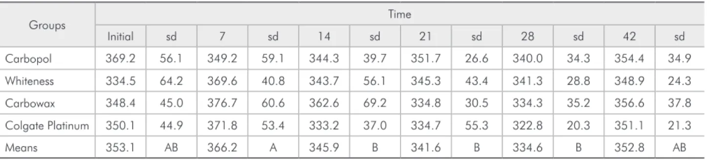
The mean values of enamel microhardness, be-fore and following the treatments, and the respective standard deviations are shown in Table 2, and the behavior of enamel microhardness may be observed in Graph 1. The ANOVA and Tukey’s tests revealed no statistically signiicant differences regarding the factor experimental treatment (F = 0.37; p = 0.78). Statistically signiicant differences were found for the factor time (F = 5.48; p < 0.01), therefore all groups had the same behavior over time. There was no statistical signiicant differences for the interac-tion between the factors experimental treatment and time (F = 1.35; p = 0.16).
Table 1 - Experimental groups, bleaching agents and com-position.
Groups Bleaching agent Composition
G1 - 2% Carbopol
G2 10% carbamide peroxide (Whiteness Perfect)
Neutralized carbopol, potassium nitratum, sodium fluoride, glycol, and deionized water.
G3 - Carbowax
G4 10% carbamide peroxide (Colgate Platinum Overnight)
Calcium pyrophosphate, water, poloxamer 407, PEG 12, glycerin, dicalcium phosphate dihydrate, polyethylene oxide, sodium acid pyrophosphate, flavor, sodium lauryl sulfate, sodium saccharin, and disodium dihydrate EDTA.
Table 2 - Mean values of initial and final Knoop microhardness measurements (KHN), and standard deviations (sd) for each group at each time.
Groups Time
Initial sd 7 sd 14 sd 21 sd 28 sd 42 sd
Carbopol 369.2 56.1 349.2 59.1 344.3 39.7 351.7 26.6 340.0 34.3 354.4 34.9
Whiteness 334.5 64.2 369.6 40.8 343.7 56.1 345.3 43.4 341.3 28.8 348.9 24.3
Carbowax 348.4 45.0 376.7 60.6 362.6 69.2 334.8 30.5 334.3 35.2 356.6 37.8
Colgate Platinum 350.1 44.9 371.8 53.4 333.2 37.0 334.7 55.3 322.8 20.3 351.1 21.3
Means 353.1 AB 366.2 A 345.9 B 341.6 B 334.6 B 352.8 AB
The Tukey test showed statistical differences among the studied times. Although at T1 all groups did not differ statistically from T0, there was a nu-merical increase in mean microhardness, except for G1.
From T1 to T2, T3, and T4, the mean values of microhardness decreased and statistically differed from that at T1. However, the data obtained at T2, T3, and T4 were statistically similar to that at T0.
From T4 to T5, all groups showed a non statistical increase in the means of microhardness. The data obtained at T5 did not differ from that obtained at the other times.
Discussion
The effects of bleaching agents and their com-pounds on teeth have been widely discussed by re-searchers.2,12-14,19 Bleaching agents can permeate the
tooth structure and break the molecules of stain.8
However, this reaction is not speciic and the matrix of enamel and dentin may be affected.
Several studies have demonstrated surface alter-ations in the enamel exposed to carbamide perox-ide bleaching agentslike pittings,10,21 erosions,15,21
porosities,10,15 waviness,15,21 surface roughness
in-crease,15,25 and dissolutions1 similar to those of
ini-tial caries.17 These effects are directly related to the
time of exposure and concentration of the active agent.11 The longer the exposure and higher the
concentration, the greater the effect of the oxida-tion process, the color change8 and also the side
ef-fects.11
The experimental design employed in this study consisted in the exposure for 28 days of the enam-el slabs to the bleaching or thickener agents for 6 hours a day, similarly to clinical night use, followed by immersion in an artiicial saliva solution for 18 hours. Immersion in the artiicial saliva solution, which contains calcium and phosphate, might mote a remineralization effect similar to that pro-duced in human saliva, which is very important to make the results closer to those obtained in clinical conditions.
A not statistically signiicant increase in enamel microhardness was found from T0 to T1. This phe-nomenon was not expected. It may, however, have happened because of the immersion of the speci-mens in the deionized distilled water, which causes some demineralization effect, before the beginning of the treatment. Thus, in the irst week, the use of artiicial saliva may have had promoted remineral-ization of the specimens.
Despite the use of artiicial saliva, the experi-mental groups of this study suffered microhard-ness loss from T1 to T2, T3, and T4, showing that the artiicial saliva solution was not able to totally revert the detrimental effects caused by the continu-ous use of the bleaching agents, and some enamel demineralization may be expected. This inding did not agree with those of several authors that showed no alterations on enamel after exposure to bleach-ing.1,4,9,12,14,21,22 However, most of them are
unani-mous in noticing the occurrence of enamel altera-tions by SEM.5,9,12,21,23
On the other hand, Basting et al.3, in 2003,
showed that an artiicial saliva solution cannot re-establish the initial enamel hardness during the use of bleaching agents for longer periods, but after the end of the treatment the remineralization potential of this solution can reestablish enamel microhard-ness. Such effect was also observed in the present study at T5 (post bleaching period) and may also be expected in clinical conditions of less intense demin-eralization because of the presence of natural saliva and luoride exposure.
Graph 1 - Means of Knoop microhardness values for each group over time.
0 50 100 150 200 250 300 350 400 450
G1 - 2% carbopol G2 - Whiteness perfect G3 - Carbowax G4 - Colgate Platinum
Time (days)
Means
o
f
K
no
o
p
micro
hardnes
s
Demineralization is also related to the viscosi-ties of the bleaching agents.2,13,19,22 Thickener agents
like carbopol cause detrimental effects on enamel over the treatment time.2,13,19,22 In this study, the
ap-plication of 2% carbopol or carbowax caused no effect on enamel microhardness that could not be ixed by the remineralizing potential of the artiicial saliva. This result is not in agreement with those of previous studies, which have observed a statistical signiicant reduction in enamel microhardness2,19
after treatment with the thickener agent (carbopol). McCraken, Haywood14 (1995) showed lower
val-ues of enamel microhardness after the use of 10% carbamide peroxide and carbopol when compared to the use of 10% carbamide peroxide without a thickener agent.14
Basting et al.3 (2003) showed a loss in dental
enamel microhardness after the in vitro treatment with the thickener agent carbopol. Rodrigues et al.19
(2005)have demonstrated a reduction in human dental enamel microhardness after in situ treatment with carbopol for 3 weeks. Furthermore, a research conducted by Basting et al.2 (2005) aimed to
com-pare the microhardness of enamel after a treatment with 10% carbamide peroxide, carbopol, and glyc-erin, and it revealed that the association of those agents produced a decrease in sound enamel micro-hardness. However, these authors used carbopol for 8 hours per day with 16 hours in artiicial saliva or in natural saliva. Thus, the detrimental effect they found may be related to the exposure time, which in the present study was of 6 hours followed by 18 hours in artiicial saliva per day.
Carbopol is an exceptional thickener, suspend-ing agent and stabilizer, utilized in a wide variety of personal care products at concentrations lower than 1%. Carbopol was added to the bleaching agents to change the carbamide peroxide formulation, that was liquid, into a gel to avoid a greater loss of the bleaching material from the tray,1 to prolong the
release of hydrogen peroxide from the carbamide peroxide and to enhance the contact between the peroxide and the tooth. It also triples or quadruples the active release time of peroxide. However, it is an acidic polymer that may cause demineralization, and it has a high calcium-binding capacity that can inhibit hydroxyapatite crystal growth.24
The carbowax applied in G3 is a polymer of eth-ylene oxide and water and their ethers (polyetheth-ylene glycol), and it is very used as a surfactant in indus-try, including foods, cosmetics and pharmaceutics. It was used here as an alternative thickener agent based on the formulation of the bleaching agent of G4, whose thickener agent is poloxamer, a co-poly-mer of polyethylene glycol used as an emulsifying, surfactant, and wetting agent similar to carbowax.
Manufacturers have made no claims about the action of the thickener agents, so these agents have generally been considered inactive ingredients. They could, however, act as demineralizing agents14 or as
an impermeable barrier, inhibiting the penetration of the artiicial saliva solution through the enamel surface and avoiding the restoration of normal mi-crohardness values.3
This study showed that the in vitro use of carbo-pol or carbowax caused no mineral loss, but further studies should be conducted to assess the effect of thickener agents for longer exposure times and also to study other options of thickener agents that may cause less effects on enamel.
Conclusions
Under the experimental conditions of this study, it can be concluded that at-home dental bleaching with 10% carbamide peroxide and the treatment with 2% carbopol or carbowax for 6 hours daily did not statistically reduce enamel microhardness. If some microhardness loss occured it could be revert-ed by the action of artiicial saliva in a post-bleach-ing period.
References
1. Akal N, Over H, Olmez A, Bodur H. Effects of carbamide peroxide containing bleaching agents on the morphology
2. Basting RT, Rodrigues AL Jr, Serra MC. The effect of 10% carbamide peroxide, carbopol, and/or glycerin on enamel and dentin microhardness. Oper Dent. 2005;30(5):608-16. 3. Basting RT, Rodrigues AL Jr, Serra MC. The effects of seven
carbamide peroxide bleaching agents on enamel microhard-ness over time. J Am Dent Assoc. 2003;134(10):1335-42. 4. Basting RT, Rodrigues Junior AL, Serra MC. The Effect of
10% carbamide peroxide bleaching material on microhardness of sound and demineralized enamel and dentin in situ. Oper Dent. 2001;26(6):531-9.
5. Bitter NC. A scanning electron microscopy study of the effect of bleaching agents on enamel: a preliminary report. J Prosthet Dent. 1992;67(6):852-5.
6. Featherstone JDB, O’Reilly MM, Shariat M. Enhancement of remineralization in vitro and in vivo. In: Leach AS. Factors relating to demineralization and remineralization of teeth. Oxford: IRL Press; 1986. p. 23-4.
7. Frysh H, Bowles WH, Baker F, Rivere-Hidalgo F, Guillen G. Effect of pH on hydrogen peroxide bleaching agents. J Esthet Dent. 1995;7(3):130-3.
8. Goldstein RE, Garber DA. Complete dental bleaching. Chi-cago: Quintessence Books; 1995. 165 p.
9. Hegedüs C, Bistley T, Flóra-Nagy E, Keszthelyi G, Jenei A. An atomic force microscopy study on the effect of bleaching agents on enamel surface. J Dent. 1999;27(7):509-15. 10. Leonard RH, Bentley CD, Haywood VB. Salivary pH changes
during 10% carbamide peroxide bleaching. Quintessence Int. 1994;25(8):547-50.
11. Leonard RH, Sharma A, Haywood VB. Use of different con-centrations of carbamide peroxide for bleaching teeth: an in vitro study. Quintessence Int. 1998;29(8):503-7.
12. Lopes GC, Bonissoni L, Baratieri LN, Vieira LCC, Monteiro S Jr. Effect of bleaching agents on the hardness and morphology of enamel. J Esthet Restor Dent. 2002;14(1):24-30.
13. McCracken MS, Haywood VB. Demineralization effects of 10 percent carbamide peroxide. J Dent. 1996;24(6):395-8. 14. McCracken MS, Haywood VB. Effects of 10% carbamide
peroxide on the subsurface hardness of enamel. Quintessence Int. 1995;26(1):21-4.
15. McGuckin RS, Babin JF, Meyer BJ. Alterations in human enamel surface morphology following vital bleaching. J Pros-thet Dent. 1992;68(5):754-60.
16. Oltu Ü, Gürgan S. Effects of three concentrations of carba-mide peroxide on the structure of enamel. J Oral Rehabil. 2000;27(4):332-40.
17. Potocnik I, Kosec L, Gaspersic D. Effect of 10% carbamide peroxide bleaching gel on enamel microhardness, microstruc-ture, and mineral content. J Endod. 2000;26(4):203-6. 18. Rodrigues JA, Basting RT, Serra MC, Rodrigues Junior AL.
Effects of 10% carbamide peroxide bleaching on enamel microhardness at different bleaching times. Am J Dent. 2001;14(2):67-71.
19. Rodrigues JA, Marchi GM, Ambrosano GMB, Heymann HO, Pimenta LAF. Microhardness evaluation of in situ vital bleaching on human dental enamel using a novel study design. Dent Mater. 2005;21(11):1059-67.
20. Serra MC, Cury JA. The in vitro effect of glass-ionomer cement restoration on enamel subjected to a demineralization and remineralization model. Quintessence Int. 1992;23(2):143-7.
21. Shannon H, Spencer P, Gross K, Tira D. Characterization of enamel exposed to 10% carbamide peroxide bleaching agents. Quintessence Int. 1993;24(1):39-44.
22. Smidt A, Weller D, Roman I, Gedalia I. Effect of bleaching agents on microhardness and surface morphology of tooth enamel. Am J Dent. 1998;11(2):83-5.
23. Tong LS, Pang MK, Mok NV, King NM, Wei SH. The effects of etching, micro-abrasion, and bleaching on surface enamel. J Dent Res. 1993;72(1):67-71.
24. van der Reijden WA, Buijs MJ, Damen JJ, Veerman EC, ten Cate JM, Nieuw Amerongen AV. Influence of polymers for use in saliva substitutes on de- and remineralization of enamel
in vitro. Caries Res. 1997;31(3):216-23.