EBV-1 and HCMV in aggressive
periodontitis in Brazilian patients
EBV-1 e HCMV na periodontite agressiva em
pacientes brasileiros
Abstract: The purpose of the present investigation was to compare the presence of Ep-stein-Barr virus type 1 (EBV-1) and of Human Cytomegalovirus (HCMV) in crevicu-lar luid samples from deep and shallow periodontal pocket sites of Brazilian patients with aggressive periodontitis. A total of 30 systemically healthy patients with aggressive periodontitis participated in the study. Paper points were inserted into 2 gingivitis sites (≤ 3 mm) and into 2 periodontitis sites (≥ 5 mm) in each patient. PCR assay was used to identify genomic copies of HCMV and EBV-1. Twenty-three patients (77%) were positive for EBV-1, while only 2 patients (6%) were positive for HCMV. The McNemar test re-vealed a positive association between EBV-1 and periodontal lesions (p = 0.043). Thirty-four (57%) out of 60 periodontitis sites were positive for EBV-1, whereas 18 (30%) gin-givitis sites were positive (p = 0.01). Only two sites (6.7%) were positive for HCMV. No positive association was found between HCMV and periodontitis or gingivitis (p = 0.479). The elevated occurrence of EBV-1 DNA in periodontal pockets of patients with aggressive periodontitis supports a possible periodontopathic role of this virus.
Descriptors: Epstein-Barr virus; Cytomegalovirus; Periodontitis.
Resumo: O objetivo do presente estudo foi comparar a presença do vírus Epstein-Barr tipo 1 (EBV-1) e do Citomegalovírus Humano (HCMV) em amostras de luido crevicu-lar de bolsas periodontais rasas e profundas de pacientes brasileiros com periodontite agressiva. Trinta pacientes sistemicamente saudáveis com periodontite agressiva partici-param deste estudo. Cones de papel foram inseridos em 2 sítios de gengivite (≤ 3 mm) e em 2 sítios de periodontite (≥ 5 mm) de cada paciente. Reações de PCR foram usadas para identiicar cópias de DNA genômico de HCMV e EBV-1. Em 23 pacientes (77%), os testes foram positivos para EBV-1, enquanto apenas 2 pacientes (6%) foram positivos para HCMV. O teste de McNemar apontou associação positiva entre EBV-1 e lesões periodon-tais (p = 0,043). Trinta e quatro (57%) dos 60 sítios de periodontites foram positivos para o EBV-1, enquanto 18 (30%) dos sítios de gengivites foram positivos (p = 0,01). Apenas 2 sítios (6,7%) foram positivos para o HCMV. Não foi encontrada associação positiva entre HCMV e periodontite ou gengivite (p = 0,479). A alta ocorrência de DNA de EBV-1 em bolsas periodontais de pacientes com periodontite agressiva corrobora a possível função periodontopática deste vírus.
Descritores: Herpesvirus 4 humano; Citomegalovírus; Periodontite. Soraia Almeida Watanabe(a)
Jeane de Fátima Correia-Silva(a) Martinho Campolina Rebello Horta(b) José Eustáquio da Costa(a)
Ricardo Santiago Gomez(a)
(a) MScs, Department of Oral Surgery and
Pathology, School of Dentistry, Federal University of Minas Gerais.
(b) PhD, School of Dentistry, Pontifical Catholic
University of Minas Gerais.
Corresponding author:
Ricardo Santiago Gomez
Faculdade de Odontologia da UFMG Depto. de Clínica, Patologia e Cirurgia Av. Antonio Carlos, 6627
Belo Horizonte - MG - Brazil CEP: 31270-901
E-mail: [email protected]
Introduction
Little is known about the conversion of gingivi-tis sites into periodontigingivi-tis sites21 or why periodontal tissue breakdown progresses in a localized and bi-laterally symmetrical pattern.4,15 Many studies have suggested that the coexistence of periodontal her-pesviruses, periodontopathic bacteria and the local host immune response are involved in periodontal disease evolution.3,6,10,12,19,20
The association of herpesviruses with oral dis-ease is not a novel issue. EBV is closely related to Burkitt’s lymphoma, nasopharyngeal carcinoma and hairy leukoplakia.8 More recently, the HCMV has been associated with periodontal abscesses.20
Genomes of two types of herpesviruses, HCMV and EBV, occur at high frequency in progressive periodontitis in adults,12,27 localized24 and general-ized aggressive periodontitis,12,20,24 HIV-periodon-titis,4,24 acute necrotizing gingivitis,3 periodontal abscesses,20 Pappilon Lefrève periodontitis26 and 21 trisomic periodontitis.9 Rones et al.18 (1983) demon-strated a signiicant positivity of both epithelial and ibroblast cells from the gingival sulcus area for the herpes simplex virus in vitro, suggesting that those cells could be a reservoir for the latent virus. The involvement of herpesviruses in the etiology of peri-odontal disease was suggested by its presence in gin-gival tissue, gingin-gival crevicular luid and subgingin-gival plaque affected by periodontal disease.6 However, the role of HCMV and EBV-1 in periodontal disease pathogenesis remains to be elucidated.
The purpose of the present investigation was to compare the presence of genomic sequences of HCMV and EBV-1 in crevicular luid samples from deep and shallow periodontal pockets sites of Bra-zilian patients with aggressive periodontitis.
Material and Methods
Patients
The study included 30 individuals with aggres-sive periodontitis (18 females, 12 males) aged 18 to 45 years (mean age: 32.4 years). Subjects were re-cruited at the Periodontics Clinic, School of Dentist-ry, Federal University of Minas Gerais, MG, Brazil. Inclusion factors were, according to Tonetti, Mom-belli25: Patients who were clinically healthy,
present-ing rapid attachment loss and bone destruction and family aggregation. We included patients who were older than 35 years of age only when they presented the three common features of aggressive periodonti-tis. Indeed, according to Tonetti, Mombelli25 (1999), this form of disease can occur at any age and is not necessarily conined to individuals under the arbi-trarily chosen age of 35. Subjects who had received antibiotics for at least 6 months prior to the study were excluded from the examination. Nineteen out of 30 patients were not submitted to any kind of periodontal treatment before sample collection. The study protocol was approved by the local Ethics Committee. Informed consent was obtained from all the subjects.
Sample collection preparation
Sample collection was obtained from each pa-tient from the 2 deepest periodontal sites of the den-tition with probing depth (PD) ≥ 5 mm and clinical attachment level ≥ 4 mm and from 2 gingivitis sites with PD ≤ 3 mm, bleeding upon probing and clini-cal absence of attachment and bone loss. Probing depth was deined as the distance from the gingi-val margin to the most apical probing depth of the gingival/periodontal pocket and was rounded to the nearest higher millimeter.
All sample sites exhibited gingival inlamma-tion and bleeding upon sampling. One examiner performed these clinical procedures. Into each gin-gival/periodontal pocket a sterile ine paper point was inserted for 30 seconds. After being imme-diately suspended in 400 ml Krebs buffer (20% NaCl, 2% KCl, 2% CaCl2 2H2O, MgSO4,KH2PO4, C6H12O6), the pellet was subjected to 10 min of cen-trifugation at 10 g, being thereafter stored at –20°C until processing. The DNA extraction was carried out as described by Boom et al.2 (1990) and stored at –20ºC.
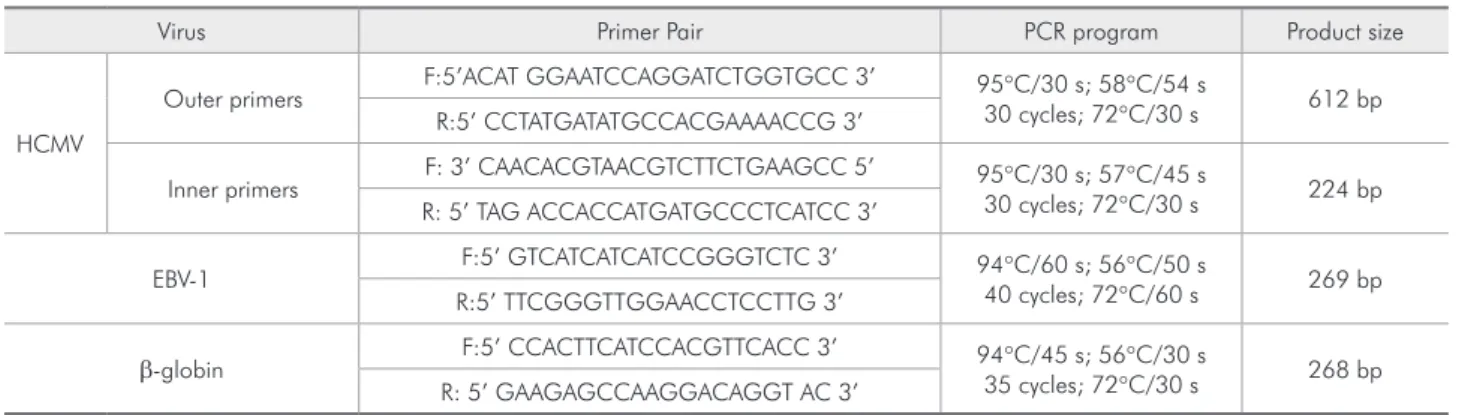
PCR ampliication was performed in a thermal cycler (Gene Amp. PCR System 2400) (Eppendorf, Westbury, NY, USA), and included an initial dena-turation, followed by cycles of denadena-turation, primer annealing, and an extension, ending with a inal extension (Table 1). Positive and negative (PCR re-agents without DNA) controls were routinely in-cluded. PCR for HCMV was carried out in a 25 µl mixture containing Taq DNA polymerase (1 unit/re-action) (Phoneutria, Belo Horizonte, MG, Brazil), PCR buffer (40 mM NaCl, 10 mM Tris-Cl pH 8.4, 2.5 mM MgCl2), 0.1 mM deoxynucleosidetriphos-phates (Amersham Biosciences, Buckinghamshire, UK), 10 pmol/reaction primers and genomic DNA. After PCR, 2 ml of the inal product were trans-ferred to the reaction mixture of the second PCR and reampliied with the inner pair of primers. For identiication of EBV-DNA, a mixture of 25 µl con-taining Taq DNA polymerase (1 unit/reaction), PCR buffer (10 mM (NH4)2SO4, 10 mM KCl, 10 mM Tris-Cl pH 8.4, 3.0 mM MgCl2), 0.1 mM deoxy-nucleosidetriphosphates, 10 pmol/reaction primers and genomic DNA was used. Primers and PCR con-ditions are shown in Table 1.
Electrophoresis
HCMV PCR products were visualized with UV light as a single band (224 bp) by staining with ethidium bromide after 1.5% agarose gel electro-phoresis. The EBV ampliied products (269 bp) were visualized by 6.5% polyacrilamide gel electrophore-sis and silver stained.
Statistical analysis
Differences in viral prevalence between gingivitis sites and periodontal pockets within the same sub-ject were determined using the McNemar test. Chi-square and Exact Fisher’s tests were used to test the independent samples. p values equal to or less than 0.05 were considered statistically signiicant.
Results
The distribution of patients according to the presence of EBV-1 in gingivitis and periodontitis sites is shown in Table 2. Patients that showed the presence of EBV-1 in at least one gingivitis or peri-odontitis site were considered positive. Twenty-three out of 30 (77%) patients demonstrated the presence of EBV-1 DNA. Ten patients presented simultane-ously positive periodontitis sites and negative gingi-vitis sites for EBV-1. In addition, only two patients presented positive gingivitis sites together with nega-tive periodontitis sites for this virus. The McNemar test revealed a positive association between EBV-1 and periodontitis lesions (p = 0.043).
The distribution of gingivitis and periodontitis sites according to the presence of EBV-1 is shown in Table 3. EBV-1 DNA was identiied in 52 (43%) gin-givitis and periodontitis sites (Table 3). Thirty-four (57%) out of 60 periodontitis sites were positive for EBV-1, whereas 18 (30%) gingivitis sites were positive for the same virus. The difference between periodontitis sites and gingivitis sites according to the presence of EBV-1 was statistically signiicant (p = 0.0057). The periodontitis sites showed an
in-Table 1 - Primers and PCR conditions.
Virus Primer Pair PCR program Product size
HCMV
Outer primers F:5’ACAT GGAATCCAGGATCTGGTGCC 3’ 95°C/30 s; 58°C/54 s
30 cycles; 72°C/30 s 612 bp R:5’ CCTATGATATGCCACGAAAACCG 3’
Inner primers F: 3’ CAACACGTAACGTCTTCTGAAGCC 5’ 95°C/30 s; 57°C/45 s
30 cycles; 72°C/30 s 224 bp R: 5’ TAG ACCACCATGATGCCCTCATCC 3’
EBV-1 F:5’ GTCATCATCATCCGGGTCTC 3’ 94°C/60 s; 56°C/50 s
40 cycles; 72°C/60 s 269 bp R:5’ TTCGGGTTGGAACCTCCTTG 3’
β-globin F:5’ CCACTTCATCCACGTTCACC 3’ 94°C/45 s; 56°C/30 s
35 cycles; 72°C/30 s 268 bp R: 5’ GAAGAGCCAAGGACAGGT AC 3’
creased prevalence of EBV-1 compared with the gin-givites sites (OR: 3.05, 95% CI: 1.43-6.47).
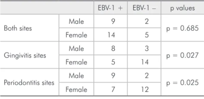
The male patients presented more positive sites for EBV-1 than female patients in both groups, gin-givitis (p = 0.027) and periodontitis (p = 0.025), as shown in Table 4.
No signiicant association was observed between the prevalence of EBV-1 and age (p = 0.371). The presence of HCMV and EBV-1 was not associated with a previous history of periodontal treatment be-fore samples’ collection (p = 0.151).
HCMV DNA was found in only 2 (6%) patients. Two sites (6.7%) were positive for HCMV: one posi-tive sample occurred in a gingivitis site and the other occurred in a periodontitis site. No positive associa-tion was found between HCMV and periodontitis or gingivitis (p = 0.479). Only one site exhibited both viruses.
Discussion
In a model for herpesviruses-related periodonti-tis, Slots, Contreras22 (2000) depicted that herpesvi-rus infection of periodontal sites may be important in a multistage pathogenesis by altering local host responses. Initially, bacterial infection of the gingiva causes inlammatory cells to enter the gingival tis-sue, with periodontal macrophages and T-lympho-cytes harboring latent EBV. Reactivation of
herpes-viruses from latency may occur spontaneously or during periods of impaired host defense, resulting from immunosuppression, infection, physical trau-ma, hormonal changes, etc. Herpesvirus activating factors are also risk factors/indicators for periodon-tal disease.16
Contreras et al.3 (1997) demonstrated an in-creased occurrence of suspected periodontal patho-gens in periodontal sites showing HCMV or EBV. A shift from latent to active virus infection of the periodontium might partly explain subgingival overgrowth of some periodontal pathogens and the burst-like pattern of periodontal breakdown. Indeed, Contreras et al.6 (1999) demonstrated an association between HCMV and EBV-1 and an increased occur-rence of various pathogenic periodontal bacteria.
To cope with hostile host environments, her-pesviruses have developed strategies to down-regu-late anti-viral host defenses to suit their replication needs. Herpesviruses aim to destroy components of the major histocompatibility complex pathways within macrophages, markedly impairing their prin-cipal role in antigen presentation, silence natural killer cells, inhibit apoptosis, and divert potent cy-tokine responses.1
In the present study, we found a signiicantly higher occurrence of EBV-1 in periodontal sites than in gingivitis sites. The higher occurrence of EBV-1 in deep periodontal sites than in gingivitis sites is consis-tent with the herpesviruses playing a role in the patho-genesis of periodontitis. The male patients presented more positive sites for EBV-1 than female patients. In contrast to our results, a previous investigation dem-Table 2 - Distribution of patients according to the presence
of EBV-1 in gingivitis and periodontitis sites.
EBV-1 + periodontitis sites
EBV-1 – periodontitis sites
EBV + gingivitis sites 11 2
EBV – gingivitis sites 10 7
McNemar Test, p = 0.0433. *Patients that showed the presence of EBV-1 in at least one gingivitis or periodontitis site were considered positive.
Table 3 - Distribution of gingivitis and periodontitis sites according to the presence of EBV-1.
EBV-1 + EBV-1 –
Gingivitis sites 18 42
Periodontitis sites 34 26
Chi square test p = 0.0057. Odds ratio: 3.05, 95% CI: 1.43 - 6.47.
Table 4 - Distribution of negative and positive samples for EBV-1 according to gender.
EBV-1 + EBV-1 – p values
Both sites Male 9 2 p = 0.685
Female 14 5
Gingivitis sites Male 8 3 p = 0.027
Female 5 14
Periodontitis sites Male 9 2 p = 0.025
Female 7 12
onstrated that the presence of EBV-1 was equally dis-tributed between men and women.13 It’s possible that boys usually initiate their sexual relationships earlier than girls, a fact that could have increased EBV-1 in-fection prevalence in male patients. Population size or ethnic differences may account for this variation. Our results demonstrated that the presence of EBV-1 showed no age predilection, conirming other previ-ous studies.13,24 Some investigators have suggested that once EBV-1 is present (or active), its herpesvirus-mediated impairment of the host defense may give rise to subgingival growth of periodontal pathogens and subsequent periodontal attachment loss.6,9,11
Some studies have associated the presence of HCMV to periodontitis sites.4,5,6,12,14,17,19,20,27 Notice-ably, our results of this experiment showed no sta-tistical relationship between HCMV and periodon-titis. This discrepancy may also be due to sample size, selection of the subjects evaluated, or ethnic differences. The absence of HCMV DNA could
de-note the stability of periodontitis sites in the sample, since a previous study observed a higher frequency of HCMV in progressing periodontitis sites com-pared to that of stable sites.10
Conclusions
In conclusion, in the current study, EBV-1 was more frequently detected in periodontitis sites than in gingivitis sites from periodontitis patients. The elevated occurrence of EBV-1 DNA in periodontal pockets of periodontitis lesions in patients with ag-gressive periodontitis supports a possible periodon-topathic role of this virus.
Acknowledgements
This study was supported by grants from the National Council for Scientiic and Technological Development (CNPq) and the State of Minas Gerais Research Foundation (FAPEMIG), Brazil. Dr. RS Gomez is a research fellow of the CNPq.
References
1. Boeckh M, Nichols WG. Immunosuppressive effects of beta-herpesviruses. Herpes. 2003;10(1):12-6.
2. Boom R, Sol CJ, Salimans MM, Jansen CL, Wertheim-van Dillen PM, van der Noordaa J. Rapid and simple method for purification of nucleic acids. J Clin Microbiol. 1990;28(3):495-503.
3. Contreras A, Falkler WA Jr, Enwonwu CO, Idigbe EO, Savage KO, Afolabi MB et al. Human herpesviridae in acute necrotiz-ing ulcerative gnecrotiz-ingivitis in children in Nigeria. Oral Microbiol Immunol. 1997;12(5):259-65.
4. Contreras A, Nowzari H, Slots J. Herpesviruses in periodontal pocket and gingival tissue specimens. Oral Microbiol Immu-nol. 2000;15(1):15-8.
5. Contreras A, Slots J. Active cytomegalovirus infection in hu-man periodontitis. Oral Microbiol Immunol. 1998;13(4):225-30.
6. Contreras A, Umeda M, Chen C, Bakker I, Morrison JL, Slots J. Relationship between herpesviruses and adult periodontitis and periodontopathic bacteria. J Periodontol. 1999;70(5):478-84.
7. Gall-Troselj K, Mravak-Stipetic M, Jurak I, Ragland Wl, Pavelic J. Helicobacter pylori colonization of tongue mu-cosa – increased incidence in atrophic glossitis and burning mouth syndrome (BMS). J Oral Pathol Med. 2001;30(9):560-3.
8. Greenspan D, De Villiers EM, Greenspan JS, De Souza YG, Zur Hausen H. Unusual HPV types in oral warts in associa-tion with HIV infecassocia-tion. J Oral Pathol. 1988;17(9/10):482-8.
9. Hanookai D, Nowzari H, Contreras A, Morrison JL, Slots J. Herpesviruses and periodontopathic bacteria in Trisomy 21 periodontitis. J Periodontol. 2000;71(3):376-84.
10. Kamma JJ, Contreras A, Slots J. Herpesviruses and periodon-topathic bacteria in early-onset periodontitis. J Clin Periodon-tol. 2001;28(9):879-85.
11. Klemenca P, Skaleri U, Artnik B, Nogra P, Marin J. Prevalence of some herpesviruses in gingival crevicular fluid. J Clin Virol. 2005;34(2):147-52.
12. Kubar A, Saygun I, Ozdemir A, Yapar M, Slots J. Real time polymerase chain reaction quantification of human Cyto-megalovirus and Epstein-Barr virus in periodontal pockets and the adjacent gingiva of periodontitis lesions. J Periodontal Res. 2005;40(2):97-104.
13. Ling LJ, Ho CC, Wu CY, Chen YT, Hung SL. Association be-tween human herpesviruses and the severity of periodontitis. J Periodontol. 2004;75(11):1479-85.
14. Michalowicz BS, Ronderos M, Camara-Silva R, Contreras A. Human herpesviruses and Porphyromonas gingivalis
15. Mombelli A, Meier C. On the symmetry of periodontal dis-ease. J Clin Periodontol. 2001;28(8):741-5.
16. Nunn M. Understanding the etiology of periodontitis: an overview of periodontal risk factors. Periodontol 2000. 2003;32:11-3.
17. Parra B, Slots J. Detection of human viruses in periodontal pockets using polymerase chain reaction. Oral Microbiol Im-munol. 1996;11(5):289-93.
18. Rones Y, Hochman N, Erlich J, Zakay Rones Z. Sensitivity of oral tissues to herpes simplex virus in vitro. J Periodontol. 1983;54(2):91-5.
19. Saygun I, Sahin S, Ozdemir A, Kurtis B, Yapar M, Kubar A
et al. Detection of human viruses in patients with chronic periodontitis and the relationship between viruses and clinical parameters. J Periodontol. 2002;73(12):1437-43.
20. Saygun I, Yapar M, Ozdemir A, Kubar A, Slots J. Human cytomegalovirus and Epstein-Barr virus type 1 in periodontal abscesses. Oral Microbiol Immunol. 2004;19(2):83-7. 21. Sheiham A. Is the chemical prevention of gingivitis necessary
to prevent severe periodontitis? Periodontol 2000. 1997;15:15-24.
22. Slots J, Contreras A. Herpesviruses: a unifying causative factor in periodontitis? Oral Microbiol Immunol. 2000;15(5):277-80.
23. Telenti A, Marshall WF, Smith TF. Detection of Epstein-Barr virus by polymerase chain reaction. J Clin Microbiol. 1990;28(10):2187-90.
24. Ting M, Contreras A, Slots J. Herpesviruses in localized ju-venile periodontitis. J Periodontal Res. 2000;35(1):17-25. 25. Tonetti MS, Mombelli A. Early-Onset Periodontitis. Ann
Periodontol. 1999;4(1):39-53.
26. Velazco CH, Coelho C, Salazar F, Contreras A, Slots J, Pa-checo JJ. Microbiological features of Papillon-Lefevre syn-drome periodontitis. J Clin Periodontol. 1999;26(9):622-7 27. Wara-Aswapati N, Boch JA, Auron PE. Activation of