Correspondence: Profa. Dra. Roseana de Almeida Freitas, Programa de Pós-Graduação em Patologia Oral, Departamento de Odontologia, Universidade Federal do Rio Grande do Norte, Avenida Senador Salgado Filho, 1787, 59056-000 Lagoa Nova, Natal, RN, Brasil. Tel/Fax: +55-84-3215-4138. e-mail: [email protected]
Immunohistochemical Evaluation of the
Inflammatory Response in Periodontal Disease
Ruthinéia Diógenes Alves Uchôa LINS1 Cláudia Roberta Leite Vieira FIGUEIREDO2
Lélia Maria Guedes QUEIROZ3 Ericka Janine Dantas da SILVEIRA4
Gustavo Pina GODOY3 Roseana de Almeida FREITAS3
1Department of Periodontics, University of the State of Paraíba, Campina Grande, PB, Brazil 2Department of Pathology, Federal University of Paraíba, João Pessoa, PB, Brazil
3Department of Oral Pathology, School of Dentistry, Health Sciences Center,
Federal University of Rio Grande do Norte, Natal, RN, Brazil
4Department of Dentistry, State University of Rio Grande do Norte, Caicó, RN, Brazil
In order to contribute to the knowledge of the pathogenesis of periodontal disease, an immunohistochemical analysis of the density of inflammatory mononucleated cells and the number of dendritic cells was performed using anti-CD4, anti-CD20, anti-CD25, anti-CD68 and anti-protein S-100 antibodies in 17 cases of chronic gingivitis (CG) and 25 of chronic periodontitis (CP). The CD4+ and CD68+ cells exhibited a diffuse distribution in the connective tissue. CD20+ cell distribution was predominantly in groups and the CD25+ cells exhibited a diffuse or focal distribution. The S-100+ cells were identified in the epithelium and the lamina propria, exhibiting distinct morphology and number. The statistical analysis showed no significant differences (p>0.05) between CG and CP regarding the density of the CD4+ and CD20+ cells and the number of S-100+ cells. However, significant differences (p<0.05) were found between the groups in the density of CD25+ and CD68+ cells . The density of macrophages was greater in CG and the level of cellular activation of the lymphocyte infiltrate was greater in CP. No differences were detected between the aforementioned conditions regarding the density of the T and B lymphocytes and to the number of the dendritic cells.
Key Words: chronic gingivitis, chronic periodontitis, immunological response, immunohistochemical.
INTRODUCTION
Periodontal disease is a chronic inflammatory infectious condition that affects the surrounding and/or supporting tissues of the teeth and, in which, like in other infections, bacteria-host interactions determine the nature of the resulting disease.
The immune response of the host organism to dental biofilm microorganisms can be either protective, destructive or both, contributing to the wide variety of tissue alterations observed in periodontal disease (1,2). Numerous studies have demonstrated variations in
the number and/or density of lymphocyte subpopulations as well as in the number of plasma cells during different stages of periodontal disease. While T lymphocytes repre-sent the predominant inflammatory cell type in early and stable periodontal lesions, B lymphocytes and plasma cells predominate in advanced and progressive lesions, such as chronic periodontitis (3,4). In addition to lymphocytes, macrophages represent another important cell type in-volved in host defense against bacterial aggression, ac-counting for about 5-30% of the inflammatory infiltrate found in periodontal lesions (4).
immunohistochemical phenotype of cells that partici-pate in the immune response in chronic gingivitis (CG) and chronic periodontitis (CP) and the profile of the cell population involved in this response in order to obtain data that might contribute to the assessment and under-standing of the multiple and complex biological steps of the etiopathogenis of periodontal disease.
MATERIAL AND METHODS
This descriptive study was based on the obser-vation, analysis and recording of the density of inflam-matory cells in chronic gingivitis and periodontitis. In addition, dependent variables were compared between the two groups studied. Sample comprised 42 paraffin-embedded tissue specimens clinically diagnosed as chronic gingivitis (17 cases) and periodontitis (25 cases), from the files of the Pathological Anatomy Service, Discipline of Oral Pathology, Department of Dentistry, Federal University of Rio Grande do Norte, Brazil.
Morphological Analysis
Initially, 5-µm thick, hematoxylin/eosin-stained histological sections, corresponding to the selected gingival specimens, were analyzed morphologically. Each case was examined by light microscopy for determination of the quality (lymphocytic, plasmacytic or lymphoplasmacytic) and intensity (discrete, moder-ate or intense) of the inflammatory infiltrmoder-ate.
Immunohistochemical Study
All paraffin-embedded gingival specimens corre-sponding to the cases selected for this study were cut into 3-µm thick histological sections, which were mounted on glass slides prepared with 3-aminopropyltriethoxy-silane adhesive (Sigma Chemical Co., St Louis, MO, USA). The sections were then submitted to immunohistochemistry by the streptavidin-biotin method (SABC) using the following primary antibodies: CD4, CD20, CD25, anti-CD68, and anti-S-100. The characteristics of the antibod-ies in terms of specificity, antigen recovery treatment, dilution and time of incubation are shown in Table 1.
Analysis of Immunostained Cells
Light microscopy was used to determine the
mean number per field of anti-S-100 protein-positive cells and the density of cells labeled for CD4, anti-CD20, anti-CD25 and anti-CD68 antibodies in all chronic gingivitis and periodontitis specimens of the sample. The anti-S-100 antibody was determined in both gingival epithelium and lamina propria, while anti-CD4, anti-CD20, anti-CD25 and anti-CD68 antibodies were analyzed in the lamina propria only.
In order to determine the intensity of cell immunostaining, scores from 0 to 3 were attributed: 0= absence of positive cells; 1 = small number of positive cells or isolated cells; 2 = moderate number of positive cells; and 3 = large number of positive cells. The labeling intensity was evaluated by two previously trained examiners in a double-blind fashion.
For quantitative analysis of S-100+ cells, 10 sequential histological fields in the epithelium and 10 in the lamina propria were examined per slide. The total number of immunostained cells in these fields was recorded and the arithmetic means corresponding to each case were calculated. The mean number of S-100+ cells per field was calculated for both the epithelial compartment (including the basal and suprabasal layers) and the lamina propria. Images of the fields were captured with an Olympus CH30 light microscope (Olympus Optical, Tokyo, Japan) at ×400 magnification under a fixed focus.
Statistical Analysis
Dependent variables of the ordinal qualitative type (density of CD4+, CD20+, CD25+ and CD68+
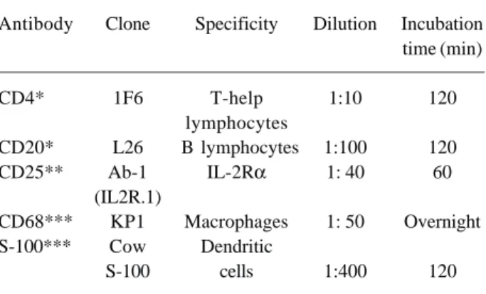
Table 1. Monoclonal antibodies used.
Antibody Clone Specificity Dilution Incubation time (min)
CD4* 1F6 T-help 1:10 120 lymphocytes
CD20* L26 B lymphocytes 1:100 120 CD25** Ab-1 IL-2Rα 1: 40 60
(IL2R.1)
CD68*** KP1 Macrophages 1: 50 Overnight S-100*** Cow Dendritic
S-100 cells 1:400 120
cells) were submitted to nonparametric analysis (Mann-Whitney test) because of the absence of normality as determined by the Kolmogorov-Smirnov test and due to the characteristic of the variable itself. However, depen-dent variables of the discrete quantitative type (quantity of S-100+ cells), which showed a normal distribution, were analyzed by a parametric test (Student t-test). Significance level was set at α=0.05 in both cases.
RESULTS
Morphological Results
All CG specimens (100%) showed an intense inflammatory infiltrate, with the infiltrate being pre-dominantly lymphocytic in 8 cases (47%), lymphoplasmacytic in 3 (17.7%) and predominantly plasmacytic in 6 (35.3%).
Twenty-two (88%) of the CP specimens exam-ined showed an intense inflammatory infiltrate, while a moderate infiltrate was observed in only 3 cases (12%). Lymphocytes were the predominant type of inflamma-tory cells in 8 cases (32%), while 17 specimens (68%) exhibited an inflammatory infiltrate ranging from lymphoplasmacytic (9 cases, 36%) to predominantly plasmacytic (8 cases, 32%).
Immunohistochemical Results
Qualitative Analysis of the Expression Pattern of CD4+ Cells in the Lamina Propria. All cases of CG
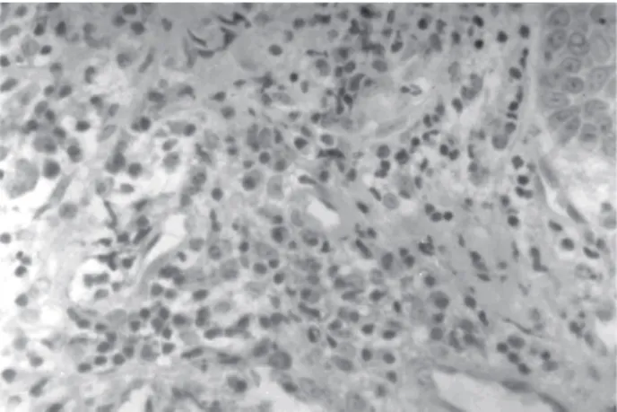
analyzed were immunoreactive to the CD4 anti-body, with positive cells being diffusely distributed in the form of isolated cells or in small foci consisting of scarce cells (score 1). Twenty-two (88%) of the CP cases were positive for the CD4 marker. However, a reduced number of CD4+ cells (score 1) distributed in an isolated manner were observed in these specimens. Qualitative Analysis of the Expression Pattern of CD20+ Cells in the Lamina Propria. Sixteen (94.1%) of the 17 cases of CG were positive for the anti-CD20 antibody, with the immunostained cells showing a predominantly focal distribution pattern. This pattern of cell distribution was characterized by the presence of foci showing variable labeling intensity consisting of a small (score 1), moderate (score 2) or large (score 3) number of positive cells. Score 2 predominated in 11 specimens (Fig. 1) and score 1 in only 5. Positive immunostaining for the anti-CD20 antibody was ob-served in 22 (88%) of the 25 CP specimens. Regarding the density of immunopositive cells, considering all cell distribution patterns analyzed, scores 2 and 1 predomi-nated in 13 and 9 cases, respectively.
Qualitative Analysis of the Expression Pattern of CD25+ Cells in the Lamina Propria. Sixteen (94.1%) of the 17 cases of CG were positive for the CD25 marker. Score 2 predominated in 7 speci-mens, score 1 in 5 specispeci-mens, and score 3 in 4 cases, with scores 2 and 1 thus predominating in the CG sample. The total CP sample demonstrated immunoreactivity to the anti-CD25 antibody. With respect to the density of immunopositive cells, 16
Figure 1. Foci of CD20+ cells/score 2 in chronic gingivitis (SABC; ×400).
specimens presented score 3 (Fig. 2), 7 showed score 2, and only 2 presented score 1, with scores 3 and 2 thus predominating in the total sample.
Qualitative Analysis of the Expression Pattern of CD68+ Cells in the Lamina Propria. All cases of CG were immunoreactive to the anti-CD68 antibody, with score 2 predominating in more than half the sample (10 cases), followed by score 1 in 5, and score 3 in only 2 cases. The total sample of CP was positive for the anti-CD68 antibody. Most speci-mens (18 cases) presented score 1, while score 2 was observed in 7 cases.
Quantitative Analysis of the Expression Pat-tern of S-100+ Cells in Epithelium and Lamina Propria. Immunoreactivity to the anti-S-100 protein antibody was determined in only of CG, with the remaining specimens being discarded because of an insufficient number of histological fields (2 cases) or because of the lack of epithelial lining (1 case). Only one of the 14 gingival specimens was negative for the 100 protein. Immunostaining for the S-100 marker was observed in the whole sample of CP (Fig. 3). However, 2 specimens were discarded because of the lack of sufficient tissue fragments for cell counting.
Statistical Analysis
Density of CD4+, CD20+, CD25+ and CD68+ Cells in the Lamina Propria. Mann-Whitney nonpara-metric test was used to determine differences in the density of CD20+, CD25+ and CD68+ cells between the two groups studied (CG and CP). The CD4
immunohis-tochemical marker was not analyzed because all values were the same, thus presenting a zero standard devia-tion. Only the anti-CD20 showed no significant differ-ence between CG and CP (p=0.4414). Significant differences (p<0.05) between groups were observed for the other antibodies (anti-CD25 and anti-CD68), with the density of CD25+ and CD68+ cells being significantly higher in chronic periodontitis and gingivi-tis, respectively.
Number of S-100+ Cells in Epithelium and Lamina Propria. Regarding the quantitative variables submitted to parametric analysis, no significant differ-ences (p>0.05) in the number of S-100+ cells were observed between the groups, irrespective of the site of analysis.
DISCUSSION
As established in the literature, the immune re-sponse to bacterial aggression plays a key role in periodontal disease (1,3). This fact was demonstrated in the present study in which inflammation was intense in 94.1% of CG cases and in 88% of CP cases. Addition-ally, an inflammatory infiltrate was observed, ranging from lymphoplasmacytic to predominantly lympho-cytic in 67.7% of the chronic gingivitis samples, and from lymphoplasmacytic to predominantly plasmacytic in 68% of chronic periodontitis specimens. These findings are consistent with those of other investiga-tions that showed the presence of lymphocytes during all phases of periodontitis and a larger number of plasma cells in more advanced stages of the disease (4-7).
The density of CD68+ cells was higher in CG than in CP, in agreement with the findings of previous studies (4-6). These investigators stated that a reduction in macrophage density in chronic periodontitis can also be attributed to the migration of these cells to regional lymph nodes for antigen presentation.
With respect to T lymphocyte density in chronic gingivitis, the present results do not agree with the literature, as most studies implicate T lymphocyte as the main cell type found in the inflammatory infiltrate during the initial stages of gingivitis (8,9). A labeling intensity score of 1 for CD4+ cells was exclusively observed in CP specimens, corroborating the studies of Kleinfelder et al. (10). The fact that this experiment was restricted to the investigation of CD4+ T lymphocytes also seems to explain the reduced density of positive T cells
observed in this sample because, even in small num-bers, CD8+ T lymphocytes are also present in CP, as reported by Séguier et al. (11).
The expression of CD20+ cells, indicative of B lymphocytes, was observed in 94.1% of CG cases and in 88% of CP cases, with a predominantly focal distri-bution. This organization of CD20+ cells can be attrib-uted to an attempt of these cells to mimic the formation of germinative centers. These findings disagree with those obtained by other authors (5,6), who reported the existence of a larger number of B lymphocytes in CP compared to CG. According to the same authors (5,6), CG is characterized by numerical variations in T and B lymphocytes, depending on their stage of development, with T lymphocytes predominating in incipient gingivi-tis and B lymphocytes in already established lesions. Therefore, the similarity in CD20+ cell density observed in the present for both groups might be explained by the fact that most CG cases were in fact established and not incipient gingivitis.
The marked presence of plasma cells in CG specimens and their quantitative increase in cases of CP observed in the present study support previous sugges-tions regarding the duration of these lesions which, on the basis of these histological criteria, can be classified as established CG and advanced CP, respectively, as proposed by Wikström et al. (12).
The monoclonal anti-CD25 antibody recognizes the a subunit of the IL-2 receptor (IL-2Rα) expressed on the surface of active B lymphocytes as well as on natural killer cells and T lymphocytes in process of activation (13), this antibody representing an excellent marker of cell activation (2).
The lymphocyte infiltrate (CD25+) was more intense in CP (score 3) than in CG (score 2), in agreement with the study of Yamazaki et al. (14). This finding explains the greater tissue destruction observed in advanced periodontal disease in view of the elevated secretion of catabolic and bone resorptive cytokines by activated T helper lymphocytes (Th1 and Th2) and the abundant production of antibodies by plasma cells. These results suggest that acute outbreaks of periodon-tal disease activity are associated with a quantitative increase in CD25+ cells.
Regarding the identification of dendritic cells, we chose to use the anti-S-100 protein immunohistochemi-cal marker because it is effective in the labeling of these cells, although it also identifies cells of neural origin.
Langerhans cells were present in the oral gingival epithelium, equally occupying the basal and suprabasal layers, and, eventually, in the sulcular epithelium. These findings agree with those reported elsewhere (15,16), who found these cells distributed within basal and suprabasal keratinocytes of the mucosal squamous epithelium, irrespective of a status of health or disease. The activation of Langerhans cells by antigen captured in epithelial tissue accompanied by the release of inflammatory mediators decreases the levels of E-cadherin (a cellular adhesion molecule), which, in turn, reduces the interaction of these cells with keratinocytes, permitting their migration to regional lymph nodes (17). This fact might explain the presence of dendritic cells in the lamina propria of these specimens. With respect to the present study, it may be suggested that the S-100+ cells found in gingival connective tissue are, in fact, dendritic cells migrating to the regional lymph nodes for antigen presentation.
In contrast, according to Dereka et al. (18), the presence of dendritic cells in gingival connective tissue, especially in papillary and perivascular regions, sug-gests that the numerical increase of Langerhans cells in gingival epithelium probably results from the migration of these cells from the blood vessels of the lamina propria to the epithelial tissue in response to greater bacterial aggression. Comparing the number of Langer-hans cells (S-100+) present in epithelial tissue with the degree of gingival inflammation. The lack of significant numerical differences in Langerhans cells between CG and CP specimens can be explained by the fact that the inflammatory infiltrate was found to be intense in all cases immunoreactive to the S-100 protein and, there-fore, no variation in the level of gingival inflammation was observed between the studied groups, in contrast to the findings of previous investigations (16,19).
This strong correlation between the expression of Langerhans cells and gingival inflammatory infiltrate might represent a reflex of the immunomodulatory activity of these cells on the magnitude of the inflamma-tory response, since this cell population is the main antigen-presenting cell in gingival tissue (20).
pathogen-esis of gingivitis, while the higher concentration of B lymphocytes compared to T lymphocytes both in gin-givitis and periodontitis suggests a greater role of the humoral immune response in the different stages of periodontal disease. In contrast, the higher density of CD25+ cells in periodontitis indicates a greater cell activation of the lymphocytic infiltrate, whereas Langer-hans cells seem to play a similar role in the two conditions studied since no difference was observed between groups.
RESUMO
Com o objetivo de contribuir para um melhor entendimento na etiopatogenia da doença periodontal, um análise imuno-histoquímica da densidade das células inflamatórias mononucleares e da quantidade das células dendríticas foi realizada utilizando os anticorpos anti-CD4, anti-CD20, anti-CD25, anti-CD68 and anti-proteína S-100 em 17 casos de gengivite crônica (GC) e 25 casos de periodontite crônica (PC). As células CD4+ e CD68+ exibiram distribuição difusa no tecido conjuntivo, enquanto que a distribuição das células CD20+ foi predominantemente em grupos, e as CD25+ exibiram distribuição ora difusa ora focal. As células S-100+ foram identificadas no epitélio e na lamina própria, exibindo morfologia e números distintos. A análise estatística não demonstrou diferenças estatisticamente significativas em relação a densidade das células CD4+ e CD20+ e no número de células S-100+ entre os casos de CG e PC. Entretanto, houve diferenças em relação a densidade das células CD25+ e CD68+ entre os grupos (p<0,05). A densidade dos macrófagos foi maior em GC e o nível de ativação celular do infiltrado linfocítico foi maior em PC, não havendo diferenças em relação a densidade de linfócitos T e B, bem como no número de células dendríticas entre as condições anteriormente mencionadas.
REFERENCES
1. Van Dike TE, Serhan CN. Resolution of inflammation: a new paradigm for the pathogenesis of periodontal diseases. J Dent Res 2003;82:82-90.
2 . Karimzadeh K, Morrison J, Zadeh HH. Comparison of gingi-val and peripheral blood T cells among patients with peri-odontitis suggests skewing of the gingival T cell antigen receptor V beta repertoire. J Periodont Res 1999;34:445-456.
3 . Offenbacher S. Periodontal diseases: pathogenesis. Ann Periodontol 1996;1:821-878.
4 . Mikhaleva LM, Barkhina TG, Shapovalov VD, Luss LV, Il’ina NI. Ultrastructure of cell populations of gingival soft tissue in chronic inflammatory processes. Arkhiv Patol 2001;63:15-21.
5 . Katz J, Michalek, SM. Effect of immune T cells derived from mucosal or systemic tissue on host responses to Porphyromonas gingivalis. Oral Microbiol Immunol 1998;13:73-80.
6 . Gamonal J, Acevedo A, Bascones A, Jorge O, Silva A. Charac-terization of cellular infiltrate, detection of chemokine re-ceptor CCR5 and interleukin-8 and RANTES chemokines in adult periodontitis. J Periodont Res 2001;36:194-203. 7 . Apsey DJ, Kaciroti N, Loesche WJ. The diagnosis of
peri-odontal disease in private practice. J Periodontol 2006;77:1572-1581.
8 . Gemmell E, Seymour GJ. Cytokine profiles of cells extracted from humans with periodontal diseases. J Dent Res 1998;77:16-26.
9 . Séguier S, Godeau G, Leborgne M, Pivert G, Brousse N. Immu-nohistologic and morphometric analysis of cytotoxic T lym-phocytes in gingivitis. J Periodontol 1999;70:1383-1391. 10. Kleinfelder JW, Lange DE, Bocker W. Some effects of
non-surgical therapy gingival inflammatory cell subsets in patients with adult and early-onset periodontitis. J Periodontol 2000;71:1561-1566.
11. Séguier S, Godeau G, Brousse N. Collagen fibers and inflamma-tory cells in healthy and diseased human gingival tissues: a comparative and quantitative study by immunohistochemis-try and automated image analysis. J Periodontol 2000;71:1079-1085.
12. Wikstrom M, Wennstrom JL, Renvert S, Jonsson R. Immu-nohistological characteristics of periodontal lesions associ-ated with Porphyromonas gingivalis and Actinobacillus actinomycetemcomitans infections. Oral Microbiol Immunol 1996;11:1-7.
13. Tkaczuk J, Yu CL, Baksh S, Milford EL, Carpenter CB, Burakoff SJ, McKay DB. Effect of anti-IL-2Ralpha antibody on IL-2-induced Jak/STAT signaling. Am J Transplant 2002;2:31-40.
14. Yamazaki K, Nakajima T, Aoyagi T, Hara K. Immunohisto-logical analysis of memory T lymphocytes and activated B lymphocytes in tissues with periodontal disease. J Periodont Res 1993;28:324-334.
15. Misery L, Dezutter-Dambuyant C. Precursors of Langerhans cells. J Europ Acad Dermatol Venereol 1995;5:124-131. 16. Seguier S, Godeau G, Leborgne M, Pivert G, Brousse N.
Quan-titative morphological analysis of Langerhans cells in healthy and disease human gingiva. Arch Oral Biol 2000;45:1073-1081.
17. Keller R. Dendritic cells: their significance in health and disease. Immunol Let 2001;78:113-122.
18. Dereka XE, Tosios KI, Chrysomali E, Angelopoulou E. Fac-tor XIIIa+ dendritic cells and S-100 protein+ Langerhans’ cells in adult periodontitis. J Periodontal Res 2004;39:447-452.
19. Bodineau A, Godeau G, Brousse N, Pellat B, Folliguet M, Seguier S. Langerhans cells express matrix metalloproteinases 9 and 2 and tissue inhibitors of metalloproteinases 1 and 2 in healthy human gingival tissue and in periodontitis. Oral Microbiol Immunol 2006;21:197-200.
20. Séguier S, Godeau G, Brousse N. Immunohistological and morphometric analysis of intra-epithelial lymphocytes and Langerhans cells in healthy and disease human gingival tis-sues. Arch Oral Biol 2000;45:441-452.