IX
Radiol Bras 2007;40(6):IX–XII
WHICH IS YOUR DIAGNOSIS?
Marcelo Souto Nacif1
, Eduardo Benchimol Saad2
, Luiz Eduardo Montenegro Camanho2
, Fernanda d’Araujo Costa Ferreira2
, Ieda Prata Costa2
, Amarino Carvalho de Oliveira Júnior3
Study developed at Hospital Pró-Cardíaco, Rio de Janeiro, RJ, Brazil. 1. Professor, Faculdade de Medicina de Teresópolis (Unifeso), Teresópolis, RJ, Sub-coordinator of Post-Graduation, Instituto de Pós-Graduação Médica Carlos Chagas (IPGMCC), Fellow PhD degree in Radiology (Cardiac MRI), Universidade Federal do Rio de Janeiro (UFRJ), MD, Radiologist, Department of Radiology and Imaging Diagnosis, Hospital Pró-Cardíaco, Rio de Janeiro, RJ, Brazil. 2. MDs, Cardiologists, Hospital Pró-Cardíaco, Unit of Cardiac Arrhythmias, Rio de Janeiro, RJ, Brazil. 3. MD, Radiologist and Coordinator for Department of Radiology and Imaging Diagnosis, Hospital Pró-Cardíaco, Rio de Janeiro, RJ, Brazil. Mailing address: Prof. Dr. Marcelo Souto Nacif. Rua Tavares de Macedo, 136, ap. 1503/A, Icaraí. Niterói, RJ, 24220-211, Brazil. E-mail: [email protected]
A male, 43-year old patient weighting 103 kg, with 1.70 m in height, presenting with frequent ventricular arrhythmias,
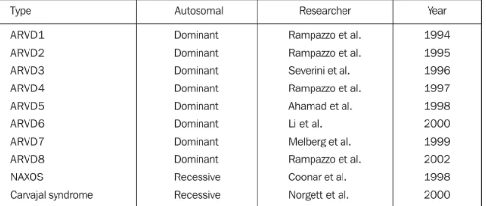
Figure 2. Images acquisition with ECG-gating. Upper images in double IR sequences, without fat suppression, and lower images demonstrating delayed enhancement, four-chamber and short axis views.
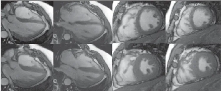
Figure 1. Images acquisition with ECG-gating, in cine-Fiesta sequences (SSFP), upper images at end-diastole, and lower images at end-systole, four-chamber and short axis views.
who had experienced three episodes quiring electrical cardioversion, was re-ferred to Hospital Pró-Cardíaco,
X Radiol Bras 2007;40(6):IX–XII Images description
Figure 1. Electrocardiographic (ECG)
gating acquisitions in cine-Fiesta se-quences (SSFP), upper images at end-diastole, and lower images at end-sys-tole, four-chamber and short axis views. Note the normal-sized atriums. The left ventricle (LV) presented with preserved global and segmented functions, with 65% ejection fraction. The right ventricle (RV) presented with global dilatation, slight dysfunction, and estimated 32% ejection fraction (Simpson). Thinned RV wall with slight parietal irregularities is observed near the outflow tract.
Figure 2. Images acquisition with
electrocardiographic (ECG) gating. Up-per images in double IR sequences, with-out fat suppression, and lower images demonstrating delayed enhancement, four-chamber and short axis views. Small foci of fibrofatty replacement are ob-served, characterized by the correlation between the two sequences where de-layed myocardial enhancement can be identified on the RV wall, near the out-flow tract.
Diagnosis: Arrhythmogenic right
ventricular dysplasia (ARVD).
COMMENTS
ARVD, also known as right ventricu-lar cardiomyopathy (RVC) because of structural alteration of the cardiac muscle, has first been identified in a group of patients submitted to surgical interven-tion for ventricular tachycardia who had not responded to the treatment with an-tiarrhythmic drugs and with no history of heart disease(1,2).
ARVD is characterized by a progres-sive fibrolipomatous replacement of myocardial cells intermingled with nor-mal myocytes(3).
A great number of cases of ARVD present a familial distribution, and, most frequently this disease is observed in some specific geographical regions of the world. Some genes implicated in the on-set of this cardiomyopathy have already been described (Chart 1).
ARVD is a rare disease with an esti-mated incidence of 1:5000, predominantly affecting young men with no history of
cardiovascular disease. It is character-ized by frequently severe ventricular ar-rhythmias associated with sudden car-diac death, particularly during physical activity. Palpitation, dizziness, and even syncope are the main symptoms of the disease as a result of ventricular tachy-cardia and, less frequently, related to ven-tricular extrasystole. Another relevant and paradoxical aspect is the frequent presence of a practically normal clinical examination during sinusal rhythm(2,4–6). ARVD is considered as a progressive condition that may potentially lead to heart failure and death over a variable period of time. The diagnosis is based on internationally accepted clinical and labo-ratory criteria. The presence of two ma-jor criteria, one mama-jor and two minor cri-teria, or four minor criteria from different categories is considered as a diagnostic proof of the disease (Chart 2)(3,5,7–9).
High-resolution ECG
As it can be seen on Figure 3, some relevant findings were observed on the high-resolution ECG of this patient, with inverted T waves in right precordial leads, late low amplitude potentials and high frequency at the end of the electrical ven-tricular activity, and left bundle-branch block-type ventricular tachycardia.
Cardiac magnetic resonance imaging (CMR)
Presently, CMR has become the stan-dard for evaluation of anatomic and both RV segmental and global functions, play-ing a significant role in the diagnosis and follow-up of ARVD(5,10).
Chart 1 Types of genes implicated in the ARVD pathogenesis.
Type ARVD1 ARVD2 ARVD3 ARVD4 ARVD5 ARVD6 ARVD7 ARVD8 NAXOS Carvajal syndrome Autosomal Dominant Dominant Dominant Dominant Dominant Dominant Dominant Dominant Recessive Recessive Researcher
Rampazzo et al.
Rampazzo et al.
Severini et al.
Rampazzo et al.
Ahamad et al.
Li et al.
Melberg et al.
Rampazzo et al.
Coonar et al.
Norgett et al.
Year 1994 1995 1996 1997 1998 2000 1999 2002 1998 2000
This method allows a morphological analysis of both ventricles and their con-tractile activities by means of ECG gat-ing acquisitions in cine-Fiesta se-quences, demonstrating cavitary diam-eters and ventricular indices(10).
In the present case, besides thinned RV wall with slight parietal irregularities observed near the outflow tract, the fol-lowing data were obtained: RV short axis – 5.2 cm (normal: 2.2 cm to 4.4 cm); 32% RV ejection fraction (normal: 40% to 60%); end-diastolic RV volume – 98 ml/ m² (normal: 62 ml/m² to 88 ml/m²); and end-systolic RV volume – 65 ml/m² (nor-mal: 10 ml/m² to 30 ml/m²); besides the normal LV ejection fraction, characteriz-ing the presence of two minor diagnos-tic criteria.
The demonstration of fatty tissue in-filtration into the ventricular wall in dark-blood sequences with and without fat-suppression (double and triple IR) may be performed and interpreted with a cer-tain degree of confidence, besides allow-ing the identification of the fibrofatty tis-sue in the analysis of the delayed-en-hancement images, demonstrating the presence of gadolinium in the fibrotic myocardium.
XI
Radiol Bras 2007;40(6):IX–XII Chart 2 Criteria for ARVD diagnosis.*
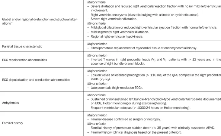
Global and/or regional dysfunction and structural alter-ations †
Parietal tissue characteristic
ECG repolarization abnormalities
ECG depolarization and conduction abnormalities
Arrhythmias
Familial history
Major criteria
– Severe dilatation and reduced right ventricular ejection fraction with no (or mild) left ventricular involvement.
– Right ventricle aneurysms (diastolic bulging with akinetic or dyskinetic areas). – Severe right ventricular dilatation.
Minor criteria
– Mild global dilatation or reduced right ventricular ejection fraction with normal left ventricle. – Mild segmental right ventricular dilatation.
– Regional right ventricular hypokinesia.
Major criterion
– Fibrolipomatous replacement of myocardial tissue at endomyocardial biopsy.
Minor criterion
– Inverted T waves in right precordial leads (V2 and V3, patients with > 12 years and in the
absence of right bundle-branch block).
Major criterion
– Epsilon waves of localized prolongation (> 110 ms) of the QRS complex in the right precordial leads (V1–V3).
Minor criterion
– Late potentials (high-resolution ECG).
Minor criteria
– Sustained or nonsustained left bundle-branch block-type ventricular tachycardia documented on ECG, Holter monitoring or during exercising testing.
– Frequent ventricular ectopias (> 1000/24 hours on Holter monitoring).
Major criterion
– Familial disease confirmed at surgery or necropsy. Minor criteria
– Familial history of premature sudden death (< 35 years) with clinically suspected ARVD. – Familial history (clinical diagnosis based on the present criterion).
* Source: McKenna et al.(3). † Detected at echocardiogram, ventriculography, MRI or radioisotopic ventriculography.
Figure 3. High-resolution ECG. Inverted T waves (A) and ventricular tachycardia (B).
A B
like Auffermann et al.(4), suggest the re-placement of angiography and, possibly, biopsy by CMR in the diagnosis of ARVD.
Because of the CMR capacity to de-tect fibrofatty tissue, this method has
been considered as capable of diagnos-ing ARVD based on the recognition of the histopathological marker of this dis-ease(8).
However, theoretical and practical is-sues should be taken into consideration:
XII Radiol Bras 2007;40(6):IX–XII with several cardiopathies, have
demon-strated that the prevalence of fatty tissue is 6% in ischemic cardiopathy, 7% in Kawasaki disease, 11% in hypertrophic cardiomyopathy, 18% in idiopathic di-lated cardiomyopathy, and 33% in cases of ARVD.
2 – In spite of the good correlation between the presence of foci of fibrosis in the RV and histopathological findings, and the value of this finding as a predic-tor of episodes of tachycardia during elec-trophysiological studies, the technical limitations associated with spatial reso-lution still do not allow the detection of small fatty or fibrofatty deposits.
3 – Patients who have recently been submitted to procedures involving abla-tion or cauterizaabla-tion of the RV wall, may present non-specific foci of delayed en-hancement as a result of these previous procedures.
Therefore, MRI can be considered as an useful tool in the diagnosis of ARVD in more advanced stages of the disease with a more diffuse and extensive RV in-volvement. It is important to note that the presence of parietal fat may represent an anatomical variation in normal myocar-diums(6).
Presently, the detection of fat and fi-brosis by CMR is less relevant than in the past, with functional and morphological alterations like the presence of aneu-rysms, “serrations” or trabecular disor-ders being more significant as far as the ARVD diagnosis is concerned(9,10).
Final considerations
Experience is an essential factor for a correct interpretation of RV findings, con-sidering that typical variations of the RV are more significant than LV variations. Therefore, isolated CMR findings should be carefully interpreted and correlated with the other diagnostic criteria in order to avoid erroneous affirmative interpre-tation.
In the present case, besides the iden-tification of fibrofatty tissue and slight RV dilatation with reduced ejection fraction, three additional minor criteria (by high-resolution ECG) were found, complement-ing the diagnostic criteria.
REFERENCES
1. Corrado D, Leoni L, Link MS, et al. Implantable cardioverter-defibrillator therapy for prevention of sudden death in patients with arrhythmogenic right ventricular cardiomyopathy/dysplasia. Circulation 2003;108:3084–3091.
2. Elias J, Tonet J, Frank R, Fontaine G. Displasia arritmogênica do ventrículo direito. Arq Bras Cardiol 1998;70:449–456.
3. McKenna WJ, Thiene G, Nava A, et al. Diagno-sis of arrhythmogenic right ventricular dysplasia/ cardiomyopathy. Task Force of the Working Group Myocardial and Pericardial Disease of the Euro-pean Society of Cardiology and of the Scientific Council on Cardiomyopathies of the International Society and Federation of Cardiology. Br Heart J 1994;71:215–218.
4. Auffermann W, Wichter T, Breithardt G, Joa-chimsen K, Peters PE. Arrhythmogenic right ven-tricular disease: MR imaging vs. angiography. AJR Am J Roentgenol 1993;161:549–555. 5. Midiri M, Finazzo M, Brancato M, et al.
Arrhyth-mogenic right ventricular dysplasia: MR features. Eur Radiol 1997;7:307–312.
6. Fontaliran F, Fontaine G, Fillette F, Aouete P, Chomette G, Grosgogeat Y. Frontières nosologi-ques de la dysplasie arythmogène. Variations quan-titatives du tissu adipeux ventriculaire droit nor-mal. Arch Mal Coeur 1991;84:33–38. 7. Kaminaga T, Naitou H, Hamada S, Takamiya M.
Detection of myocardial fatty components with ultrafast CT. Nippon Igaku Hoshasen Gakkai Zasshi 1993;53:28–34.
8. Castillo E, Tandri H, Rodriguez ER, et al. Arrhyth-mogenic right ventricular dysplasia: ex vivo and in vivo fat detection with black-blood MR imag-ing. Radiology 2004;232:38–48.
9. Tandri H, Friedrich MG, Calkins H, Bluemke DA. MRI of arrhythmogenic right ventricular cardiomy-opathy/dysplasia. J Cardiovasc Magn Reson 2004; 6:557–663.