Effects of biolavonoid ternatin on liver regeneration and oxidative stress in rats
1José Ulisses de Souza Melo
I, Radamés Bezerra Melo
II, Jefferson Menezes Viana Santos
III, Manoel Messias Campos Júnior
III,
Sérgio Botelho Guimarães
IV, Paulo Roberto Leitão Vasconcelos
VIPhD, Associate Professor, Department of Surgery, UFC, Ceara, Brazil. Conception and design of the study, technical procedures, acquisition of data. IIFellow Master degree, Postgraduate Program in Surgery, Department of Surgery, UFC, Ceara, Brazil. Technical procedures, acquisition of data. IIIGraduate student, UFC, Ceara, Brazil. Animal care, application and maintenance of inhalatory anesthesia, post-operative animal care.
IVPhD, Associate Professor, Department of Surgery. Head, LABCEX, UFC, Ceara, Brazil. Manuscript writing and critical revision.
VPhD, Associate Professor. Coordinator, Postgraduate Program in Surgery, Department of Surgery, UFC, Ceara, Brazil. Conception, design, intellectual
and scientiic content of the study, inal approval of manuscript.
ABSTRACT
PURPOSE:
To evaluate the effect of biolavonoid ternatin (TRT) on rat liver regeneration and oxidative stress after 70% partial
hepatectomy (PH).
METHODS
: Thirty six young male Wistar rats were randomly assigned to two groups of 18 animals each – control (G1) and experimental
(G2) – and were submitted to PH under inhalatory diethylether anesthesia. G1 rats received daily intraperitoneal (ip) injections of saline
(NaCl 0.9% solution) 0.1 mL/kg for 14 days; G2 animals received daily ip injections of TRT 0.1% 1.0mg/kg for 14 days. At 36h (T1),
168h (T2) and 336h (T3) post-PH timepoints, a subgroup of six rats in each group was chosen in a randomized way to complementary
hepatectomy (CH) and blood samples haversting. Collected material was saved for laboratory analysis (total bilirubin (TB), D-Glucose,
glutathione (GSH) and thiobarbituric acid reactive substances (TBARS) and assessment of liver regeneration.
RESULTS
: TRT induced a signiicant decrease in liver and plasma GSH concentrations; liver regeneration process was not affected.
TRT promoted a signiicant decrease in blood glucose levels 168h after partial hepatectomy compared with controls. TB levels remained
unchanged.
CONCLUSION
: Intraperitoneal biolavonoid ternatin injection in partially hepatectomized rats induces a decrease in oxidative stress
and a signiicant hypoglycemic state, but does not promote any change in the evolution of liver regeneration.
Introduction
The liver exhibits a remarkable regenerative capacity
after tissue damage, including partial hepatectomy (PH)¹.
Following removal of the two major lobes of the rat liver (70%
partial hepatectomy, Higgins-Anderson partial hepatectomy), the
remaining minor lobes rapidly undergo an essentially hyperplastic
response to repopulate loss of tissue and cells² attaining its original
size by three to 14 days
2-4.
Reactive oxygen species (ROS), antioxidant substances
and lipid peroxidation have been implicated in control mechanisms
of cellular growth and proliferation
5-7. The administration of
exogenous antioxidants such as
a
-tocopherol (vitamin E) and
reduced gluthatione (GSH) retards liver regeneration
7,8. Moreover,
several studies have reported the genesis and/or formation of
free radicals as an important factor on the liver regeneration
phenomenon, necessary to its natural outcome
6,9.
Flavonoids are claimed to have protective effects against
free radicals induced lipid peroxidation of living cell membranes
10.
Ternatin (TRT), 4’.5-dihydroxy-3.3’,7.8-tetramethoxylavone, a
natural antioxidant, was evaluated under the classical experimental
model of partial hepatectomy due to Higgins and Anderson¹ to
assess its effects on rat liver regeneration and oxidative stress.
TRT was isolated from the lowering tops of
Egletes
viscosa
L. (Asteraceae)
11, popularly known as “macela da terra”,
which is a small empirical medicinal herb that grows abundantly
in northeast of Brazil and others areas of South America. Previous
studies have demonstrated anti-inlammatory, antianaphylactic,
antihrombotic and antihepatotoxic properties of TRT
12-14;
these
pharmacological effects have been related to its free radical
scavenging and antioxidant properties
15.
Methods
All procedures were approved by the Commission of
Ethics on Animal Research (now Committee of Ethics on the
Use of Animals (CEUA), Federal University of Ceara, protocol
14/06 on August 11, 2006. Surgical procedures and animal
handling were conducted in accordance with the Brazilian Federal
Law No. 11794 of October 8, 2008 (http://www.planalto.gov.
br/ccivil_03/_Ato2007-2010/2008/Lei/L11794.htm). Rats were
raised under controlled conditions, housed in polypropylene cages
at temperature of 23
+
4ºC on a light schedule of 12h light/dark
cycle and fed regular rat chow and potable water
ad libitum.
Thirty
six young (70
+
10 days) male Wistar rats weighting 100-235g were
included in the study and randomly assigned to two groups (n=18)
– control (G1) and experiment (G2). All rats were submitted to the
classical Higgins-Anderson 70% partial hepatectomy¹ (PH).
G1
rats received daily intraperitoneal (ip) injections of saline (NaCl
0.9% solution) 0.1 mL/kg for 14 days; G2 animals received daily
ip injections of TRT 0.1% 1.0mg/kg for 14 days. At 36h (T1), 168h
(T2) and 336h (T3) post-PH, a subgroup of six rats in each group
was chosen in a randomized way to complementary hepatectomy
(CH) and blood samples haversting. All surgical procedures were
performed under inhalatory diethylether anesthesia. Collected
material was stored for laboratory analysis
Thiobarbituric acid reactive substances (TBARS),
glutathione (GSH), glycemia and total bilirubin (TB) were assayed
.
Tissue samples were snap-frozen in liquid nitrogen and stored in
glass tubes at -70ºC until subsequent preparation and analysis of
liver tissue homogenate. Plasma samples were obtained from
heparinized blood after 10 minutes of refrigerated centrifugation
(4.000 rotations/minute) and were stored as well.
Chemicals and drugs
Saline (NaCl 0.9%) was obtained from Química
Farmacêutica Gaspar Viana, Brazil. TRT was isolated from
dried lower buds of
E. viscosa
and its chemical identiication
was conirmed by Prof. E. R .Silveira of the UFC. The voucher
specimen (#16327) is deposited in the
Herbarium
Prisco Viana
(UFC). TRT 100mg was dissolved in 3% dimethylsulfoxide
(DMSO) 100mL in order to achieve a 0.1% TRT solution. DMSO
was purchased from Cromato Produtos Químicos Ltda, Brazil. All
other chemicals were of analytical grade.
Biochemical determinations
The amount of GSH was determined from a standard
curve simultaneously obtained under the same conditions with
various concentrations of GSH. Liver regeneration was estimated
by weighting the rat liver residual lobes. TB content was valuated
following Meites modiication of Malloy and Evelyn procedure
16.
D-glucose was determined according to the Slein´s
method
17.
Lipid peroxidation, a measure of free radical damage, was
assayed by measuring malondialdehyde (MDA) as thiobarbituric
acid-reactive substances (TBARS) levels using the thiobarbituric
acid method
18. In brief, H
3PO
4(1%, 3ml) and aqueous TBA solution
(0.6%, 3 mL) were added to the 10% homogenate (0.5 ml). The
assay medium was shaken and heated on a boiling-water bath for
45 min. After cooling, 4 ml of
n
-butanol was added and the mixture
at 1200 g for 15 min its optical density was determined in a
spectrophotometer (Beckman DU 640 B) with 535 and 520nm as
absorption wavelengths, respectively. The difference between the
results of the two optical density determinations was taken as the
TBA value and the amount of malondialdehyde (MDA) in the testis
was calculated, comparing with MDA standards and expressed as
µmol MDA per gram of wet tissue. GSH levels were estimated by
the method of Sedlak and Lindsay
19which is based on the reaction
between thiol groups and 5-5’-dithiobis-(2-nitrobenzoic acid) to
give a compound that absorbs light at 412 nm. The amount of GSH
was determined from a standard curve simultaneously obtained
under the same conditions with various concentrations of GSH.
Statistical analysis
GraphPad Prism 4.0 (GraphPad Software, San Diego,
California, USA, www.graphpad.com
) was used for computation
and statistical analysis.
To ensure the appropriateness of parametric
testing, all data were examined for normality, using
Kolmorogov-Smirnov test (with Dalal-Wilkinson-Lilliefor P Value).
Non-parametric data were analyzed using ANOVA (Kurskal-Wallis/
Dunn) tests. Values of p<0.05 were accepted as statistically
signiicant.
Results
No animal died during the experiment.
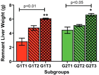
Liver regeneration
Figure 1 depicts the evolution of residual liver lobes in
each subgroup throughout the experiment. There was a signiicant
increase in liver weight at T3 timepoint in both groups.
FIGURE 1 - Liver regeneration in G1 (control) and G2 (experiment)
groups. Bars represent mean ± SD of G1 (red bars) and G2 (green bars) subgroups at 36h (T1), 168h (T2) and 336h (T3) timepoints post-PH. Values were signiicantly different (G1:p<0.01, G2:p<0.05) at T3 timepoint compared with T1 timepoint, in both groups, by ANOVA (Kruskal-Wallis/Dunn) test.
Total billirubin
Although there was an apparent decrease of TB
concentrations in both groups (G1 / G2), no signiicant differences
were demonstrated, as illustrated in Figure 2.
FIGURE 2 - Blood total billirubin concentrations in G1 (control) and G2
(experiment) groups. Bars represent mean ± SD of G1 (red bars) and G2 (green bars) subgroups at 36h (T1), 168h (T2) and 336h (T3) timepoints post-PH. Values were not signiicantly different by ANOVA (Kruskal-Wallis/Dunn) test.
D-Glucose
D-Glucose levels decreased signiicantly (p<0.01) in
TRT-treated rats in T2 timepoint compared with control T2 (Figure
3).
G1T1 G1T2 G1T3 G2T1 G2T2 G2T3 0
2 4 6
8 p<0.01
**
*
p<0.05
Subgroups
R
e
m
n
a
n
t
L
iv
e
r
W
e
ig
h
t
(g
)
G1T1 G1T2 G1T3 G2T1 G2T2 G2T3 0.0
0.1 0.2 0.3
SubGroups
T
o
ta
l
B
il
li
ru
b
in
(
m
g
%
FIGURE 3 - D-Glucose levels in G1 (control) and G2 (experiment)
groups. Bars represent mean ± SD of G1 (red bars) and G2 (green bars) subgroups at 36h (T1), 168h (T2) and 336h (T3) timepoints post-PH. Values were signiicantly different (G1 vs. G2, p<0.01) at T2 timepoint by ANOVA (Kruskal-Wallis/Dunn) test.
Plasma GSH
Plasma GSH concentrations decreased signiicantly in
TRT treated rats, 336h post-PH (T3) compared with T1 (p<0.001)
and T2 (p<0.05) (Figure 4).
FIGURE 4 - Plasma GSH concentrations in G1 (control) and G2
(experiment) groups. Bars represent mean ± SD of G1 (red bars) and G2 - TRT subgroups at 36h (T1), 168h (T2) and 336h (T3) timepoints post-PH. Values were signiicantly different at T2 and T3 compared with T1 timepoint in TRT-treated rats by ANOVA (Kruskal-Wallis/Dunn) test.
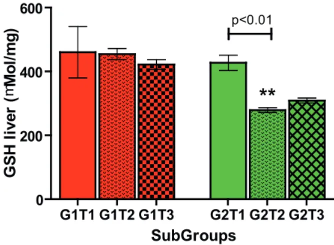
Liver GSH
Liver GSH concentrations decreased signiicantly
(p<0.01) in TRT treated rats, 168h post-PH (Figure 5).
FIGURE 5 - Liver GSH concentrations in G1 (control) and G2 (TNT)
groups. Bars represent mean ± SD of G1 (red bars) and G2 (green bars) subgroups at 36h (T1), 168h (T2) and 336h (T3) timepoints post-PH. Values were signiicantly different (p<0.01) at T2 compared with T1 timepoint in TRT-treated rats by ANOVA (Kruskal-Wallis/Dunn) test.
Plasma TBARS
Plasma TBARS values were not different in G1 and G2
rats (Figure 6).
FIGURE 6 - Plasma TBARS concentrations in G1 (control) and G2
(experiment) groups. Bars represent mean ± SD of G1 - control (red bars) and G2 - TRT (green bars) subgroups at 36h (T1), 168h (T2) and 336h (T3) timepoints post-PH. Values were not signiicantly different in control and TRT-treated rats by ANOVA (Kruskal-Wallis/Dunn) test.
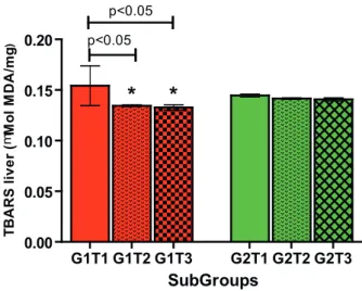
Liver TBARS
Liver TBARS values decreased in T2 and T3 subgroups
compared with T1 in control rats. No differences were found in
TRT-treated rats (Figure7).
G1T1 G1T2 G1T3 G2T1 G2T2 G2T3 0
50 100 150 200
**
p<0.01
SubGroups
B
lo
o
d
g
lu
c
o
s
e
(
m
g
%
)
G1T1 G1T2 G1T3 G2T1 G2T2 G2T3 0
50 100 150 200
p<0.001
p<0.01
SubGroups
G
S
H
P
la
s
m
a
-m
M
o
l/
m
L
G1T1 G1T2 G1T3 G2T1 G2T2 G2T3 0
200 400 600
**
p<0.01SubGroups
G
S
H
l
iv
e
r
(
m
M
o
l/
m
g
)
G1T1 G1T2 G1T3 G2T1 G2T2 G2T3 0.000
0.005 0.010 0.015 0.020
SubGroups
T
B
A
R
S
P
la
s
m
a
(
mM
o
l
M
D
A
/m
L
FIGURE 7 - Liver TBARS concentrations in G1 (control) and G2
(experiment) groups. Bars represent mean ± SD of G1 - control (red bars) and G2 - TRT (green bars) subgroups at 36h (T1), 168h (T2) and 336h (T3) timepoints post-PH. TBARS concentrations decreased signiicantly different in control rats in T2 and T3 timepoints compared with T1 by ANOVA (Kruskal-Wallis/Dunn) test.
Discussion
The rat was chosen for this study because it is the
most studied animal in hepatic regeneration research
20. Male
young animals were used whereas older rats may compromise
the regenerative process
21. Besides, steroids present in greater
quantity in females may inluence hepatic regeneration
22. The
maximum period of sample collecting (14 days) was chosen
based on previous studies that show that liver regeneration in rats
subjected to HP is completed in two weeks
1,20.
Considering that TB concentrations did not show
any signiicant change at any time we can perceive that TRT
administration had no effects on TB levels. The absence of
signiicant differences in the weight of the remnant liver in G1 and
G2 groups suggests that TRT administration does not interfere in
a signiicant way with the natural post-HP rat liver regeneration.
Some antioxidants have different effects on liver
regeneration. In fact, the administration of omega-3 polyunsaturated
fatty acids (PUFA), inhibits the liver regeneration
23and the
administration of a
-tocopherol (vitamin E) and GSH retards liver
regeneration evolution
7,8.
Partial hepatectomy is related to the formation
of free radicals
24-26. Indeed, many studies have shown an
increased production of free radicals production measured by
malonyldialdehyde in liver mitochondria following partial
hepatectomy
24-26. Decreased liver and plasma GSH observed
in G1 and G2 rats could be related to the utilization of GSH in
order to attenuate the oxidative stress generated by the partial
hepatectomy.
The lipid peroxidation inhibitory effects of several
lavonoids such as luteolin, apigenin, galangin, gardenin D, (+)
catechin
27, quercitin
28, rutin (quercetin-3-rhamnosyl glucoside)
29ant TRT
30have been previously reported. In the present study,
liver TBARS values decreased in T2 and T3 subgroups compared
with T1 in control rats. This could be explained by the reduction
of GSH concentration in G1 rats. The absence of reduction of
TBARS concentration in rats subjected to HP and treated with
TRT suggests that the antiperoxidative effect of TRT was not
observed here.
In the present study, blood glucose concentrations
decreased signiicantly in TRT-treated group. Studies have
demonstrated that insulin levels decrease
31,32while glucagon
levels increase
33in partially hepatectomized rats. These changes
may relect part of homeostatic mechanism to maintain glucose
levels within normal limits. Moreover, antioxidants improve
insulin action, due in part by the protection of b-cells from free
radicals injuries
32.
Conclusions
Biolavonoid ternatin administration induces an
inhibitory effect on oxidative stress but does not change the liver
regeneration evolution. Moreover, TRT promotes a signiicant
hypoglycemic state in TRT-treated rats.
References
1. Higgins GM, Anderson RM. Experimental pathology of the liver: I. Restoration of the liver of the white rat following partial surgical removal. Arch Pathol. 1931;12:186-202.
2. Michalopoulos G. Liver regeneration: molecular mechanisms of growth control. FASEB J. 1990;4:176-87.
3. Steer CJ. Liver regeneration. FASEB J. 1995;9:1396-2000.
4. Hockings PD, Roberts T, Campbell SP, Reid DG, Greenhill RW, Polley SR, Nelson P, Bertram TA, Kramer K. Longitudinal magnetic resonance imaging quantiication of rat liver regeneration after partial hepatectomy. Toxicol Pathol. 2002;30(5):606-10.
5. Van Norden CJ. Effects of n-3 and n-6 polyunsaturated fatty
acid-enriched diets on lipid metabolism in periportal and pericentral compartments of female rat liver lobules and the consequences
for cell proliferation after partial hepatectomy. J Lipid Res. 1995;36(8):1708-20.
6. Nakatani T, Inouye M, Mirochnitchenko O. Over expression of antioxidants enzymes in transgenic mice decreases cellular ploidy during liver regeneration. Exp Cell Res. 1997;236(1):137-46.
7. Holocec M, Skopec F, Sprongl L. Inluence of buthionine,
s-adenosylmethionine and glutathione on liver regeneration following
partial hepatectomy. Arzneimittelforschung. 2000;50(12);1093-8. G1T1 G1T2 G1T3 G2T1 G2T2 G2T3
0.00 0.05 0.10 0.15 0.20
*
*
p<0.05 p<0.05
SubGroups
T
B
A
R
S
l
iv
e
r
(
mM
o
l
M
D
A
/m
g
8. Trejo-Solis C, Sanchez VC, Fraustro AA, Sevilla LS, Ruiz CG, Munoz RH. Inhibitory effect of vitamin E administration on the progression
of liver regeneration induced by partial hepatectomy in rats. Lab
Invest. 2003;83(11):1669-79.
9. Kurir TT, Markotić A, Katalinić V, Bozanić D, Cikes V, Zemunik T, Modun D, Rincić J, Boraska V, Bota B, Salamunić I, Radić S. Effect of hyperbaric oxygenation on the regeneration of the liver after partial hepatectomy in rats. Braz J Med Biol Res. 2004;37(8):1231-7.
10. Czaplińska M, Czepas J, Gwoździński K. Structure, antioxidative and anticancer properties of lavonoids. Postepy Biochem. 2012;58(3):235-44.
11. Lima MAS, Silveira ER, Marques MSL, Santos RH, Gambardela MPT. Biologically active lavonoids and terpenoids from Egletes viscosa. Phytochemistry. 1996;41;217-23.
12. Souza MF, Rao VSN, Silveira ER. Antianaphylactic and anti-inlammatory effects of ternatin, a lavonoid isolated from Egletes viscosa Less. Braz J Med Biol Res. 1992;25:1029-32.
13. Rao VSN, Figueiredo EG, Melo CL, Viana GSB, Menezes DB, Matos MSF, Silveira ER. Protective effect of ternatin, a lavonoid
isolated from Egletes viscose Less., in experimental liver injury. Pharmacology. 1994;48:392-97.
14. Souza MF, Cunha GMA, Fontenele JB, Rao VSN, Silveira ER. Antithrombotic activity of ternatin, a tetramethoxy lavone from Egletes viscosa Less. Phytother Res. 1994;8:478-81.
15. Souza MF, Rao VSN, Silveira ER. Inhibition of lipid peroxidation by ternatin, a tetrametoxylavone from Egletes viscosa L.
Phytomedicine. 1997;4:25-9.
16. Malloy HT, Evelyn KA. The determination of bilirubin with the photoelectric colorimeter. J Biol Chem. 1937;119:481-90.
17. Slein MW. Determination with hexokinase and glucose-6-phosphate
dehydrogenase. In: Bergmeyer HU, editor. Methods of enzymatic
analysis. New York: Verlag Chemie, Weinheim/Academic Press; 1963. p.117-23.
18. Uchiyama M, Mihara M. Determination of malonaldehyde precursor in tissues by thiobarbituric acid test. Anal Biochem. 1978;86(1):271-8.
19. Sedlak J, Lindsay RH. Estimation of total, protein-bound, and nonprotein sulfhydryl groups in tissue with Ellman’s reagent. Anal Biochem. 1968;25(1):192-205.
20. Ramalho FS, Ramalho LNZ, Zucoloto S, Castro e Silva Jr O. Regeneração hepática: algumas indeinições num universo de incertezas. Acta Cir Bras. 1993;8:177-89.
21. Bucher NL, Swafield MN, Ditroia JF. The inluence of age upon the incorporation of thymidine-2-c14 into the DNA of regenerating rat liver. Cancer Res. 1964;24:509-12.
22. Francavilla A, Gavaler JS, Makowka L, Barone M, Mazzaferro V, Ambrosino G, Iwatsuki S, Guglielmi FW, Dileo A, Balestrazzi
A, Van Thiel DH, Starzi TE. Estradiol and testosterone levels in patients undergoing partial hepatectomy. A possible signal for
hepatic regeneration? Dig Dis Sci. 1989;34(6):818-22.
23. Melo JUS, Santos JMV, Kimura OS, Campos Junior MM, Melo RB, Vasconcelos PRL. Effects of fatty acids on liver regeneration in rats. Rev Bras Cir. 2010;37(5):351-7.
24. Aguilar-Delin I, Lopez-Barrera F, Hernandez-Munoz R. Selective enhancement of lipid peroxidation in plasma membrane in two experimental models of liver regeneration: partial hepatectomy and acute CC14 administration. Hepatology. 1996;24:657-62.
25. Guerrieri F, Vendemiale G, Grattagliano I, Cocco T, Pellecchia G, Altomare E. Mitochondrial oxidative alterations following partial hepatectomy. Free Radic Biol Med. 1999;26:34-41.
26. Hernandez-Munoz R, Sanchez-Sevilla L, Martinez-Gomez A,
Dent MA. Changes in mitochondrial adenine nucleotides and in permeability transition in two models of rat liver regeneration.
Hepatology. 2003;37:842-51.
27. Cholbi MR, Paya M, Alcaraz MJ. Inhibitory effects of phenolic compounds on CCl4-induced microsomal lipid peroxidation. Experientia. 1991;47(2):195-9.
28. Kanter M. Protective effect of quercetin on liver damage induced by biliary obstruction in rats. J Mol Histol. 2010;41(6):395-402. 29. Korkmaz A, Kolankaya D. Protective effect of rutin on the
ischemia/ reperfusion induced damage in rat kidney. J Surg Res. 2010;164(2):309-15.
30. Guimarães SB, Santos JM, Aragão AA, Kimura OS, Silveira ER, Vasconcelos PR. Ternatin pretreatment attenuates testicular injury induced by torsion/detorsion in Wistar rats. Acta Cir Bras. 2011;26(4):325-8.
31. Facchini FS, Humphreys MH, Nascimento CA, Abbasi F, Reaven GM. Relation between insulin resistance and plasma concentrations of lipid hydroperoxides, carotenoids, and tocopherols. Am J Clin Nutr. 2000;72(3):776-9.
32. Koksal C, Bozkurt AK, Cangel U, Ustundag N, Konukoglu D, Musellim B, Sayin AG. Attenuation of ischemic/reperfusion injury by n-acetylcysteine in a rat hind limb Model. J Surg Res. 2003;111(2):236-9.
33. Michalopoulos GK, DeFrances MC. Liver regeneration. Science. 1997;276(5309):60-6.
Acknowledgement
The authors thank PhD João Aragão Ximenes Filho for his
valuable help with statistical analysis.
Correspondence:
Prof. Paulo Roberto Leitão de Vasconcelos Rua Professor Costa Mendes, 1608/ 3º andar 60430-140 Fortaleza – CE Brasil
Tel.: (55 85)3366-8083 Fax: (55 85)3366-8064
Received: February 11, 2013 Review: April 12, 2013 Accepted: May 14, 2013 Conlict of interest: none
Financial source: none
1Research performed at Experimental Surgery Laboratory (LABCEX),
Faculty of Medicine, Federal University of Ceara (UFC), Fortaleza-CE,
Brazil. Part of PhD degree thesis, Postgraduate Program in Surgery, UFC.