7-valent conjugate vaccine:
epidemiological studies and testing
pathogenic potential in animal models
Nelson Frazão
Universidade Nova de Lisboa
Instituto de Tecnologia Química e Biológica
Financial support from Fundação para a Ciência e a Tecnologia, Portugal through grant SFRH/BD/30103/2006 awarded to Nelson Frazão.
Cover by Nelson Frazão First edition, October 27, 2010 Second edition, January 03, 2011 © Nelson Frazão, Oeiras 2010
Massive shift in the pneumococcal nasopharyngeal flora after the 7-valent conjugate vaccine: epidemiological studies and testing pathogenic potential in animal models All rights reserved
Supervisors
Hermínia de Lencastre, PhD Alexander Tomasz, PhD
Co-Supervisor
Raquel Sá Leão, PhD
Chairman of Examiners
Luís Paulo S. N. M. Rebelo, PhD
Examiners
Peter Hermans, PhD Josefina Liñares, PhD
José A. G. Melo Cristino, MD-PhD
First of all, I would like to thank my main supervisor, Professor Hermínia de Lencastre, for having pushed me “on board” for this adventure. Thank you for having confidence in me and letting me pursue my ideas, for your guidance and fantastic critical sense, and for making it possible for me to attend so many exciting conferences and meetings. Just for the record, besides all I have learned scientifically speaking, now I also know how to get a coffee.
I am deeply grateful to Professor Alexander Tomasz, also my supervisor. Your remarkable knowledge and crystal clear thinking helped me immensely when I was stuck, thinking everything was going wrong. Thank you for teaching me see the big picture. Your experience, astuteness and great sense of humor were crucial in not giving up during the hardest moments.
To Dr. Raquel Sá Leão, my co-supervisor, for her guidance, many fruitful discussions, and also for providing me with the opportunity to participate in interesting conferences.
Thanks to all friends and colleagues at the Laboratory of Genetics at the Instituto de Tecnologia Química e Biológica and the Laboratory of Microbiology at The Rockefeller University, who were vital in the development of my PhD thesis.
Thanks to the day-care center team, including directors, staff, parents, children, and all those who participated in the isolation and characterization of the pneumococcal
The work presented in this thesis would not have been possible without the excellent conditions provided at the Instituto de Tecnologia Química e Biológica and The Rockefeller University, and the financial support of Fundação para a Ciência e a Tecnologia (SFRH/BD/30103/2006).
Finally, I want to thank all my family, especially my parents, brother, and grandparents, who support me no matter what.
Although it exists mostly as a commensal bacterium colonizing the human nasopharynx,
particularly in children, the Gram-positive bacterium Streptococcus pneumoniae, is also
a major human pathogen that can cause a wide range of diseases, which include otitis media, sinusitis, pneumonia, and such life-threatening afflictions as bloodstream infection and meningitis.
Created to protect children against pneumococcal disease, the 7-valent pneumococcal conjugate vaccine (PCV7) showed high efficacy in preventing disease caused by the serotypes included in the vaccine, the so-called vaccine types (VTs). Since colonization is an essential first step to develop pneumococcal disease, it is of importance to investigate the effect of this vaccine on the degree of colonization, on changes in the composition of the nasopharyngeal flora and the virulence potential of the non vaccine type (NVT) strains.
The present thesis is aimed at evaluating the impact of PCV7 on single and multiple colonization among Portuguese children by determining the serotypes and clonal types of pneumococci replacing the original flora under the influence of the conjugate vaccine. The thesis also includes experiments testing the virulence potential of the non vaccine serotype strains of pneumococci through the use of animal models.
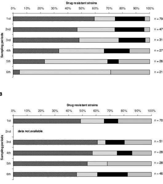
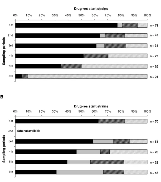
First, we conducted a prospective study to evaluate the PCV7 effect on the nasopharyngeal colonization, with particular emphasis on drug resistant strains in children attending day-care centers in Lisbon, Portugal. Comparison of vaccinated and control groups revealed that PCV7 caused replacement of strains expressing vaccine serotypes by novel clonal types of pneumococci that produced capsular polysaccharides not included in the vaccine. On the other hand, the general rate of carriage of pneumococci and the rate of carriage of drug resistant strains has remained unaltered.
and after vaccination) has allowed assessment of the actual mechanism of the vaccine’s effect. In conclusion, in children immunized with a single PCV7 dose a serotype replacement phenomenon occurred, both at the population and individual levels.
Moreover, the PCV7 mode of action relied on the prevention of the de novo acquisition
of VTs on one hand, and unmasking NVTs on the other.
In a final study, murine models of colonization and virulence were used to characterize the most common penicillin nonsusceptible non vaccine serotypes colonizing the nasopharynx of Portuguese children vaccinated with PCV7. Additionally, the role of the
capsule versus the genetic background in colonization and disease was also
A bactéria Gram-positiva Streptococcus pneumoniae existe normalmente como um microrganismo comensal, colonizador da nasofaringe humana, especialmente em crianças. Contudo esta bactéria é também um importante agente patogénico humano capaz de causar um vasto leque de doenças incluindo otite média, sinusite, pneumonia, e ainda patologias que colocam em risco a vida humana como a infecção do sangue e a meningite.
A vacina pneumocócica conjugada 7-valente (PCV7) foi criada para proteger crianças
contra a doença provocada por S. pneumoniae revelando enorme eficácia na
prevenção de doença causada pelos serótipos incluídos na vacina, também chamados serótipos vacinais. Sendo a colonização um primeiro passo fundamental para o desenvolvimento da doença pneumocócica, reveste-se da maior importância a investigação do efeito desta vacina no grau de colonização, nas alterações da composição da flora da nasofaringe e no potencial de virulência das estirpes não vacinais.
Os objectivos propostos para esta tese levaram à execução de estudos epidemiológicos e de experimentação animal. Os estudos epidemiológicos que realizámos permitiram avaliar o impacto da vacina PCV7 em colonização simples e múltipla em crianças portuguesas identificando os serótipos e os tipos clonais que substituem a flora pneumocócica original sob a influência da vacina. Por outro lado, estudou-se o potencial de virulência das estirpes pneumocócicas não vacinais seleccionadas pela vacina usando modelos animais de colonização e doença.
colonização geral por S. pneumoniae e a taxa de colonização por estirpes resistentes a antimicrobianos permaneceu inalterada.
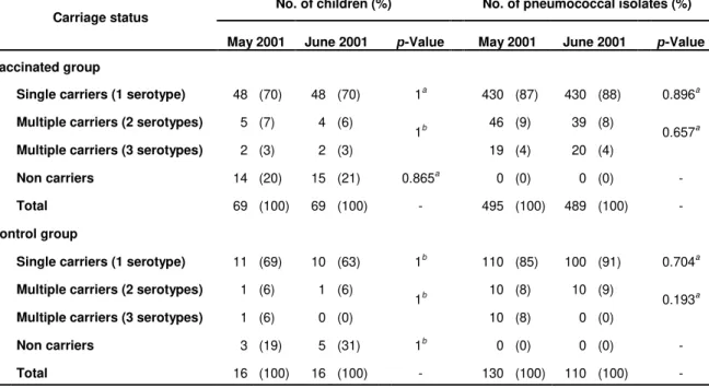
De modo a estudar o efeito de apenas uma dose da vacina PCV7, foi realizado um estudo-piloto no qual se estudou além da colonização pneumocócica simples a colonização pneumocócica múltipla. O estudo envolveu crianças que foram analisadas durante dois períodos de colheita com apenas um mês de intervalo (i.e., antes e depois da vacinação), o que permitiu identificar o mecanismo de acção da vacina. Os resultados obtidos neste estudo permitiram concluir que em crianças imunizadas com apenas uma dose de vacina ocorre um efeito de substituição de serótipos vacinais por não vacinais, tanto a nível da população como a nível do indivíduo. Foi possível ainda
concluir que o mecanismo de acção da PCV7 consiste na prevenção da aquisição de
novo dos serótipos vacinais por um lado e no “unmasking” de serótipos não vacinais por
outro.
No último trabalho apresentado nesta tese foram utilizados modelos murinos de colonização e doença. Estes modelos animais serviram para caracterizar os serótipos não vacinais e não susceptíveis à penicilina mais comuns, que colonizam a nasofaringe
de crianças portuguesas vacinadas com a PCV7. O papel da cápsula versus o
The work presented in this Doctoral Thesis describes the evaluation of the impact of the 7-valent pneumococcal conjugate vaccine (PCV7) on colonization among Portuguese children attending day care. This general objective was attained firstly by investigating the epidemiology of the pneumococcus, and secondly by assessing the virulence
potential of non vaccine serotypes selected in vivo by the PCV7 vaccine.
Chapter I – General introduction. This chapter provides an outline of the Streptococcus
pneumoniae epidemiology in colonization and disease. The pivotal role of the
pneumococcal capsule and antibiotic resistance on the development and impact of the pneumococcal vaccines is also addressed. Moreover, mouse models are described as important and valuable systems to study pneumococcal colonization and pathogenesis.
Chapter II – Effect of the seven-valent conjugate pneumococcal vaccine on carriage
and drug resistance of Streptococcus pneumoniae in healthy children attending
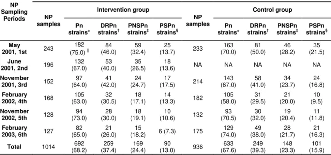
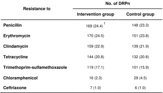
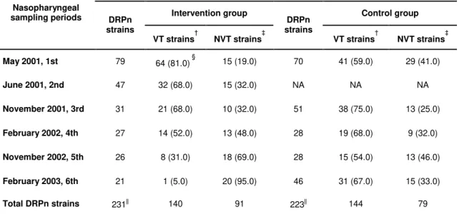
day-care centers (DCCs) in Lisbon. This chapter reports, for the first time in Portugal, the impact of the PCV7 in colonization and drug resistance among DCC attendees.
Chapter III – Impact of a single dose of the 7-valent pneumococcal conjugate vaccine
on colonization. This chapter describes the effect of one PCV7 dose on single and
multiple carriers, assessing the actual mechanism of the vaccine’s effect.
Chapter IV – Virulence potential of serotypes selected in vivo by the 7-valent
pneumococcal conjugate vaccine (PCV7) in Portugal. This chapter presents a study on
the virulence potential of colonizing non vaccine types selected in vivo by the PCV7
vaccine using several mouse models.
Chapter V – Conclusions and perspectives. This chapter combines the major findings of
this Doctoral thesis, suggesting future follow-ups of these studies.
Chapter II – Frazão, N., A. Brito-Avô, C. Simas, J. Saldanha, R. Mato, S. Nunes, N. G. Sousa, J. A. Carriço, J. S. Almeida, I. Santos-Sanches, and H. de Lencastre.
2005. Effect of the seven-valent conjugate pneumococcal vaccine on carriage and drug
resistance of Streptococcus pneumoniae in healthy children attending day-care centers
in Lisbon. Pediatr Infect Dis J 24:243-52.
Chapter III – Frazão, N., R. Sá-Leão, and H. de Lencastre. 2010. Impact of a single
dose of the 7-valent pneumococcal conjugate vaccine on colonization. Vaccine 28:
A
AOM – Acute otitis media
C
CDC – Centers for Disease Control
CFU – Colony forming units
CSF – Cerebrospinal fluid
D
DCC – Day-care center
DR – Drug resistant
DRPn – Drug resistant Streptococcus pneumoniae
I
i.cist. – Intracisternal
i.n. – Intranasal
IPD – Invasive pneumococcal disease
i.p. – Intraperitoneal
i.t. – Intratracheal
i.v. – Intravenous
M
MIC – Minimum inhibitory concentration
N
NCCLS – National Committee for Clinical Laboratory Standards
NCKP – Northern California Kaiser Permanente
NP – Nasopharyngeal
NT – Non typeable
NVT – Non vaccine type
P
PCV7 – 7-valent pneumococcal conjugate vaccine
PCV13 – 13-valent pneumococcal conjugate vaccine
PFGE – Pulsed-field gel electrophoresis
PMEN – Pneumococcal Molecular Epidemiology Network
Pn – S. pneumoniae
PNSPn – Penicillin nonsusceptible S. pneumoniae
PP23 – 23-valent pneumococcal polysaccharide vaccine
PS – Capsular polysaccharides
PSPn – Penicillin susceptible S. pneumoniae
R
RTI – Respiratory tract infections
S
SXT – Trimethoprim-sulfamethoxazole
V
ACKNOWLEDGMENTS ...V
ABSTRACT ...VII
RESUMO ...IX
THESIS OUTLINE...XI
LIST OF ABBREVIATIONS ...XIII
CHAPTER I ... 1
1. General introduction ... 3
1.1. Streptococcus pneumoniae... 3
1.2. Historical background ... 4
1.3. Epidemiology of Streptococcus pneumoniae... 5
1.3.1. Pneumococcal colonization... 5
1.3.2. Pneumococcal disease ... 6
1.3.3. Capsule, antibiotic resistance, and vaccines ... 7
1.4. Pneumococcal conjugate vaccine... 10
1.4.1. Immunological basis ... 10
1.4.2. Impact on colonization ... 11
1.4.3. Impact on disease... 15
1.5. Mouse models ... 17
1.5.1. Mouse model of colonization... 18
1.5.2. Mouse models of pneumococal disease ... 19
1.6. Aim of the work ... 25
1.7. References ... 27
CHAPTER II ... 47
2. Effect of the seven-valent conjugate pneumococcal vaccine on carriage and drug resistance of Streptococcus pneumoniae in healthy children attending day-care centers in Lisbon. ... 47
2.3. Materials and Methods ... 51
2.4. Results ... 54
2.5. Discussion ... 65
2.6. Acknowledgments... 67
2.7. References ... 69
CHAPTER III ... 75
3. Impact of a single dose of the 7-valent pneumococcal conjugate vaccine on colonization ... 75
3.1. Abstract ... 77
3.2. Introduction... 77
3.3. Materials and Methods ... 79
3.4. Results ... 82
3.5. Discussion ... 91
3.6. Acknowledgments... 94
3.7. References ... 95
CHAPTER IV... 101
4. Virulence potential of serotypes selected in vivo by the 7-valent pneumococcal conjugate vaccine... 101
4.1. Abstract ... 103
4.2. Introduction... 103
4.3. Materials and Methods ... 105
4.4. Results ... 108
4.5. Discussion ... 121
4.6. Acknowledgments... 124
4.7. References ... 125
CHAPTER V ... 129
5. Concluding remarks and future perspectives ... 131
1. General introduction
Streptococcus pneumoniae (the pneumococcus) has been studied for more than a
century and has so far bypassed all the human initiatives targeted to eliminate this apparently unbeatable pathogen. The widespread belief that antimicrobial drugs would cure all pneumococcal infections waned the interest in using vaccines to prevent infections. Only the emergence of antimicrobial resistant strains led to a recurring interest in developing new pneumococcal vaccines that aim to eventually eradicate this bacterium. To attain this long-term goal it is essential to be aware of the epidemiological dynamics of the pneumococcus.
Since colonization is the first step to disease development, an in-depth understanding of the major pneumococcal serotypes and clones carried in the nasopharyngeal mucosa together with data on their virulence potential is critical. Based on this information, it may be possible to predict future pneumococcal disease serotypes/genotypes and include them in future vaccine formulations.
1.1.
Streptococcus pneumoniae
Streptococcus pneumoniae, also known as the pneumococcus, is a lancet-shaped
gram-positive bacterium which can grow in liquid medium as single cells, diplococci or in
chains. It is a facultative anaerobic microorganism which shows α-hemolysis when
growing in blood agar plates, solubility when in presence of bile salts, catalase negativity and usually susceptibility to optochin. All these characteristics together with common positive agglutination with specific antipneumococcal polysaccharide capsule antibodies are taken into consideration when classifying a bacterium as belonging to the
Chapter I
1.2. Historical background
In 1875, while searching for a proof of the infectious nature of pneumonia, Edwin Klebs
was probably the first to recognize S. pneumoniae in pneumonic lung tissue,describing
the phenomenon as nonmotile, sometimes linked “monads” (White et al., 1938).
Six years later, in 1881, Louis Pasteur in France and Georg Sternberg in the USA
reported for the first time the isolation of S. pneumoniae in the laboratory (Pasteur,
1881; Sternberg, 1881).
After the discovery of the microorganism, its name changed several times. Pasteur called his isolate the “microbe septicémique du saliva” while Sternberg called his
Micrococcus pasteuri. In 1886, Albert Fraenkel performed the first complete description
of the microorganism and gave us the familiar name “pneumokokkus”. In the same year
Anton Weichselbaum suggested the name Diplococcus pneumoniae (White et al.,
1938), which became the official name until 1974 when the organism was reclassified
Streptococcus pneumoniae according to its characteristic property of growing as chains
of cocci in liquid media (Bergey, 1974).
Isolated and characterized more than 120 years ago, S. pneumoniae has been one of
the most extensively studied microorganisms leading to seminal discoveries in several scientific areas, including the putative use of polysaccharide antigens as vaccines
(Avery et al., 1917), bacterial gene transfer (Griffith, 1928), the isolation and chemical
characterization of the first polysaccharide antigen (Goebel & Adams, 1943), the identification of the “transforming principle” (later named DNA) as the genetic material
(Avery et al., 1944), the therapeutic efficacy of penicillin (Tillett et al., 1944), the role of
the bacterial capsule in resistance to phagocytosis (Felton et al., 1955), and the first
1.3. Epidemiology of
Streptococcus pneumoniae
Pasteur and Sternberg, although independently, performed similar experiments when
they isolated for the first time S. pneumoniae. Both injected human saliva, from
asymptomatic carriers, into rabbits that died shortly after from blood infection (Pasteur, 1881; Sternberg, 1881). Altogether, these pioneering experiments showed for the first
time that S. pneumoniae on one hand can be asymptomatically carried and on the other
has the potential to cause disease.
1.3.1. Pneumococcal colonization
The pneumococcus colonization is described as a commensal relationship with its principal host, man (Austrian, 1997). Asymptomatic pneumococcal colonization of the
human nasopharyngeal mucosa starts immediately after birth (Aniansson et al., 1992)
and, at any given time, up to 30% of adults and 60% of children are colonized (Austrian, 1986). The children’s nasopharynx constitutes the major reservoir for the pneumococcus, specially day-care center (DCC) attendees where pneumococcal colonization rate is particularly high. In Portugal up to 71% of the children up to 6 years
old attending day-care centers are colonized with pneumococci (Mato et al., 2005).
Depending on the host’s age and the pneumococcal colonizing strain, the duration of
carriage ranges between 1 to 17 months (Gray et al., 1980). Pneumococci can present
two patterns of colonization: i) single colonization, where the host carries only one pneumococcal strain or ii) multiple colonization, also called co-colonization, where the host carries simultaneously more than one pneumococcal strain, which can differ in
phenotypic and/or genotypic characteristics (Auranen et al., 2010; Frazão et al., 2010;
Gray et al., 1980; O'Brien et al., 2007; Sá-Leão et al., 2002).
Chapter I
a sibling and day-care center attendance. Among adults, recurrent contact with children and crowdness constitute the major risk factors to be colonized with pneumococci (Kristinsson, 1997).
Pneumococcal colonization is usually studied by performing the characterization of a single pneumococcal isolate for each colonized individual. However, studies show that
individuals can be co-colonized with different pneumococcal strains (Auranen et al.,
2010; Frazão et al., 2010; Gray et al., 1980; O'Brien et al., 2007; Sá-Leão et al., 2002).
Epidemiological studies based on a single isolate characterization are important and valuable tools to detect differences in the pneumococcal dynamics at a population level. However, studies where co-colonization is detected are crucial to identify differences at
the individual level, such as de novo acquisition, clearance and unmasking of
pneumococcal strains (Dagan et al., 2003; Lipsitch, 1999; Rinta-Kokko et al., 2009).
1.3.2. Pneumococcal disease
The pneumococcus is described as being a commensal human pathogen and
colonization of the nasopharyngeal mucosa is often a transient process (Hill et al., 2008;
Hogberg et al., 2007), constituting the initial event in the progression to disease
(Henriques-Normark & Normark, 2010). As a pathogen S. pneumoniae can give rise to a
wide variety of diseases that can be divided into invasive and non-invasive. In invasive
pneumococcal disease (e.g. meningitis, bacteremic pneumonia and bloodstream
infections – bacteremia and septicemia), pneumococcus can be isolated from blood or
other normally sterile body fluids. In non-invasive pneumococcal disease, also known as mucosal infections such as sinusitis, conjunctivitis, non-bacteremic pneumonia and otitis
media, pneumococcus can be isolated from mucosal excretions only (Bogaert et al.,
2004a; Feldman & Klugman, 1997; Musher, 1992).
The spectrum of pneumococcal diseases differs in different age groups and different
populations (Bogaert et al., 2004a; Hausdorff et al., 2005; Musher, 1992; O'Brien &
underlying illness, socio-economic status, previous antibiotic therapy and day-care attendance have been reported (O'Brien & Santosham, 2004).
Pneumococcus is a leading pathogen, causing infections with high mortality and
morbidity (Austrian, 1977; Greenwood et al., 2007; Hausdorff et al., 2000a; Hausdorff et
al., 2000b; Mulholland, 2007; O'Brien & Santosham, 2004; Scott et al., 1996). Up to one
million children die annually from pneumococcal diseases, and most of them are young
children in developing countries (O'Brien et al., 2009; WHO, 1999; Williams et al., 2002).
In industrialized countries invasive pneumococcal infections occur, especially among
children and elderly people (Eskola et al., 1992; Hausdorff et al., 2005; Sankilampi et al.,
1997; Scott et al., 1996).
1.3.3. Capsule, antibiotic resistance, and vaccines
In the overwhelming majority of pneumococcal isolates the outside surface of the bacterium is covered by a polysaccharide capsule, which plays a major role in
colonization and disease and constitutes the main virulence factor of S. pneumoniae.
The chemical nature of the capsule and the amount of capsule produced are major factors inhibiting complement activity, neutrophil phagocytosis, and bacterial killing by
neutrophil extracellular traps (Hyams et al., ; Kim & Weiser, 1998; Kim et al., 1999;
Macleod & Krauss, 1950; Magee & Yother, 2001; Nelson et al., 2007; Paton et al., 1993;
Quin et al., 2007). The capsule also plays a major role in bacterial interactions withthe
epithelium in colonization (Bootsma et al., 2007; Macleod & Krauss, 1950; Magee &
Yother, 2001; Nelson et al., 2007; Quin et al., 2007; Wartha et al., 2007).
The pneumococcal capsules are very diverse in chemical structure and a nomenclature system was created based on the antigenic differences of the capsular polysaccharides (PS) grouping the capsules into more than 90 serotypes (Austrian, 1981; Henrichsen, 1999; Sorensen, 1995). In recent years additional new serotypes 6C, 6D and 11E were discovered, suggesting that the diversity of pneumococcal capsules is even greater than
was previously recognized (Calix & Nahm, 2010; Henrichsen, 1995; Jin et al., 2009;
Chapter I
Shortly after the pneumococcal capsule was identified as a major immunogen in the 1930’s, vaccine development focused on purified PS culminating with the commercialization of two hexavalent polysaccharide vaccines in 1946 in the United States (Felton, 1938; Francis & Tillett, 1930). Concomitantly, sulfonamides and penicillin had become readily available and new antimicrobial drugs were on the way. The discovery of antimicrobials paved the way for better health for millions around the world. Before penicillin became a viable medical treatment in the early 1940's, no true cure for gonorrhea, strep throat, or pneumonia existed. Patients with infected wounds often had to have a wounded limb removed, or face death from infection. The dawn of antimicrobial drugs enabled all these fierce medical conditions to be cured with a short course of antimicrobial treatment. Thus, the vaccines were forgotten and were
withdrawn from the market in 1954 for lack of demand (Fedson et al., 1994). However, it
did not take long before it became clear that antimicrobials had not eliminated pneumococcal disease.
Resistance to antimicrobials is closely linked to the fact that S. pneumoniae is a
naturally transformable bacterium. Pneumococci, when in a competent state, have the capacity of taking up DNA and incorporate it into their genome (Claverys & Havarstein, 2007). Whenever the DNA incorporated into the genome has resistance genes the pneumococci become resistant to drugs.
In 1964, a study by Austrian and Gold reported that nearly one in four patients admitted with pneumococcal bacteremia died even with antimicrobial drug treatment (Austrian & Gold, 1964). In 1967, the first intermediately penicillin resistant pneumococcal isolate was reported in Australia (Hansman & Bullen, 1967). In the following years penicillin remained the drug of choice to treat pneumococcal infections as resistance to other drugs such as tetracyclines, chloramphenicol, macrolides, and trimethoprim-sulfamethoxazole (SXT) had emerged (Klugman, 1990). In 1978, the first pneumococcal strains resistant to all the above mentioned antimicrobial classes, as well as fully
resistant to penicillin, were isolated (Jacobs et al., 1978). It was the beginning of the
(Klugman, 1990). In the 1990s, multidrug resistance continued to increase such that in the United States in 2000 nearly half of all invasive pneumococcal disease (IPD) was
caused by pneumococcal isolates resistant to penicillin and/or macrolides (Whitney et
al., 2000).
The widespread belief that antimicrobials alone would “solve” all pneumococcal infections was definitely put aside as soon as pneumococcal drug resistance became a reality causing treatment failures (Kaplan & Mason, 2002). Consequently, a renewed interest in the prevention through vaccination was regained and led to clinical trials of a PS vaccine. In 1977 the Merck’s 14-valent pneumococcal polysaccharide vaccine (Pneumovax) was licensed in the United States. Similar 14-valent vaccines were later produced by Lederle (Pnu-Immune) and by Pasteur Mérieux. The 14 PS types included in these vaccines (1, 2, 3, 4, 6A, 7F, 8, 9N, 12F, 14, 18C, 19F, 23F, and 25F) were selected based on epidemiological information from the United States, parts of Europe, and South Africa, being the most common serotypes causing pneumococcal disease
(Robbins et al., 1983). In 1983 the 14-valent vaccine was expanded to include 23 PS
types, and is currently the only approved formulation available for adults in the United States (Fedson & Musher, 2004; Siber, 1994).
The main disadvantage of the 23-valent vaccine is the fact that it is not effective in children less than two years old, who are at highest risk of life-threatening pneumococcal infection and acute otitis media (CDC, 2000; Makela & Butler, 2008). To overcome this major weakness the concept of pneumococcal conjugate vaccine was developed. The novelty of these conjugate vaccines is the fact that PS types instead of being “presented” alone to the child’s immune system are attached to an immunogenic carrier protein that allows a highly efficient antibody production and induces immunological memory.
Chapter I
PCV7 was introduced in June 2001 and, despite widespread vaccination, it is still not part of the national vaccination plan.
Very recently, higher valency pneumococcal conjugate vaccines were licensed such as PCV10 (10-valent pneumococcal conjugate vaccine), which includes the PCV7 serotypes plus serotypes 1, 5, and 7F. PCV13 (13-valent pneumococcal conjugate vaccine) was the last to be licensed and includes PCV10 serotypes plus serotypes 3, 6A, and 19A.
1.4. Pneumococcal conjugate vaccine
The pneumococcal conjugate vaccine was a big step forward to win the apparently
never-ending war against the S. pneumoniae pathogen, especially in children under two
years of age who are at higher risk of contracting pneumococcal infections. This age group is not immunologically responsive to the 23-valent pneumococcal polysaccharide vaccine, but is protected by a pneumococcal conjugate vaccine.
1.4.1. Immunological basis
The primary pneumococcal antigens eliciting a host immune response are the pneumococcal capsular polysaccharides, which induce a T-cell independent immune response that is virtually absent in children until around two years of age. Conversely, when capsular polysaccharides are covalently coupled to immunogenic proteins such as
the mutant diphtheria toxin CRM197 used in PCV7, a T-cell dependent response is
The pneumococcal conjugate vaccine can confer both systemic and mucosal immunity
(Korkeila et al., 2000; Nieminen et al., 1999; Nurkka et al., 2001a).The immunological
protection is mediated through the production of specific antibodies. Mucosal immunity is mainly mediated through the production of serotype-specific immunoglobulin A (IgA)
antibodies, which are locally produced at the nasopharyngeal mucosa (Choo et al.,
2000; Korkeila et al., 2000; Nurkka et al., 2001a). Serotype-specific immunoglobulin G
(IgG) antibodies are mainly found in serum, conferring systemic immunity. However, they can also be detected in the nasopharyngeal mucosa, although more rarely than IgA
(Choo et al., 2000; Kauppi et al., 1995; Korkeila et al., 2000; Nurkka et al., 2001a;
Nurkka et al., 2001b). It is believed that circulating IgG antibodies can passively cross
the nasal mucous membrane and help prevent pneumococcal colonization, though
mucosal IgG production has been suggested to happen as well (Berneman et al., 1998;
Ogra, 2000).
1.4.2. Impact on colonization
The impact of the 7-valent pneumococcal conjugate vaccine on colonization can have both a direct effect — protecting those successfully immunized from carriage — and an
indirect effect — providing protection against carriage among unimmunized individuals
by reducing transmission of the organism within the community.
The vaccination schedule varies between countries, but all the primary PCV7 immunization series include two or three vaccine doses between 6 weeks and 6 months of age. A booster dose in the second year of life is the recommended practice in economically developed countries, but is not included in the schedules of developing countries due to the high price of the vaccine (ACIP, 2000). Studies of the effect in colonization of fewer than the recommended doses are scarce. A mathematical model of vaccination suggested that a single dose given between 5 and 7 months of age could
prevent up to one-third of invasive pneumococcal disease (Barzilay et al., 2006).
Chapter I
development of less expensive vaccine schedules that eventually may foster the introduction of pneumococcal vaccines in developing countries.
The major conclusion concerning the direct effect of the conjugate vaccines is that in vaccinated individuals there is a reduction in the prevalence of nasopharyngeal colonization by the serotypes included in the pneumococcal conjugate vaccines. This general effect has been shown in children after a primary PCV immunization series
(Dagan et al., 1997; Mbelle et al., 1999; O'Brien et al., 2007), or after a boosting dose of
either the 23-valent polysaccharide or conjugate vaccines (Dagan et al., 1997; Dagan et
al., 2000; Kilpi et al., 2001; Obaro et al., 1996).
In the specific case of children attending DCCs, where colonization rates are normally
very high (Dagan et al., 1996a; Dagan et al., 2002; Dagan et al., 2005; Frazão et al.,
2005), the pneumococcal conjugate vaccine also showed efficacy in reducing the colonization by vaccine serotype strains. The same effect of reducing vaccine type colonization was also reported when immunization was carried out with fewer than the
recommended conjugate vaccine doses (Frazão et al., 2010; Jones et al., 2005; van
Gils et al., 2009)
The above mentioned effect has been reported for all serotypes in the pneumococcal conjugate vaccine formulation. However, in some studies, the effect on serotype 19F is
reported as being the lowest (Dagan et al., 2002; Dagan et al., 2005; Huang et al., 2005;
Millar et al., 2006; Veenhoven et al., 2003). Concerning the vaccine related serotypes,
the ones belonging to the same serogroups, 6A prevalence decreases in vaccinated
individuals whereas 19A and 23A prevalence increases (Huang et al., 2005; Millar et al.,
2006; Sá-Leão et al., 2009; Veenhoven et al., 2003). Regarding the non vaccine
serotypes, there is an increase in vaccinated children when compared to non vaccinated
children (Dagan et al., 1996a; Dagan et al., 1997; Dagan, 2002; Frazão et al., 2005;
Frazão et al., 2010; Sá-Leão et al., 2009). The immune response to the pneumococcal
studies. However, there are strong indications that the primary mechanism of the
vaccines’ effect is the prevention of the de novo acquisition of VTs, rather than
clearance, and additionally the reduction of the density of colonization by the vaccine
serotypes (Dagan et al., 2005; Frazão et al., 2010; O'Brien et al., 2007).
The reduction in the VT pneumococcal carriage due to the PCV thus opens a biological niche that is filled by non vaccine type (NVT) strains leading to a phenomenon which has been termed as serotype replacement colonization. The colonization of the nasopharynx with serotypes not included in the vaccine (NVTs) can happen through i) capsular serotype switching, such that previous vaccine type clones exhibit now NVT capsules, ii) the expansion and propagation of existing NVT clones in the community, and iii) the introduction of new NVT clones into the community.
Worth to be investigated should be the “unmasking” phenomenon in which a minority clone/serotype is masked by a dominant clone/serotype in hosts that carry more than one type of pneumococci at the same time. This can only be addressed if multiple colonization (co-colonization) studies are undertaken. These studies may clarify whether the new NVT clones/serotypes are in fact new or simply types already present but masked and unidentified by approaches where only a single pneumococcal isolate per sample was recovered.
Chapter I
eligible for vaccination, pointing at the same time to an increased prevalence of non
vaccine serotypes colonizing the nasopharynx of unvaccinated individuals (Givon-Lavi et
al., 2003; Hammitt et al., 2006; Hennessy et al., 2005; Moore et al., 2004; O'Brien et al.,
2007).
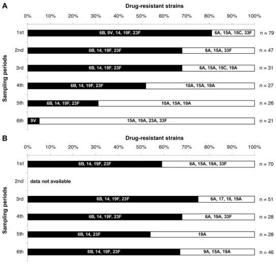
The effect of the pneumococcal conjugate vaccine on antibiotic resistance is explained by the fact that the global pandemic of pneumococcal resistance was dominated by a relatively small number of resistant and multidrug resistant clones that express mainly five (6B, 9V, 14, 19F, and 23F) of the seven serotypes included in the PCV7 vaccine
(Dagan & Klugman, 2008; Kyaw et al., 2006; McGee et al., 2001). Since both the direct
and indirect effects of the pneumococcal conjugate vaccine on colonization lead to the decrease of the VTs, which are strongly associated with resistance, particularly high-level penicillin resistance and multidrug resistance, one can expect that vaccination with
a pneumococcal vaccine will reduce the prevalence of antibiotic resistant S.
pneumoniae strains. In fact, studies in Israel, where multi-valent pneumococcal
conjugate vaccines were used to immunize children, reported a reduction in the drug
resistant pneumococcal strains colonizing the nasopharynx (Dagan et al., 1996a; Dagan
et al., 1997; Dagan et al., 2000; Dagan et al., 2003; Mbelle et al., 1999). Nevertheless, in some locations the increasing prevalence of NVTs, also drug resistant, as a result of the serotype replacement phenomenon led to a compensatory effect that allows the maintenance of pneumococcal drug resistance in the populations. In Portugal, after vaccination with the PCV7 the carriage rate of drug resistant pneumococcal strains exhibiting vaccine capsular types decreased, but was compensated by a gradual increase in the prevalence of drug resistant NVTs, nonsusceptible to penicillin (6A, 6C,
10A, 15A, 15B/C, 19A, 24F and 33F) (Frazão et al., 2005; Sá-Leão et al., 2009). In the
United States, a similar trend was found (Huang et al., 2005). In PCV7 vaccinated
children, parallel to the decrease of drug resistant VTs, a substantial increase in NVTs was seen, namely in the most commonly carried penicillin nonsusceptible serotypes: 6A,
Moore and co-workers also found the same serotype dynamics in vaccinated children where the carriage of penicillin nonsusceptible strains was maintained after vaccination
with PCV7 (Moore et al., 2004).
Possible explanations for the rise of drug resistant strains after vaccination with a
pneumococcal conjugate vaccine include the de novo acquisition of resistance, serotype
switching, the introduction of new clones, and the expansion of existing clones. In Portugal the increase of drug resistance among the non vaccine type population (6A,
10A, 15A, 15B/C, 19A, 23A, and 33F) seems to be due to the de novo acquisition of
resistance (a rare phenomenon only observed for serotype 10A) and, most frequently,
the introduction of new clones and the expansion of existing ones (Frazão et al., 2007).
Hanage and co-workers, observed a similar scenario where resistance increased mainly through the expansion of pre-existent clones of non vaccine serotypes, particularly 15A,
19A, and 35B (Hanage et al., 2007).
1.4.3. Impact on disease
The direct effect of the 7-valent pneumococcal conjugate vaccines translates into a
sharp decline of invasive disease among immunized children caused by S. pneumoniae
serotypes included in its formulation (Aguiar et al., 2008; Aguiar et al., 2010; Bettinger et
al., 2010; Black et al., 2004; Dagan et al., 2001; Hennessy et al., 2005; Hsu et al., 2005;
Kellner et al., 2005; Munoz-Almagro et al., 2008; Pilishvili et al., 2010).
Concerning the efficacy of PCV7, data from the Northern California Kaiser Permanent health system (NCKP), which conducted the first clinical trial to evaluate PCV7, demonstrated an efficacy of 97% against invasive pneumococcal disease (IPD) caused
by the vaccine serotypes (Black et al., 2000). After the PCV7 licensure in 2000 in the
United States, a population-based study from the Active Bacterial Core Surveillance of the Centers for Disease Control (CDC) and Prevention reported a large IPD reduction from 24.3 cases in 1998-1999 to 17.3 per 100000 in 2001. The main decline was observed in children younger than two years old showing a 78% decrease in the rate of
Chapter I
Regarding the pneumococcal non-invasive mucosal disease, the impact of the pneumococcal conjugate vaccine was confirmed although considered moderate. The estimated efficacy of PCV7 in preventing first episodes of radiographic pneumonia was
30% (Hansen et al., 2006). Additionally, studies based on data from the NCKP showed
a 7% efficacy in preventing episodes of otitis media, a disease with high burden and
associated related costs. The same was found in the FinOM trial (Black et al., 2000;
Eskola et al., 2001; Kilpi et al., 2001).
Just like in the colonization phenomenon, in disease too, the pneumococcal conjugate vaccine was also accompanied by an indirect effect. The rationale for this relies on the fact that by immunizing a subset of a population (children) one can reduce significantly the circulation of vaccine types, reducing carriage and even more the disease caused by these serotypes, among the non-vaccinated individuals. Evidence for this indirect or herd immunity effect, described early after PCV7 licensure, highlights an important component of the public health profit of pneumococcal conjugate vaccine use. A report from the NCKP study indicated that the reduction in disease was greater than the
percentage of children vaccinated (Black et al., 2001). A similar pattern was observed in
the CDC data where the rate of vaccine type disease among children less than 5 years old dropped 94%, even though only 68% of children had been vaccinated (CDC, 2005). Reported decrease of IPD caused by vaccine types among children too young to be immunized (under 2 months) and older children and adults who are not eligible for vaccination further confirm the herd immunity effect of the pneumococcal conjugate
vaccine (Black et al., 2004; Poehling et al., 2006; Whitney et al., 2003).
The overall reduction of the disease-causing vaccine serotypes in settings where pneumococcal conjugate vaccines were administered raised an expectancy of decreasing the overall pneumococcal drug resistance. This expectation is based on the fact that five of the serotypes included in the conjugate vaccine (6B, 9V, 14, 19F, and 23F) were responsible for the prevalence of drug resistance among pneumococci. Indeed, several studies reported large decreases in disease episodes caused by drug
resistant pneumococci (Hennessy et al., 2005; Kaplan et al., 2004; Klugman et al., 2003;
and that caused by drug resistant pneumococci in particular, were observed, an increase in disease caused by non vaccine types, namely resistant to drugs, was
reported (Byington et al., 2005; Eskola et al., 2001; Hicks et al., 2007; Porat et al.,
2004). The substitution of vaccine by non vaccine serotypes in disease due to the selective pressure exerted by the conjugate vaccine is called replacement disease. Early studies of the impact of vaccination found that, among children, increases in the rate of disease due to non vaccine serotypes were either small in magnitude or limited to
a single geographic location (Byington et al., 2005; Flannery et al., 2004; Kaplan et al.,
2004). It was hoped that IPD caused by non vaccine types such as 6A, and 19A could be prevented through cross protection with serotypes 6B and 19F included in the PCV7.
Although this reduction has been reported for serotype 6A (Millar et al., 2006; Whitney et
al., 2003), an increase rather than a decrease in serotype 19A IPD has been observed
(Beall et al., 2006; Hennessy et al., 2005; Pai et al., 2005). More recent data showed
larger increases in the incidence of serotype 19A and serogroup 15 strains among children and of multiple non vaccine serotypes among adults, specially with HIV/AIDS,
where the infection with S. pneumoniae occurs up to 100 times more frequently than
among the general population (Aguiar et al., 2010; Dworkin et al., 2001; Flannery et al.,
2006; Hicks et al., 2007; Munoz-Almagro et al., 2008; Nuorti et al., 2000; Pai et al.,
2005).
In just a few years after the licensing of the PCV7 vaccine, serotype 19A has become the predominant cause of invasive disease in children and, not less importantly as it can
lead to medical treatment failures, increasingly drug resistant (Aguiar et al., 2010; Hicks
et al., 2007; Munoz-Almagro et al., 2008; Pai et al., 2005).
1.5. Mouse models
Experimental animal models have been used extensively to understand vital processes
of the pneumococcal colonization and disease (Chiavolini et al., 2008; Malley & Weiser,
Chapter I
1.5.1. Mouse model of colonization
The pneumococcal colonization is a crucial step for invasive disease development,
rendering the in vivo study of this phenomenon fundamental to complement
epidemiological and in vitro studies.
The mouse model is the most widely used for studying pneumococcal colonization, for a number of reasons: i) mice are colonized by pneumococci of multiple serotypes, ii) there is availability of reagents and diversity of mice strains (such as CD1, C57BL/6, BALB/c, and CBA/n), iii) the intranasal inoculation of anesthetized mice with selected strains can subsequently lead to disease, in a way that mimics natural pneumococcal infections in
humans (Balachandran et al., 2002; Takashima et al., 1997; Wu et al., 1997), and iv) the
persistence of colonization in mice was shown to last several weeks, which again may
be viewed as representative of what may happen in humans (Gray et al., 1980; Hogberg
et al., 2007; McCool & Weiser, 2004). Moreover, the characterization of experimental nasal colonization in humans provided a basis for assessing the applicability of the mouse models of colonization. An example of this is the fact that the same isolate used in human studies was found to colonize mice in association with a similar inoculum dose, duration, and immune response (McCool & Weiser, 2004).
Several variations of the model have been used, but in the majority of cases, a 10-µl bacterial inoculum is intranasally dropped atraumatically into the nares of the mice. After inoculation the presence and density of pneumococcal colonization can be investigated over the subsequent days, generally by obtaining nasopharyngeal washes from recently
euthanized animals (Wu et al., 1997).
Several limitations have been attributed to the mouse model, such as the fact that different laboratories have used different mouse strains, which can vary in susceptibility to colonization and may prevent reproducibility of results. More specifically, much of the
research work has been carried out using mainly the D39 isolate (Iannelli et al., 1999),
when compared to other pneumococcal isolates (Lipsitch et al., 2007; van Ginkel et al.,
2003; Wu et al., 1997).
Mouse models have provided important information on the pneumococcal commensal state, colonization. For example, using a mouse model it was shown that a minimum amount of capsular polysaccharide is needed for efficient colonization, and that unencapsulated mutants remain capable of colonizing but at a lower density and duration when compared with the capsulated parental strains (Magee & Yother, 2001;
Nelson et al., 2007). Regarding prevention strategies to avoid colonization by using
protein vaccines, it was shown in a mouse model that the choline-binding protein A (CbpA), also named PspC or SpsA, is crucial for pneumococcal colonization and that another choline-binding surface protein, PspA, can elicit a mucosal immune response
that may confer protection against colonization (Balachandran et al., 2002; Rosenow et
al., 1997).
Also the ease of use of genetically modified mice allows the characterization of host factors essential in colonization. Studies which examined the time course of colonization using genetically modified mice showed that pneumococcal clearance in the nasopharynx is dependent on members of a family of Toll-like receptors, which recognize pathogen associated patterns and lead to efficient clearance of colonization
(van Rossum et al., 2005). In recent years, McCool and Weiser showed, using a mouse
model of colonization, that pneumococcal colonization occurs in an antibody-independent manner (McCool & Weiser, 2004).
1.5.2. Mouse models of pneumococal disease
Pneumococcal disease can range from mild to life-threatening infections. Similarly to what happens with colonization, the most commonly used animal model for pneumococcal disease is the mouse.
Chapter I
diverse scenarios of pneumococcal disease, such as pneumonia, septicemia,
meningitis, and otitis media (Briles et al., 1981; Canvin et al., 1995; Iizawa et al., 1996;
Melhus & Ryan, 2003; Shapiro et al., 2000; Wang et al., 2001; Zwijnenburg et al., 2001).
The mouse outbred strains have become increasingly popular because they maintain the maximum heterozygosity, which leads to a phenotypic diversity resembling that of humans. This feature is important as it mimics the natural variation in response to infection in humans. Moreover, the lower cost of these outbred mice strains makes them attractive alternatives to inbred strains.
1.5.2.1. Pneumonia model
Mouse models of pneumococcal pneumonia allow the analysis of various parameters such as survival after challenge, bacterial presence in lungs and blood, inflammation levels, and histology of lung tissue. Additionally, quantification of antibody titers and antimicrobials performed in vaccine and drug pharmacokinetic studies, respectively, are also feasible. Two main routes of infection are currently used to induce pneumonia: the intratracheal (i.t.) and the intranasal (i.n.) route.
The i.t. model is a complex and invasive technique, but has the advantage of allowing the delivery of the entire bacterial inoculum into the lower respiratory tract directly
causing pneumonia (Rubins et al., 1996). Briefly, the bacterial suspension is injected
trough the mouse oropharynx or through the mouse exposed or cannulated trachea into
the lungs (Azoulay-Dupuis et al., 1991; Iwasaki et al., 1999; Tasaka et al., 2002).
The i.t. model is still used in studies of drug efficacy, host response to infection, and the
role of pneumococcal virulence factors in the disease process (Abgueguen et al., 2007;
Mohler et al., 2003; Rubins et al., 1996).
The i.n. model is a simple and non-invasive technique which includes both the standard
aspiration method (Canvin et al., 1995) and the aerosol nebulizer system (Nuermberger
route of infection in humans. Briefly, a volume of bacterial inoculum higher than 10 µL is intranasally dropped atraumatically into the nares of anesthetized mice. The model of i.n. aerosol system requires an exposure chamber equipped with a nebulizer. This aerosol technique less closely resembles the development of pneumococcal pneumonia in humans, where the disease generally follows aspiration of bacteria from the upper respiratory tract.
Studies on the host immune response to lung infection, the role of virulence determinants, mice susceptibility and resistance to disease, the efficacy of antibiotics and anti-inflamatory drugs and even studies on protection from i.n. challenge after immunization with vaccine candidates have been carried out using the pneumonia
mouse model (Alexander et al., 1994; Bergeron et al., 1998; Blue et al., 2003; Coil et al.,
1978; Gingles et al., 2001; Wang et al., 2000; Wang et al., 2005).
1.5.2.2. Septicemia model
The mouse is the most common experimental animal used to study septicemia induced by S. pneumoniae. The occurrence of pneumococcal septicemia in mice is assessed mostly by determining the presence of bacteria in the blood and by observing post-challenge survival. Pneumococcal septicemia is induced in the mouse either by injecting bacteria directly into the bloodstream (i.v.) or by injection into the peritoneal cavity (i.p.).
In brief, the i.v. model consists of an injection of the bacterial suspension into the mouse tail vein. Due to the reduced size of the mouse vein, an infrared lamp is used to allow vasodilatation and facilitate the procedure. The i.p. route of infection is one of the earliest techniques used to induce septicemia and consists of an injection of the bacterial suspension into the peritoneal cavity, which leads to posterior infection of the blood. The i.v. model of infection is a more direct system than the i.p. model. However, the former can be time-consuming and difficult to perform comparing to the i.p. route, which is technically easier but presents disadvantages including the risk of tissue
Chapter I
Studies that aimed to investigate the role of virulence factors during septicemia, the efficacy of potential vaccines and of novel antimicrobials, as well as blood clearance
mechanisms have been performed using the septicemia mouse model (Benton et al.,
1995; Briles et al., 1981; Briles et al., 1992; Brown et al., 2001; Cao et al., 2007; Casal
et al., 2002; Iannelli et al., 2004; Loeffler et al., 2003; Ogunniyi et al., 2007; Swiatlo et
al., 2003).
1.5.2.3. Meningitis model
Only recently has the mouse become an experimental system for studying meningitis
induced by S. pneumoniae.
There are two main types of procedures to induce pneumococcal meningitis in mice – a direct infection by intracerebral or the intracisternal (i.cist.) route and infection via the blood route (i.p. or i.n.). A third route was recently described by van Ginkel and co-workers, in which pneumococci colonize the nasopharynx and enter the central nervous system along the olfactory nerves without causing blood infection. The latter contradicts the assumption that pneumococci induce meningitis only by invasion via the blood (van
Ginkel et al., 2003).
The direct infection of the central nervous system mimics the contiguous spread of pneumococci infecting the sinuses or middle ear or the direct infection due to trauma. It is an experimental system that allows the study of host-pneumococci interactions once the disease is established, but does not allow the study of the previous steps that occur
from colonization to disease (Koedel et al., 2002). Conversely, meningitis induced via
i.n. or i.p. routes allows the analysis of pathogenesis according to what is believed to be the natural route of infection. However, a major disadvantage is the fact that using this technique ~50% of the mice die due to septicemia even before developing meningitis
(Tsao et al., 2002; Zwijnenburg et al., 2001).
presence in the brain and cerebrospinal fluid, histological analysis, and determination of
leukocyte and cytokine levels (Gerber et al., 2001; Grandgirard et al., 2007; Koedel et
al., 2001; Marra & Brigham, 2001; Tan et al., 1995).
Studies on the efficacy of antimicrobials against meningitis, host and pneumococcal virulence factors involved in meningitis pathogenesis, and the assessment of postinfectious sequelae have been performed using the meningitis mouse model
(Bottcher et al., 2003; Echchannaoui et al., 2002; Iizawa et al., 1998; Kostyukova et al.,
1995; Nau et al., 1999; Shapiro et al., 2000; Wellmer et al., 2000).
1.5.2.4. Otitis media model
The interest in using the mouse as a model for otitis media has been growing in recent years as mice present advantages when compared to other animal systems, including lower price and a better characterization regarding immunological and genetic information. Drawbacks of using the mouse as an otitis media model include its small size together with the reduced accessibility of the middle ear for inoculation and sampling purposes.
In the mouse model of otitis media two different routes are used: direct injection (intratympanic or intrabullar) into the middle ear, and the i.n. route that mimics the natural mode of middle ear infection. Briefly, in the direct injection there is a direct application of the inoculum into the middle ear cavity either through the tympanic membrane or by exposing the bulla and injecting the bacterial suspension through the
bony wall using a thin needle (Ryan et al., 2006; Sabirov & Metzger, 2006). In the i.n.
route, an intranasal inoculation is performed to establish colonization of the nasopharynx, which is followed by invasion of the middle ear cavity by the pneumococci
in about 50% of the cases (Ryan et al., 2006; Sabirov & Metzger, 2008).
Chapter I
applied to translocate pneumococci from the nasopharyngeal cavity into both mouse
middle ears (Stol et al., 2009)
Studies on potential vaccines and new therapies, pneumococcal virulence factors, inflammation, and histology have been carried out using the otitis media mouse model
(MacArthur et al., 2006; McCoy et al., 2005; McCullers et al., 2007; Melhus & Ryan,
1.6. Aim of the work
S. pneumoniae is a colonizer of the nasopharyngeal mucosa in young children.
1.7. References
Abgueguen, P., Azoulay-Dupuis, E., Noel, V., Moine, P., Rieux, V., Fantin, B. &
Bedos, J. P. (2007). Amoxicillin is effective against penicillin-resistant Streptococcus
pneumoniae strains in a mouse pneumonia model simulating human pharmacokinetics.
Antimicrob Agents Chemother51, 208-214.
ACIP (2000). Preventing pneumococcal disease among infants and young children:
recommendations of the Advisory Commitee on Immunization Practices (ACIP). MMWR
Morb Mortal Wkly Rep49, 1-35.
Aguiar, S. I., Serrano, I., Pinto, F. R., Melo-Cristino, J. & Ramirez, M. (2008).
Changes in Streptococcus pneumoniae serotypes causing invasive disease with
non-universal vaccination coverage of the seven-valent conjugate vaccine. Clin Microbiol
Infect14, 835-843.
Aguiar, S. I., Brito, M. J., Gonçalo-Marques, J., Melo-Cristino, J. & Ramirez, M.
(2010). Serotypes 1, 7F and 19A became the leading causes of pediatric invasive
pneumococcal infections in Portugal after 7 years of heptavalent conjugate vaccine use.
Vaccine28, 5167-5173.
Alexander, J. E., Lock, R. A., Peeters, C. C., Poolman, J. T., Andrew, P. W.,
Mitchell, T. J., Hansman, D. & Paton, J. C. (1994). Immunization of mice with
pneumolysin toxoid confers a significant degree of protection against at least nine
serotypes of Streptococcus pneumoniae. Infect Immun62, 5683-5688.
Aniansson, G., Alm, B., Andersson, B., Larsson, P., Nylen, O., Peterson, H.,
Rigner, P., Svanborg, M. & Svanborg, C. (1992). Nasopharyngeal colonization during
the first year of life. J Infect Dis165 Suppl 1, S38-42.
Antao, V. C. & Hausdorff, W. P. (2009). Global epidemiology of pneumococcal
disease--new prospects for vaccine control. Adv Exp Med Biol634, 19-29.
Auranen, K., Mehtala, J., Tanskanen, A. & M, S. K. (2010). Between-strain
competition in acquisition and clearance of pneumococcal carriage--epidemiologic
evidence from a longitudinal study of day-care children. Am J Epidemiol171, 169-176.
Austrian, R. & Gold, J. (1964). Pneumococcal Bacteremia with Especial Reference to
Bacteremic Pneumococcal Pneumonia. Ann Intern Med60, 759-776.
Austrian, R. (1977). Pneumococcal infection and pneumococcal vaccine. N Engl J Med
297, 938-939.
Austrian, R. (1981). Pneumococcus: the first one hundred years. Rev Infect Dis3,
Chapter I
Austrian, R. (1986). Some aspects of the pneumococcal carrier state. J Antimicrob
Chemother18 Suppl A, 35-45.
Austrian, R. (1997). The enduring pneumococcus: unfinished business and
opportunities for the future. Microb Drug Resist3, 111-115.
Avery, O. T., Chickering, H. T., Cole, R. & Dochez, A. R. (1917). Acute lobar
pneumonia, prevention and serum treatment. Monogr Rockefeller Inst7, 1-110.
Avery, O. T., Macleod, C. M. & McCarty, M. (1944). Studies on the Chemical Nature of
the Substance Inducing Transformation of Pneumococcal Types : Induction of Transformation by a Desoxyribonucleic Acid Fraction Isolated from Pneumococcus Type
III. J Exp Med79, 137-158.
Azoulay-Dupuis, E., Bedos, J. P., Vallee, E., Hardy, D. J., Swanson, R. N. &
Pocidalo, J. J. (1991). Antipneumococcal activity of ciprofloxacin, ofloxacin, and
temafloxacin in an experimental mouse pneumonia model at various stages of the
disease. J Infect Dis163, 319-324.
Balachandran, P., Brooks-Walter, A., Virolainen-Julkunen, A., Hollingshead, S. K.
& Briles, D. E. (2002). Role of pneumococcal surface protein C in nasopharyngeal
carriage and pneumonia and its ability to elicit protection against carriage of
Streptococcus pneumoniae. Infect Immun70, 2526-2534.
Barzilay, E. J., O'Brien, K. L., Kwok, Y. S., Hoekstra, R. M., Zell, E. R., Reid, R.,
Santosham, M., Whitney, C. G. & Feikin, D. R. (2006). Could a single dose of
pneumococcal conjugate vaccine in children be effective? Modeling the optimal age of
vaccination. Vaccine24, 904-913.
Beall, B., McEllistrem, M. C., Gertz, R. E., Jr., Wedel, S., Boxrud, D. J., Gonzalez, A. L., Medina, M. J., Pai, R., Thompson, T. A., Harrison, L. H., McGee, L. & Whitney, C.
G. (2006). Pre- and postvaccination clonal compositions of invasive pneumococcal
serotypes for isolates collected in the United States in 1999, 2001, and 2002. J Clin
Microbiol44, 999-1017.
Benton, K. A., Everson, M. P. & Briles, D. E. (1995). A pneumolysin-negative mutant
of Streptococcus pneumoniae causes chronic bacteremia rather than acute sepsis in
mice. Infect Immun63, 448-455.
Bergeron, Y., Ouellet, N., Deslauriers, A. M., Simard, M., Olivier, M. & Bergeron, M.
G. (1998). Cytokine kinetics and other host factors in response to pneumococcal
pulmonary infection in mice. Infect Immun66, 912-922.
Bergey, D. H. (1974). Bergey's manual of determinative bacteriology, 8th edn.