Cátia Cristina Moreira Proença
Dissertation presented to obtain the Ph.D degree in Biology
Instituto de Tecnologia Química e Biológica | Universidade Nova de LisboaInsert here an image
with rounded corners
function: Lessons from “old” and “new” protein families
density
Cátia Cristina Moreira Proença
Dissertation presented to obtain the Ph.D degree in Biology
Instituto de Tecnologia Química e Biológica | Universidade Nova de Lisboa
Oeiras, September, 2011
Molecular modulation of brain development
and function: Lessons from “old” and “new”
protein families
Aos meus pais e irmã
ao Pedro,
e à minha família.
4
Acknowledgements
The work presented in this thesis, would not have been possible if it wasn’t
for the help, support and friendship of many people surrounding me for the past
years. With the aim of acknowledging everyone that supported me during this time, I
would like to state here how thankful I am.
First of all, I would like to thank my mentor Francis Lee. Starting with my
joining process and throughout my PhD, Francis was always extremely supportive. I
was lucky enough to be one of the few students who learned my very first cell culture
skills directly from Francis, who put on his gloves and showed me around. Francis is a
great scientist and his passion for science is contagious. He also has an incredible eye
for what key experiment is missing that will solve the puzzle. Our discussions during
my PhD were extremely helpful for the development of my projects but, more
importantly, they contributed for me to grow as a scientist. Nonetheless, besides
being a great scientist, Francis has a great heart and his good humor and personality
always kept a great environment in the lab. Overall this was a great experience and
my mentor had a huge impact on making it so positive. The best I can say with all my
heart is that I could not have asked for a better supervisor.
I would like to thank the current and pass members of the Lee lab and
especially to the “Lee lab girls”, Iva and Siobhan, you really made these years special!
We went through some rough times together but we always kept motivating each
other, and in the end, the fun times are the ones we will remember. Overall, everyone
in the lab was extremely supportive and helpful which made it a great environment
to work in.
I am also very thankful for our joint lab meetings with Moses Chao and
Barbara Hempstead and everyone in these two labs, whose critical questions and
help with expertise and reagents, was very important during these years.
I have to thank my past professors and mentors, especially prof Carlos
6
Neuroscience and somehow contribute to where I am today. The first for providing
me my first contact with this field in a very critical and passionate way and, the
second for allowing me work in his lab and being a great mentor during that time.
I would like to thank the PGDB program for selecting me and giving me this
great opportunity to basically chose a lab wherever I wanted. Also, the first year of
advance courses really prepared us for what was still to come. I would also like to
thank Fundação para a Ciência e Tecnologia for financial support.
My experience in New York wouldn’t have been the same if it weren’t for all
the great people I met during these years. From my first and favorite roommate, who
was also my colleague and thought me invaluable lessons in lab, to all the “Happy
Hour crowd”, the “Annex crowd”, the “Snowboard crowd”… To all my friends from
back home that visited and to others that didn’t. Special thanks to Stephan who is a
friend and also made helpful comments for this work.
I would also like to thank the financial support provided by the “Fundação
para a Ciência e Tecnologia” (FCT) and Weill Cornell Medical College.
À minha família que me é tão próxima, valiosa e sempre encorajadora.
À minha irmã que está lá sempre que é preciso, principalmente no chat todos
os dias.
Um agradecimento muito especial à minha mãe, que me ouve literalmente
todos os dias, os meus queixumes, as minhas alegrias, as minhas frustações e os
meus sucessos, e que sempre me encorajou, desde a difícil decisão de vir trabalhar
para tão longe até ao presente.
E finalmente à minha cara metade, o Pedro, que esteve comigo todos estes
anos e sempre me apoiou e ajudou nestas etapas.
8
Table of Contents
Resumo ... 14
Abstract ... 17
Chapter 1 – General Introduction ... 20
General Introduction ... 21
1.
Neurotrohphin Family ... 22
1.1 Neurotrophin family and receptors ... 23
1.2 Neurotrophin expression and functions ... 24
1.3 Neurotrophin-‐mediated signaling ... 26
1.4 Regulation of signal specificity ... 28
2. Slitrk family of proteins and function ... 33
2.1 Slitrk gene family ... 34
2.2 LRR protein families and their function in the CNS ... 36
2.3 Slitrks and their functions in the CNS ... 40
2.4 Slitrk1 and OCD-‐spectrum disorders ... 42
2.5 Glutamate receptors implicated in psychiatric disorders ... 47
3. Research question and goal of the thesis ... 50
Chapter 2: Endocytic trafficking of TrkB receptor mediated by BDNF
and NT4 ... 52
Author’s contribution for Chapter 2 ... 53
Introduction ... 54
Materials and Methods ... 58
Reagents and antibodies ... 58
Cell culture ... 58
Degradation assay ... 59
10
Western blotting and immunoprecipitation ... 60
Immunocytochemistry ... 60
Endocytosis assay ... 61
Fluorescence Microscopy ... 61
SILAC (stable isotope labeling by amino acids in cell culture) ... 62
Results ... 64
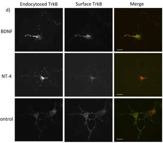
NT4 leads to efficient endocytosis of TrkB receptor and targets TrkB to the early endosome compartment ... 64
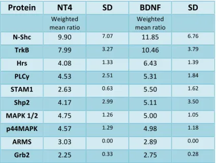
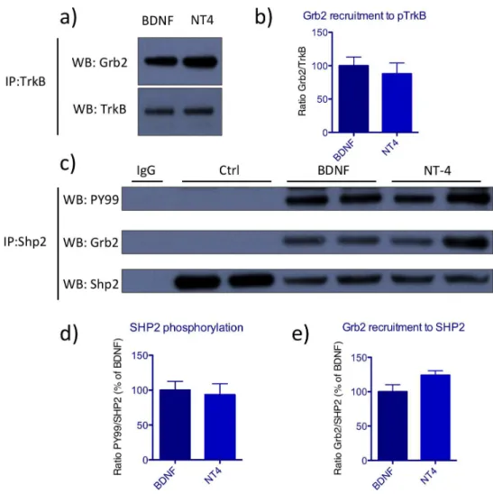
BDNF and NT4 activate TrkB receptor with similar kinetics ... 68
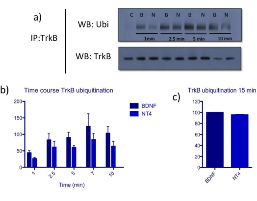
BDNF leads to more efficient ubiquitination of TrkB receptor ... 74
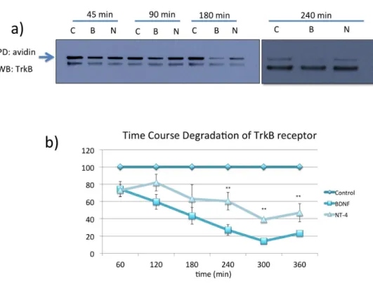
BDNF targets TrkB receptor efficiently to the degradative pathway whereas NT4 leads to sustained TrkB activation and decreased degradation ... 75
Discussion ... 87
Chapter 3: Slitrk5 as a new key player for CNS function ... 96
Author’s contribution ... 97
Introduction ... 98
Methods ... 99
Reagents ... 99
Animals. ... 99
Generation of the Slitrk5 knockout lacZ knock-‐in mice and determination of Slitrk5 expression pattern ... 100
Fluoxetine treatment ... 100
Cell culture ... 101
Lentiviral vector production and constructs ... 101
Western blotting ... 103
Synaptosomal fractionation ... 103
Elevated plus maze ... 104
Marble burying test ... 105
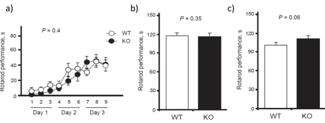
Rotarod test ... 105
Cylinder test ... 106
Electrophysiology ... 106
Endocytosis assay ... 107
Surface biotinylation and degradation assay ... 108
Coculture assay ... 109
Object placement test of spatial memory ... 109
Morris Water Maze ... 110
Results ... 111
Bioinformatic analysis of the Slitrk protein family ... 111
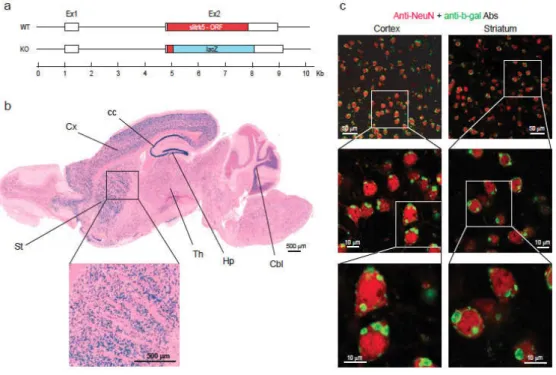
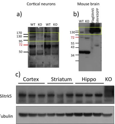
Generation of Slitrk5-‐/-‐ mouse and Slitrk5 expression ... 116
Slitrk5-‐/-‐ mice display anxiety-‐like behaviors ... 120
Slitrk5-‐/-‐ mice have impaired striatal function ... 122
Slitrk5-‐/-‐ mice have impaired cortico-‐striatal transmission ... 125
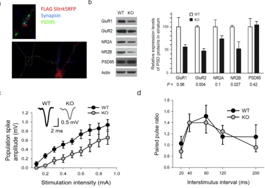
Slitrk5 modulates surface expression of AMPARs ... 128
Slitrk5 interacts with PICK1, a known modulator of GluA2 trafficking .... 132
Slitrk5-‐deficient mice are impaired in spatial reference memory ... 137
Future directions, Slitrk5 a multitalented protein ... 140
Slitrk5 induces synapse formation ... 140
Slitrk5 modulates TrkB trafficking and signaling ... 142
Discussion ... 146
Chapter 4 -‐ General Discussion ... 152
General Discussion ... 153
References ... 171
12
14
R
ESUMOprimeira explicação molecular para os efeitos distintos observados em estudos comparando as funções de BDNF e NT4.
16
localizado em dendrites e sinapses e regula o tráfego dos receptores de glutamato AMPA, reduzindo os níveis membranares da subunidade GluA2. Estudos feitos com ratinhos Slitrk5-‐/-‐ novos revelaram que a aprendizagem e memória dependente do hipocampo estava afectada. Por outro lado, também observámos que o Slitrk5 modula a degradação do receptor TrkB e de proteínas sinal ajusante. Estes resultados sugerem que o Slitrk5 é uma proteína que desempenha múltiplas funções no cérebro e consequentemente, a eliminação deste gene no ratinho leva a graves fenótipos neurológicos. Mais estudos serão necessários para a compreendermos completamente a relevância desta proteína em estados fisiológicos normais bem como em doença (incluíndo OCD). No entanto, os resultados aqui apresentados claramente favorecem uma papel activo do Slitrk5 para a função normal do SNC e que o nosso conhecimento sobre a função deste sistema é ainda limitado.
Em suma, com estes estudos providenciamos uma explicação molecular para as funções distintas mediadas por BDNF e NT4. Por outro lado, descobrimos um candidato novo que pode agora ser estudado em pacientes com OCD e criámos um modelo animal novo onde esta doença pode mais facimente ser estudada e manipulada.
A
BSTRACTUnderstanding how protein families interact to coordinate the development and function of the central nervous system (CNS) is critical for the understanding of this structure in normal conditions as well as in disease. Endocytic trafficking of membrane proteins to alternate intracellular targets can have significantly different biological outcomes. The neurotrophin family of proteins, composed of three receptors (TrkA, TrkB and TrkC) and four ligands (BDNF, NGF, NT3 and NT4), has a well-‐established role in the development and maintenance of the nervous system. TrkA receptor and its ligand NGF, are expressed mostly in the peripheral nervous system. In the CNS, TrkB and its two ligands, BDNF and NT4, are dominant, whereas TrkC and NT3 are expressed in both systems. Our current knowledge on the mechanisms of receptor activation, internalization and downstream targeting and signaling are mostly based on studies performed with sensory neurons relying on the TrkA/NGF system. On the other hand, it is still puzzling that in the CNS two ligands (BDNF and NT4) with similar affinity to the same receptor (TrkB) are expressed but seem to fulfill distinct biological responses. We hypothesized that these differences could be due to differential regulation of the endocytic trafficking pathway. In fact we observed that, even though NT4 and BDNF can equally activate TrkB receptor and its downstream signaling, BDNF promoted more efficient ubiquitination of the receptor than NT4. As a consequence, BDNF led to more efficient lysosomal degradation. On the other hand, even after long continuous treatment, NT4 did not induce efficient downregulation of TrkB. Consequently, NT4-‐induced activation of TrkB signaling was efficiently sustained, as seen by prolonged activation of the Akt and MAPK pathways. This data provides a new mechanism that can potentially explain the differential functions reported for these two ligands and adds to the current understanding of neurotrophin regulation and function in the CNS.
18
Nevertheless, our pioneer work clearly supports an active role of Slitrk5 for CNS function and that this system is still poorly understood.
Thus, with these studies we uncovered the mechanisms mediated by BDNF and NT4 modulation of TrkB endocytic trafficking and we also discovered a new candidate molecule for obsessive-‐compulsive disorder, and provide a new mouse model that can be used to study this condition.
Chapter 1 – General Introduction
G
ENERALI
NTRODUCTIONThe human brain is composed of approximately 100 billion neurons connected by up to 10 000 synaptic sites [1]. Understanding how neuronal numbers are controlled and how the connections between different brain structures are established are key questions in the field of neuroscience.
Chapter 1 – General Introduction
22
The continuous search for factors regulating central nervous system development led to the discovery of a new protein family named Slitrk [8]. Slitrks were discovered in a screen for molecules that were differentially expressed in mice with neural tube defects. Slitrks are tansmembrane proteins primarily expressed in the brain of both mice and humans. They can modulate neurite outgrowth and synapse formation. Interestingly, it was found that mutations in the Slitrk1 gene, a member of the Slitrk family, are associated with psychiatric disorders. Similarly to neurotrophins, it seems that this protein family can play important roles not only during CNS development but also for adult CNS maintenance [9]. We are still at early stages in terms of understanding the main functions of this protein family. It is plausible to speculate that, as opposed to neurotrophins, a set of receptors was identified before their ligands. Discovery of Slitrk’s binding partners, such as a secreted ligand, other transmembrane protein, or even cytoplasmic partners, will provide invaluable tools to explore this family and extend our understanding of CNS development and function.
The goal of this thesis work was to contribute to a better understanding of the mechanisms of action of polypeptides critical for CNS function. On the one hand I was interested in consolidating our knowledge of neurotrophin’s downstream signaling pathways in the CNS. On the other, we aimed to explore the newly identified Slitrk protein family, which is highly enriched in the brain.
1.
N
EUROTROHPHINF
AMILY[12,13]. Altered neurotrophin levels have been implicated in the development of several neurodegenerative disorders including Alzheimer’s and Hungtington’s disease, as well as in psychiatric disorders, such as depression [14].
1.1 Neurotrophin family and receptors
The neurotrophin family is composed of four polypeptides: NGF, brain-‐ derived neurotrophic factor (BDNF), neurotrophin 3 (NT3) and neurotrophin 4 (NT4). Even though NGF was the first neurotrophin to be identified, very few neurons in the CNS require NGF for survival. The search for a functional analog to NGF in the CNS led to the discovery BDNF [11,15].
The p75 receptor belongs to the tumor necrosis factor (TNF) receptor superfamily and, neurotrophin binding to this receptor has been shown to lead to apoptosis [16]. P75 receptor expression is downregulated during postnatal development in the CNS but is rapidly induced after seizure or neuronal lesion. Although p75-‐mediated signaling has been shown to be important for nervous system development the discussion of the mechanisms involved in these pathways is outside the scope of this thesis. For detailed information about this subject, please refer to the following reviews [17,18,19].
Chapter 1 – General Introduction
24
Different splice variants have been described for all Trk receptors that differ in neurotrophin affinity and function. A TrkB splice variant lacking exon9 in the extracellular domain that has reduced affinity for NT4 and NT3 but not for BDNF, was found in the developing nervous system of the chick [22]. A TrkB splice variant lacking exon9 in the extracellular domain that has reduced affinity for NT4 and NT3 but not for BDNF, was found in the developing nervous system of the chick [23]. Moreover, splice variants of TrkC, and especially TrkB, lacking the catalytic kinase domain, are highly expressed in the mature CNS [24]. This truncated TrkB variant is thought to act as a dominant negative since it can bind and internalize BDNF and possibly restrict its availability [25].
1.2 Neurotrophin expression and functions
Target-‐derived NGF plays critical roles for cell survival and differentiation of peripheral neurons. NGF expression in the CNS is more restricted and it promotes survival of cholinergic neurons in the basal forebrain. BDNF and NT4 are more widely expressed in the CNS with particular high levels in the cortex and hippocampus [2,26]. Their expression is developmentally regulated increasing during the first postnatal weeks [27,28,29]. NT3 is broadly expressed throughout the nervous system. In the periphery, it acts in coordination with NGF to support the survival of sympathetic neurons, being expressed in intermediate targets at high levels [30].
The best-‐studied system showing neurotrophin-‐dependance for survival and development has been the PNS. All peripheral sensory neurons express one of the Trk receptors at some point in development and, presence or absence of corresponding neurotrophins at their innervated targets, determines survival or death. Ablation of individual Trk receptors leads to elimination of specific neuronal subpopulations. For example, NGF/TrkA ablation in mice leads to a complete loss of nociceptive neurons, whereas, null mutations of NT3/TrkC, leads to a lack of propioceptive neurons [31]. Moreover it was shown that mice null for both TrkB and TrkC have decreased synaptic density and lower numbers of pre-‐synaptic vesicles in the hippocampus [32].
Chapter 1 – General Introduction
26
numbers. It did however have defects in dendritic arborization specifically in striatal neurons, suggesting that BDNF is negligible for mature hippocampal maintenance [27].
1.3 Neurotrophin-‐mediated signaling
Trks form single pass transmembrane proteins containing 3 leucine-‐rich repeats (LRR) flanked by cysteine-‐rich domains, and two immunoglobulin-‐like domains involved in ligand binding. Non-‐covalent homodimers of neurotrophins bind to Trks inducing receptor dimerization and activation. Dimeric Trks phosphorylate each other in the cytoplasmic auto-‐regulatory loop of the kinase domain and in other cytoplasmic tyrosines, further strengthening the catalytic activity of the kinase. Trks are highly homologous in their intracellular domain which is 73% conserved. Most research on Trk-‐mediated signaling has focused mainly on two phosphorylated tyrosines: Y496 in human TrkA (homologous to Y516 in TrkB) that is localized in the juxtamembrane domain within the NPxY motif and tyrosine Y791 in human TrkA (Y817 in TrkB), that is located at the carboxy-‐terminal (C-‐terminal). Phosphorylation of these tyrosines creates docking sites for adaptor proteins that couple to downstream signaling cascades including the Ras/mitogen activated protein kinase (a MAPK also known as extracellular signal regulated kinase -‐ Erk) pathway, phosphatidyl inositol-‐3 kinase (PI3K)/Akt kinase pathway and the PLC-‐γ/cAMP response element binding protein (CREB) pathways [2,19,39]. For a summary of neurotrophin mediated signaling see figure 1.
nucleotide exchange factor Son of Sevenless (SOS) that converts Ras into its GTP bound form. This active form of Ras activates the PI3K, the p38, MAPK and c-‐Raf pathways [43]. One of the targets of MAPK is the ribosomal protein kinase 2 that phosphorylates CRE-‐binding proteins modulating transcription of critical genes.
Trk receptor mediated activation of Ras through Grb2 and Shc promotes MAPK signaling in a transient rather than sustained fashion [39]. Prolonged MAPK activation depends on the recruitment of Frs2 to Y490. Frs2 phosphorylation in turn provides binding sites for several other signaling players including the protein tyrosine phosphatase 2 (Shp2), Grb2 and the adaptor protein Crk. Association with Crk results in activation of the small GTPase Rap1 that that initiates the MAPK cascade. Recruitment of Grb2 provides an independent mechanism for Ras activation. Signaling through Y490 leads to survival and differentiation of cells [39,41,42,44,45,46]. Shp2 is a rare protein phosphatase implicated in signaling amplification. It acts as an adaptor protein recruiting Grb2/SOS complex, and it also mediates dephosphorylation of Sprouty, reversing its general receptor tyrosine kinase (RTK) inhibitory effects [47].
Phosphorylation of Y791 in human TrkA recruits and activates PLC-‐γ that triggers hydrolysis of phosphatidyl-‐inositol (4,5) 2 phostate (PIP2) to generated
inositol (1,4,5) triphosphate (IP3) and diacylglycerol (DAG). IP3 mobilizes Ca 2+
Chapter 1 – General Introduction
28
Figure 1: Summary of neurotrophin signaling. Neurotrophin binding to Trk receptors triggers dimmerization and activation of different signaling pathways. Depicted here is human TrkB receptor and recruitment of adaptor proteins to the two main phosphorylated tyrosines. Phosphorylation of the tyrosine residue located in the juxtamembrane region of Trk receptor recruits two complexes of adapter proteins the Shc/Grb2/SOS and the FRS2/Shp2/Grb2/SOS complexes. Activation of Ras triggers the MAPK/Erk signaling pathway, which stimulates neuronal differentiation including neurite outgrowth. Activation of PI3K through Ras/Gab1 promotes cell survival and growth. Phosphorylation of the c-‐terminal tyrosine recruits PLC-‐γ that results in increase in intracellular Ca2+ and modulation of synaptic plasticity.
1.4 Regulation of signal specificity
similar: all contain a ligand-‐binding domain in the extracellular region, a single transmembrane domain and a cytoplasmic domain that contains the tyrosine kinase plus regulatory sequences at the juxtamembrane region and the C-‐terminus. The signaling cascades previously described (RAS/MAPK and MAPK) are the canonical pathways that can be activated by virtually all RTKs [49], thus raising the question of how Trk-‐induced signaling is different from other RTKs and within the Trk family. Differential temporal and spatial distribution of signaling partners amongst different cell types provides one level of specificity. Specific neuronal populations express particular types of adaptors, and different Trks are expressed at different levels in the same cell type which can lead to diverse outcomes. A recent study reported that expression of TrkA or TrkC in mouse embryonic stem cells which were later differentiated into glutamatergic neurons, leads to neuronal death, whereas TrkB does not [50]. This suggests that even in similar contexts, different Trks are capable of engaging alternative signaling networks.
Differential sorting of adaptor proteins within intracellular membrane compartments also contribute to signal specificity and regulation. Recently, it was shown that transport of BDNF-‐activated TrkB to lipid rafts, through Fyn, was critical for efficient PLC-‐γ activation. In Fyn knockout mice, BDNF was unable to direct TrkB to intracellular lipid raft compartments and phopshorylation of PLC-‐γ was reduced. On the other hand, Akt and MAPK were excluded from lipid rafts and phosphorylation of these proteins occurred outside these domains [51].
Chapter 1 – General Introduction
30
proteins such as AP2. CCP fuse with a specialized intracellular organelle the early endosome, also named sorting endosome. This process is regulated by Rab5 and early endosome antigen 1 (EEA1). The early endosome is characterized by a mildly acidic pH that leads to the uncoupling or receptor-‐ligand complex. Membrane proteins rapidly exit the early endosome and can follow two opposing routes that result in different signaling outcomes: recycling to the plasma membrane or degradation through the lysosome [52,53,54,55]. Phosphorylation, mono-‐ ubiquitination and receptor-‐ligand uncoupling are key mechanisms modulating endocytic sorting. Transferrin receptor (TfnR) is the canonical constitutive recycled receptor. From the early endosome TfnR can either recycle directly to the membrane (rapid recycling) or it can be targeted to the recycling endosome and then to the membrane (slow recycling). EGFR is the best described RTK to undergo ligand-‐induced degradation through the lysosome. From the early endosome, EGFR is sorted to a late endosome, also known as multivesicular body, and subsequently, to the hydrolytic interior of the lysosome, where proteins are degraded [56,57,58,59,60,61].
neurotrophin treatment TrkA promotes sustained Akt phosphorylation as well as survival responses whereas TrkB does not [63].
In PC12 cells it has been shown that blocking NGF-‐dependent endocytosis of TrkA disrupts differentiation, even though survival was unaffected. Since Frs2 signaling is required for sustained MAPK activation and differentiation, this study suggests that efficient Frs2 activation requires endocytosis of the neurotrophin-‐ receptor complex [64]. Endocytosis also seems to facilitate activation of the small G and attenuates Ras. On the other hand, PI3K activation occurs independently of endocytosis and this mechanism appears to attenuate this signaling pathway [65].
Chapter 1 – General Introduction
32
amounts of ligands to study downstream signaling effects. However, this probably does not mimic endogenous contexts in which limiting amounts of ligands might induce specific signaling pathways.
2.
S
LITRK FAMILY OF PROTEINS AND FUNCTIONThe advances in gene sequencing and cloning techniques of the past decades have contributed to the discovery of several protein families key for the development and function of the CNS. An example of this is the neurotrophin family that was covered in the previous section. However, our current knowledge of the molecular players involved in these processes is still limited and not sufficient to explain the complexity and diversity of this system. On the one hand, many open questions remain regarding the pathways and mechanisms of actions mediated by known protein families. On the other, the discovery and characterization of new molecular players is still necessary. This knowledge will allow us not only to understand the function of the normal nervous system, but also what fails in disease conditions.
Understanding the molecular mechanisms underlying pathological conditions are key question in neuroscience, being critical for the discovery of new molecular targets for pharmacological treatments. In this context, we were interested in studying new protein families that play critical roles for CNS maintenance and function. Slitrks are a recently discovered family of proteins primarily expressed in the brain that were discovered in a screen for proteins de-‐ regulated in mice with neural tube defects [8]. So far, the few research studies addressing the function of these proteins in the CNS suggest that Slitrks mediate basic functions in neuronal development and also that they might be associated with psychiatric disorders ranging from obsessive-‐compulsive spectrum disorders to schizophrenia. Reminiscent of the neurotrophin family, it seems that the Slitrk family is key not only for the elaboration and development of the CNS but also in disease conditions.
Chapter 1 – General Introduction
34
generated a Slitrk5 knockout mouse line and found that this protein plays critical roles for CNS function (see chapter 3).
2.1 Slitrk gene family
Slitrks are composed of six members, Slitrk1 through Slitrk6. They were initially identified in a screen for genes that were differentially expressed in mice with neural tube defects [8]. Slitrk5 had been previously discovered as a gene expressed in early hematopoietic progenitors but not in mature hematopoietic cells [70]. All Slitrks form single pass (type I) transmembrane proteins with an intracellular domain that varies in length [8] (Figure 2). At the extracellular domain, Slitrks contain two leucine-‐rich repeat (LRR) domains, which are each composed of 6 LRRs, flanked by cysteine rich capping domains [8]. Sequence analysis has revealed that the extracellular LRR domains of Slitrks resembled Slit proteins, and a conserved region in their intracellular carboxyl terminus (C-‐terminus) has a high degree of consensus with the last 16 amino acids of the neurotrophin receptor (Trk) [8]. Based on their similarity with Slits and Trks, these proteins were named Slitrks [8].
Figure 2: Schematic representation of the Slitrk protein family. Slitrks are composed of six members, Slitrk1 through Slitrk6. All contain extracellular LRR motifs that vary in numbers, flanked by cysteine-‐rich motifs, followed by a single transmembrane domain and an intracellular domain. All but Slitrk1 contain long intracellular domains that share some homology with Trk receptors at a C-‐terminus tyrosine. Phosphorylation of intracellular residues has been showed for Slitrk2, Slitrk5 and Slitrk6, however, the function is still unknown.
Slit is one of the most well-‐known LRR-‐containing proteins. These proteins have a tandem of four LRR domains at the N-‐terminus (D1-‐D4), each containing an array of five to seven LRRs [71]. Slit was originally discovered in Drosophila as a protein secreted by midline glia of the developing CNS [72,73]. Three Slit homologues (Slit1-‐ 3) have been discovered in mammals [74]. Later it was shown that Slit is a ligand for Robo receptors [75]. Slit-‐Robo signaling is involved in a variety of processes, key for proper nervous system development, such as repulsion of axons from the midline, axon guidance and repulsion, orchestrating tangential neuronal migration, regulating cytoskeletal dynamics and modifying cell adhesion properties [76,77].
LRR domains are composed of tandem repeats of LRR motifs and have curved solenoid structures that are suitable for mediating protein-‐protein interactions [78,79,80]. The LRR is a widespread structural motif of 20-‐30 amino acids with a defining sequence LxxLxLxxN/GxL (x being any amino acid). Given their extracellular LRR domains, Slitrks are considered part of the LRR superfamily. LRR-‐ containing proteins are emerging as key regulators of the CNS functions, including neurite outgrowth, neuronal survival, myelin-‐based axon growth inhibition, synapse formation, dendritic morphogenesis, etc [81,82,83]. Given below is a brief summary of some LRR protein families and their main roles in CNS, including those in synapse formation.
Chapter 1 – General Introduction
36
2.2 LRR protein families and their function in the CNS
Several LRR proteins act as synaptic cell adhesion molecules (CAMs). These molecules play important roles in the establishment, maintenance and activity-‐ dependent changes of synapses. For a protein to be classified as a synaptic CAM, it should be able to mediate cell adhesion through interacting with other proteins in trans; and it should interact in cis with scaffolding proteins that recruit synaptic proteins to the synapse. Many LRR-‐containing proteins meet these criteria and have been shown to regulate synapse formation and maturation.
The Amphoterin-‐induced gene and ORF (Amigo) family is composed of three members that are almost exclusively expressed in the CNS [81]. They contain six LRRs at the extracellular domain and an immunoglobulin-‐like domain located next to the transmembrane segment [84]. When the ectodomain of Amigo is expressed as an Fc fusion protein and substratum-‐attached, it promotes prominent neurite extension of cultured hippocampal neurons [84]. Moreover Amigo2 has also been implicated in activity-‐dependent survival of cerebellar neurons [85].
excitatory and inhibitory synapses in contacting axons [89]. The ligands for the SALM proteins are still unknown.
The netrin-‐G ligand (NGL) family form type one membrane proteins with nine LRRs at the extracellular domain, flanked by cysteine rich capping domains (LRRCT and LRRNT) and a C2 imunoglobulin domain [90]. NGLs are synaptic CAMs, primarily localized to the postsynaptic compartment of excitatory synapses. There are three known members: NGL-‐1, NGL-‐2 and NGL-‐3 and they interact with the pre-‐ synaptic ligands, netrin-‐G1, netrin-‐G2 and LAR, respectively [81,90,91,92]. NGLs associate with main components of the synapse such as PSD-‐95 and NMDA receptors [86,93]. NGL-‐1 is highly expressed in the hippocampus, striatum and cerebral cortex and it promotes outgrowth of thalamocortical axons [94]. Both NGL-‐ 2 and NGL-‐3 induce pre-‐synaptic differentiation in a co-‐culture assay, increase the excitatory synapse density when over-‐expressed in cultured neurons and, when directly aggregated on dendrites, they recruit PSD-‐95 post-‐synaptically [93,95]. Disruption of NGL-‐2 or NGL-‐3 expression in cultured hippocampal neurons decreases the number and function of excitatory synapses [93,95].
Chapter 1 – General Introduction
38
attenuates the strength of evoked excitatory synaptic currents in vivo. LRRTM2 also interacts with PSD-‐95 and regulates surface expression of AMPA receptors [97,98].
In summary, LRR domains are involved in protein-‐protein interactions and LRR-‐containing proteins mediate a myriad of functions key for CNS development and function.
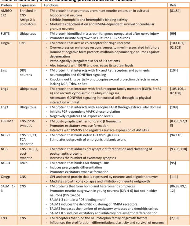
Table 1: Summary of LRR-‐containing proteins and their functions
Protein Expression Functions Refs AMIGO
1/2
Enriched in CNS Amigo 2 is ubiquitous
-‐ TM protein that promotes prominent neurite extension in cultured hippocampal neurons
-‐ Exhibits homophilic and heterophilic binding activity
-‐ Modulates depolarization and NMDA-‐dependent survival of cerebellar granule neurons
[81,84]
FLRT3 Ubiquitous -‐ TM protein identified in a screen for genes upregulated after nerve injury -‐ Promotes neurite outgrowth in cultured DRG neurons
[99] Lingo-‐1 CNS -‐ TM protein that acts as co-‐receptor for Nogo receptor
-‐ Over-‐expression enhances responsiveness to myelin-‐associated inhibitors -‐ Dominant negative form protects midbrain dopaminergic neurons against
degeneration
-‐ Pathologically upregulated in SN of PD patients -‐ Also interacts with EGFR and decreases its protein levels
[100,101,1 02,103]
Linx DRG neurons
-‐ TM protein that interacts with Trk and Ret receptors and augments neurotrophin and GDNF/Ret signaling
-‐ Knocking out Linx partially photocopies axonal projection defects in mice lacking NGF, TrkA, or Ret
[104]
Lrig1 Ubiquitous; -‐ TM protein that interacts with ErbB receptor family members (EGFR, ErbB2-‐ 4) and recruits cytoplasmic E3 ubiquitin ligases
-‐ Attenuates GDNF/Ret signaling in neuronal cells through its physical interaction with Ret
[105,106,1 07,108]
Lrig3 Ubiquitous -‐ TM protein that interacts with Xenopus FGFR through extracellular domain -‐ Inhibits FGF-‐dependent MAPK phosphorylation
-‐ Negatively regulates FGF expression levels
[109]
LRRTM2 CNS, post-‐ synaptic
-‐ TM post-‐synaptic partner for α and β Neurexins -‐ Promotes excitatory synapse formation
-‐ Interacts with PSD-‐95 and regulates surface expression of AMPARs
[83,96,97,9 8]
NGL-‐1 CNS: ST, CT, TCA, dendritic
-‐ TM protein that binds netrin G-‐1 through LRRs -‐ Stimulates outgrowth of embryonic thalamic axons
[94,110]
NGL-‐ CNS, HC, CT, post-‐ synaptic
-‐ TM protein that induces presynaptic differentiation and clustering of postsynaptic proteins
-‐ Increases the number of excitatory synapses
[93,95,110]
NGL-‐3 Brain -‐ TM protein that binds LAR through LRRs -‐ Induces presynaptic differentiation -‐ Promotes excitatory synapse formation
[95]
Omgp CNS -‐ GPI-‐anchored protein that is expressed by neurons and oligodendrocytes -‐ Mediates growth cone collapse and inhibition of neurite outgrowth
[111]
SALM 1-‐ 3
CNS -‐ TM proteins that form homo and heteromeric complexes
-‐ Promotes neurite outgrowth in young neurons (DIV 4-‐6) but not in older neurons (DIV 14-‐16)
-‐ SALM1-‐3 contain a PDZ-‐binding motif
-‐ SALM1 induces the dendritic clustering of NMDA receptors
-‐ SALM2 increases the number of excitatory synapses and dendritic spines -‐ SALM3 & 5 induces excitatory and inhibitory pre-‐synaptic differentiation
[86,88,89,1 12]
Trks CNS -‐ TM receptors that bind the neurotrophin family of growth factors
-‐ Influences the proliferation, differentiation, plasticity and survival of neurons [2,19]
Chapter 1 – General Introduction
40
TCA, thalamocortical axons; ST, striatum; CT, Cortex; PD, Parkinson’s disease; DA, dopamine; TM, transmembrane; DRG, dorsal root ganglia; GDNF, glial cell line-‐derived neurotrophic factor; SN, substantia nigra; TM, transmembrane; HC, hippocampus, AMPAR, α-‐amino-‐3-‐hydroxy-‐5-‐methyl-‐4-‐ isoxazolepropionic acid receptor; FGF, fibroblast growth factor; ErbB/EGFR, epidermal growth factor receptor; LAR, leukocyte antigen-‐related; GPI, glycosylphosphatidylinisotol; DIV, developmental day in vitro; PSD-‐95, synapse associated protein 95 kDa
2.3 Slitrks and their functions in the CNS
The Slitrk gene family has been implicated in psychiatric disorders, such as Tourrete Syndrome (TS), trichotillomania (TTM), obsessive-‐compulsive disorder (OCD) and schizophrenia [113,114,115,116]. Mechanistically, Slitrks can modulate neurite outgrowth, dendritic complexity, neuronal survival, and synapse formation [83,113,116,117]. Slitrks are highly expressed in the CNS, with its expression starting prenatally both in the mouse and humans [8,118,119].
Initial studies performed by over-‐expressing each of the Slitrk members in PC12 cells revealed that treatment of the transfected cells with NGF resulted in a decreased number of neurites per cell [8]. Neurite length was also decreased except for SLitrk1 and Slitrk4 [8]. Subsequently, the neurite outgrowth function of Slitrk1 has been analyzed in mouse cortical neurons [113,120]. Cortical neurons cultured from mouse embryos previously electroporated with human Slitrk1 cDNA showed increased dendritic length [113], suggesting that this Slitrk member plays an important role in promoting neurite outgrowth. More recently, it has been demonstrated that phosphorylation of Slitrk1 on Ser695 by casein kinase II is critical for the interaction of Slitrk1 with 14-‐3-‐3 proteins [120], which are ubiquitously expressed phosphorylation-‐binding proteins that regulate a number of important cellular processes including cell proliferation, neuronal migration and membrane excitability [120,121]. Mutation of this serine residue to an alanine residue abolished the interaction of Slitrk1 with 14-‐3-‐3 proteins as well as the induction of neurite outgrowth in cultured mouse cortical neurons [120].
analysis of this organ in a mouse model that ubiquitously lacks Slitrk6 expression [117]. Results from this study demonstrated that Slitrk6 promotes the survival and neurite outgrowth of sensory neurons of the inner ear [117]. Histological examinations revealed that vestibular innervation was markedly decreased and sometimes misguided in the Slitrk6 null mice. The mutant mice also showed significant cell death in the spiral and vestibular ganglia. Furthermore, cochlear sensory epithelia taken from these mice and co-‐cultured with wild-‐type spiral ganglion neurons were less effective in promoting neurite outgrowth of the spiral ganglion neurons compared to co-‐cultures in which both types of cells were taken from wild-‐type mice [117]. Evidence for a trophic role of Slitrk6 was further strengthened by the observation that both BDNF and neurotrophin 3 (NT3) mRNA levels, as well as their corresponding receptor protein levels, TrkB and TrkC respectively, were down-‐regulated in the inner ear of Slitrk6 null mice. Interestingly, this was observed at an early developmental stage of the cochlea, when cell death had not occurred [117]. This study suggests that, at least in part, Slitrk6 may exert trophic actions by modulation of the neurotrophin system.Several LRR containing proteins have been shown to interact and modulate RTKs. For example Linx, a leucine-‐rich repeat and immunoglobulin (LIG) family protein, physically interacts with both Trk and Ret receptors, resulting in increased neurotrophin and GDNF signaling, respectively [104]. Besides Slitrk6, it would be interesting to investigate if other Slitrks could also modulate Trk receptor signaling.
Chapter 1 – General Introduction
42
neurite outgrowth that are endogenously expressed in neurons are lacking in PC12 cells.
Slitrks have also been implicated in synapse formation. A recent study performing an expression screen for synaptogenic proteins revealed the abundance of LRR containing proteins capable of inducing synapse formation [83]. Slitrk2 was one of the LRR proteins identified in this screen, and it was shown to induce excitatory pre-‐synaptic neuronal differentiation in a cellular co-‐culture system, suggesting that it could be a postsynaptic protein [28]. It is possible that Slitrks could interact with a pre-‐synaptic partner such as the Neurolgin-‐Neurexin system, via their extracellular LRR domains and could recruit intracellular post-‐synaptic proteins. Identifying the intracellular binding partners for Slitrks would contribute significantly to understanding the molecular basis of Slitrk functions in the nervous system.
2.4 Slitrk1 and OCD-‐spectrum disorders
trichotillomania (TTM), that is characterized by a compulsive hair pulling behavior that leads to visible patches, and body dysmorphic disorder, characterized by obsession and extreme body image dissatisfaction and engaging in repetitive behaviors in order to change owns appearance, [125,128,129,130].
Chapter 1 – General Introduction
44
Figure 3. Schematic representation of the direct and indirect pathway, which are two opposing circuit loops in the basal ganglia. The direct pathway originates in the frontal cortex and projects to the striatum. Medium spiny neurons in the striatum, expressing D1 dopamine receptor, project to the globus pallidus interna/substantia nigra pars reticulata (GPi/SNr) complex, which projects to the thalamus that in turn has reciprocal, excitatory projections to and from the cortical site of origin. The indirect pathway also originates in the frontal cortex and projects to medium spiny neurons expressing D2 dopamine receptor in the striatum, then projects to the globus pallidus externa (GPe), then to the subthalamic nucleus, then back to GPi/SNr, before returning to the thalamus and finally back to frontal cortex. The direct pathway contains two excitatory and two inhibitory connections, resulting in a net positive circuit back to the cortex. The indirect pathway has three inhibitory connections resulting in a net negative feedback loop to the cortex.
breakpoint. Subsequent sequencing of Slitrk1 gene in a cohort of 174 individuals with TS, identified one proband (diagnosed with TS and attention-‐deficit hyperactive disorder) with a single base deletion, that led to a frame-‐shift and the expression of a truncated protein (named varCDf). The same mutation was also found in the patient’s mother that suffered from TTM but not in unaffected relatives. Functional analysis showed that truncated Slitrk1 was ineffective in inducing dendritic outgrowth of cultured mouse cortical neurons as compared with wild type Slitrk1. An additional non-‐coding sequence variant (var321) in the 3’UTR was found in two unrelated individuals. This variant altered binding to the microRNA hsa-‐miR-‐189 and it negatively modulated Slitrk1 mRNA expression. These mutations were absent in over 3600 control samples, indicating that they are rare variants [113].
Subsequent studies were unable to find associations between these two reported variants in larger TS populations [134,135,136,137,138], arguing against a segregation of Slitrk1 and TS. Furthermore, it was reported that var321 was over-‐ represented in the Ashkenazi Jew population, raising the possibility that population stratification might have led to a false positive result [136,137]. The largest screen for Slitrk1 and TS performed so far, which involved sequencing 1048 samples from the Tourette Syndrome Association International Consortium for Genetics, did not find association between var321 and TS [137]. Only two individuals were found who carried var321: one was diagnosed with TS, OCD and TTM, and the other with OCD but not TS. None transmitted the mutation to their affected offspring [137]. VarCDf was not analyzed. Additional studies provided more information regarding the ethnicity of the initially reported cases, and five additional subjects carrying var321 were reported [139]. Fine mapping of all seven carriers of the Slitrk1 region argued against population stratification confounding the original data [139].
Chapter 1 – General Introduction
46
a common TS risk factor of low penetrance in linkage disequilibrium (LD) with the associated marker/haplotypes [140]. Furthermore, novel mutations in the Slitrk1 gene have been found to co-‐segregate with OCD-‐spectrum disorders [114,140,141]. Two non-‐synonymous mutations in the Slitrk1 extracellular region were discovered in two independent individuals of European descent, in a set of 44 families with TTM, and were absent in a group of almost 3000 healthy controls [114]. TTM is an OCD-‐spectrum disorder thought to be genetically associated with TS [130,142]. Moreover, a mutation in the 3’UTR of Slitrk1 gene was found in a group of 92 Austrian patients, that segregated in two additional family members with tic disorders, and which was absent in 192 control subjects [141].
may occur with traditional knockout mouse models. Furthermore, it would be interesting to determine the consequences of the introduction of human Slitrk1 variants (eg. var321 or varCDfs) in future knock-‐in mouse models, and to assess these mice for their subsequent neuroanatomical and behavioral phenotypes. Such studies will help to directly evaluate the in vivo consequences of specific mutants in the Slitrk1 gene, and they would avoid possible compensatory mechanisms from other Slitrk members that could have occurred in the Slitrk1 mouse leading to a fairly modest phenotype and no tics.
2.5 Glutamate receptors implicated in psychiatric disorders