Vesna Bozanic
Dissertation presented to obtain the Ph.D degree in biochemistry Instituto de Tecnologia Química e Biológica | Universidade Nova de Lisboa
Oeiras, January, 2013
F
rom
Fundação para Ciência e Tecnologia funded PhD grant SFRH/BD/18746/2004 for Vesna Bozanic and research project grant POCTI/BIO/58465 (PI: Cláudio M. Gomes).
This thesis is the result of research work done at Protein Biochemistry Folding and Stability Laboratory at Instituto de Tecnologia Química e Biológica (ITQB), Universidade Nova de Lisboa (UNL), Portugal, under the supervision of Professor Doctor Cláudio M. Gomes.
The studies here reported were performed during the term of a four year PhD fellowship from Fundação para a Ciência e Tecnologia (FCT), from February 2005 to February 2009.
The thesis comprises six chapters. The first chapter introduces the reader to the life at high temperatures and addresses mechanisms through which proteins acquire enhanced stability.
The second chapter addresses methodologies that are used in proteomics studies of protein stability.
Chapter Three outlines the results regarding a proteomic study of selected hyperstable cytosolic proteins originating from the thermophile Sulfurisphaera sp.
Chapter Four presents results regarding a comparative study of thermostable proteins selected from the cytosolic proteome of the thermophile Sulfolobus solfataricus and the mesophile Escherichia coli.
Chapter Five discusses the subset of results regarding hyperstable cytosolic proteome from Escherichia coli discussing relationships between protein thermostability and solubility.
The last chapter of the thesis presents in brief concluding remarks regarding the work presented.
I would like to express my gratitude to the following people and institutions that have contributed to my PhD thesis, among them especially:
• My supervisor, Professor Doctor Cláudio M. Gomes
• All my colleagues, past and present members of the Protein Biochemistry Folding and Stability Laboratory: Dr. Bárbara Henriques, Catarina Silva, Dr. Hugo Botelho, Dr. João Rodrigues, Dr. Patrícia Faísca, Dr. Raquel Correia, Dr. Sónia S. Leal.
• Instituto de Tecnologia Quimica e Biológica (ITQB) and Prof. Dr. L.P. Rebelo
Collaborating laboratories:
• Prof. Dr. Phillip Wright, head of Department of Chemical and Biological Engineering, University of Sheffield, UK with Dr. Trong Khoa Pham, for enabeling iTRAQ experiments and MS data
analysis.
• Dr. José P. Leal, head of Computational Genomics Laboratory, Instituto Gulbenkian de Ciência, with Renato Alves, for providing computational algorithms.
Funding of PhD grant and research project:
• Fundação para Ciência e Tecnologia for funding my PhD grant SFRH/BD/18746/2004 and research project grant POCTI/BIO/58465 (PI: Cláudio M. Gomes).
My deepest gratitude goes to:
The work presented in this thesis is based on the following publications in international peer reviewed journals:
Vesna Prosinecki, Hugo M. Botelho, Simona Francese, Guido Mastrobuoni, Gloriano Moneti, Tim Urich, Arnulf Kletzin and Cláudio M. Gomes, A Proteomic Approach toward the Selection of Proteins with Enhanced Intrinsic Conformational Stability, Journal of Proteome Research, 2006, 5 (10): 2720‐2726
Vesna Prosinecki, Patrícia F.N. Faísca and Cláudio M. Gomes, Conformational States and Protein Stability in a Proteomic Perspective, Current Proteomics, 2007, 4 (1): 44‐52
Additional publications resulting from the work during the term of PhD grant from Fundação para a Ciência e Tecnologia (FCT):
Hugo M. Botelho, Sónia S. Leal, Andreas Veith, Vesna Prosinecki, Christian Bauer, Renate Frohlich, Arnulf Kletzin, Cláudio M. Gomes Role of a novel disulfide bridge within the all‐beta fold of soluble Rieske proteins, J Biol Inorg Chem, 2010, 15(2):271‐81
This work involved the identification and analysis of the properties of the most stable proteins present within proteomes, aiming at obtaining a general perspective of the factors that determine protein stability. As models we have focused on ensembles of proteins with high intrinsic stability, and for this purpose we have studied proteomes from the hyperthermophilic archaeon Sulfolobus solfataricus and Sulfurisphaera sp., whose properties were compared to those of the mesophilic bacterium Escherichia coli.
To carry out this study, we have implemented a novel approach aimed at profiling a soluble proteome for its most intrinsically stable proteins. For this purpose the hyperthermophilic archaeon Sulfurispharea sp., which is able to grow between 70‐97°C, was used as a model organism. We have thermally and chemically perturbed the cytosolic proteome as a function of time (up to 96h incubation at 90°C), and proceeded with analysis of the remaining proteins by combining one and two dimensional gel electrophoresis, liquid chromatography fractionation, protein identification by N‐terminal sequencing, and mass spectrometry methods. A total of 14 proteins with enhanced stabilities which are involved in key cellular processes such as detoxification, nucleic acid processing and energy metabolism were identified. We demonstrate that these proteins are biologically active after extensive thermal treatment of the proteome. This method has thus illustrated an experimental approach aimed at mining a proteome for hyperstable proteins, a valuable tool for target selection in protein stability and structural studies, and biotechnological applications.
each organism) corresponding to the subset of proteins with above average stability (labeled ‘survivors’), unchanged stability, and less stable than the average. These sequences were investigated using bioinformatics tools, in order to determine relationships between thermostability, physicochemical properties, structural folds, amino acid type and biological function.
We concluded that per se the prevalence of certain types of amino acids is not essential to make a protein more stable and that SCOP folds are also not strongly biased in respect to thermostability. The group of thermostable proteins was slightly enriched in smaller proteins (<50kDa), with slightly negative GRAVY scores and higher aliphatic indexes. Regarding COG functional categories, the identified sequences with increased stability and solubility belonged to the following categories: Information storage and processing group (27%), cellular processes group (30%) and metabolism group (40%). This could suggest that these particular processes have evolved so as to preserve stable folds. In any case our results show that enhanced thermal stability results from a combination of properties, and not from a single exclusive factor.
Another interesting finding is that enhanced stability seems to be correlated with increased solubility, as shown from the comparison of our results with those obtained in an independent study that compared the solubility/aggregation ratio for soluble Escherichia coli proteins expressed in a cell‐free and chaperone‐free system. Indeed, analysis of the solubility/aggregation profile of the superstable cytosolic proteins that survived harsh thermal treatment shows that these are mostly overlapping with those which rank soluble. This denotes that protein stability and solubility are intertwined properties grounded in comparable physical principles, as selection for stability yields increased solubility as a read‐out.
Based on this, further development of experimental, theoretical and computational approaches on entire ensembles of proteins from a given organism will result in a better understanding of the physical principles underlying protein folding, stability and solubility.
O trabalho apresentado nesta tese envolveu a identificação e análise das propriedades de proteínas com estabilidade elevada no contexto do proteoma onde se inserem, visando a obtenção de uma perspetiva geral acerca dos determinantes da estabilidade proteica. Como modelos selecionamos conjuntos de proteínas com elevada estabilidade intrínseca, pelo que se estudaram os proteomas dos Archaea hipertermófilos Sulfolobus solfataricus e Sulfurisphaera sp., cujas propriedades foram comparadas com as da bactéria mesófila Escherichia coli.
A implementação deste estudo envolveu o desenvolvimento de uma nova metodologia que permitiu selecionar as proteínas constituintes de um proteoma de acordo com a sua estabilidade relativa. Para o efeito estudou‐se o hipertermófilo Sulfurisphaera sp., que cresce entre 70‐97°C, como modelo. O protocolo desenvolvido consistiu na perturbação química e térmica do proteoma citosólico em função do tempo (até 96h de incubação a 90°C), seguido da análise das proteínas remanescentes usando técnicas cromatográficas, eletroforese de proteinas (1D e 2D), identificação por sequenciação do N‐terminal e espectrometria de massa.
Um conjunto de 14 proteínas foi identificado neste estudo piloto, estando estas sobretudo implicadas em processos celulares essenciais como destoxificação, processamento de ácidos nucleicos e metabolismo energético. Verificou‐se igualmente que estas proteínas permaneciam biologicamente ativas após o processo de perturbação térmica. Este método ilustra assim um modo de pesquisar proteínas híper estáveis, uma valiosa ferramenta na seleção de alvos para estudos estruturais e de estabilidade proteica, assim como aplicações biotecnológicas.
definição de três grupos de proteínas (cerca de 300 proteínas distintas em cada organismo) correspondendo a subconjuntos de proteínas com 1) estabilidade acima da média (designados ‘sobreviventes’), 2) inalteradas e 3) com estabilidade decrescida. Estas sequências foram analisadas recorrendo a ferramentas de bioinformática de modo a estabelecer correlações entre termo‐estabilidade, características físico‐ químicas, tipos de estrutura, amino ácidos e função biológica.
Concluiu‐se que per se, a prevalência de um certo tipo de amino ácido(s) não determina a estabilidade de uma proteína e que os tipos de estrutura de acordo com a classificação SCOP não denotam igualmente uma preponderância. O grupo de proteínas hiperestáveis denotou um ligeiro enriquecimento em proteínas relativamente pequenas (< 50 kDa), com scores GRAVY negativos e índices alifáticos mais elevados. No que diz respeito às categorias funcionais COG, a maioria das proteínas pertence a um dos grupos seguintes: grupo armazenamento e processamento de informação, processos celulares e metabolismo. Isto sugere que estes processos em particular podem ter evoluido de modo a preservar proteínas estáveis. Globalmente os resultados obtidos sugerem que a estabilidade acrescida resulta de uma combinação de factores.
Uma outra observação muito relevante foi a de uma correlação entre estabilidade e solubilidade, que resultou da comparação do rácio solubilidade/agregação determinado num estudo independente para o proteoma solúvel de E. coli a partir da expressão individual de cada uma das proteínas num sistema cell free. A análise das proteínas que resistiram ao tratamento de perturbação térmica de acordo com este índice revelou que estas são predominantemente solúveis. Esta observação indica que estabilidade e solubilidade proteica são propriedades conexas e fundamentadas nos mesmos princípios físicos e químicos, na medida em que o resultado da seleção para o factor ‘estabilidade’ é o factor ‘solubilidade’.
metodologia pode ser facilmente expandida para o estudo de qualquer outro proteoma ou mistura complexa de proteínas. A partir destes resultados, espera‐se que o desenvolvimento de métodos experimentais, teóricos e computacionais com base nestes princípios possa levar a uma melhor compreensão dos princípios físicos subjacentes ao folding, estabilidade e solubilidade proteica.
1DE One dimensional 2DE Two dimensional Å Angstrom AI Aliphatic index
ASCA Complete Set of E.coli K‐12 ORF Archive ATP Adenosine triphosphate
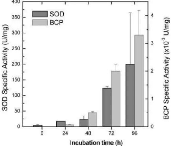
BCP Bacterioferritin co‐migratory protein Bis‐ANS 4,4'‐bis(1‐anilinonaphthalene 8‐sulfonate)
CATH Hierarchical domain classification of protein structures CD Circular Dichroism
CHAPS 3‐[(3‐cholamidopropyl)dimethylammonio]‐1‐propanesulfonate CID Collision Induced Dissociation
COG Clusters of Orthologous Groups Da Dalton
DLS Dynamic Light Scattering DNA Deoxyribonucleic acid DTT 1,4‐Dithiothreitol E. Coli Escherichia coli
EDTA Ethylenediaminetetraacetic acid eg. example given (for example) et al. And others
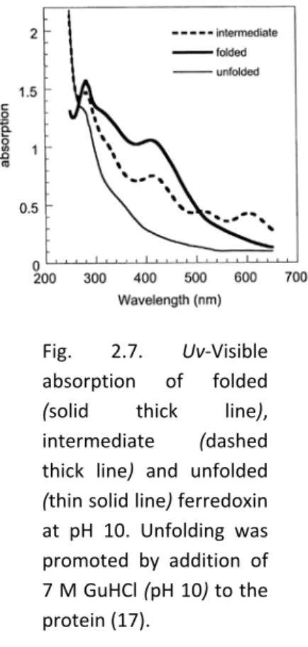
Fd Ferredoxin
GDP Guanosine diphosphate
GRAVY Grand average of hydropathicity index GTP Guanosine triphosphate
GuHCl Guanidine hydrochloride IEF Isoelectric focusing IPG Imobilizad pH gradient IR Infrared spectroscopy
iTRAQ Isobaric Tags for Relative and Absolute Quantitation
M Molar
MALDI Matrix Assisted Laser Desorption Ionization mRNA Messenger RNA
NL Non linear
NMR Nuclear Magnetic Resonance OGT Optimal growth temperature ORF Open reading frames
PAGE Polyacrilamide gel electrophoresis PDB Protein Data Bank
pI Isoelectric point
PMSF Phenylmethylsulfonyl fluoride Prx Peroxiredoxin
RNA Ribonucleic acid
ROS Reactive oxygen species rRNA Ribosomal ribonucleic acid S. solfataricus Sulfolobus solfataricus
SCOP Structural Classification of Proteins SDS Sodium dodecyl sulfate
SOD Superoxide dismutase TCA Trichloroacetic acid TF Transcription factor TFA Trifluoroacetic acid
Tm Midpoint transition temperature Topt Optimal temperature
Tris‐HCL Tris(hydroxymethyl)aminomethane hydrochloride UV Ultraviolet
vs. Versus
A Ala Alanine C Cys Cysteine D Asp Aspartate E Glu Glutamate F Phe Phenylalanine G Gly Glycine H His Histidine I Ile Isoleucine K Lys Lysine L Leu Leucine M Met Methionine N Asn Asparagine P Pro Proline Q Gln Glutamine R Arg Arginine S Ser Serine T Thr Threonine V Val Valine W Trp Tryptophane Y Tyr Tyrosine
INTRODUCTION……….……….……….………...23
U
1.1.U UBiodiversity of thermophilesU... 24
U
1.2.U UModel organismsU... 26
U
1.2.1.U UThermophilic model organisms: SulfolobalesU... 26
U
1.2.2.U UMesophilic model organism: Escherichia coliU... 28
U
1.3.U UConvergent Evolution Theory of Thermal AdaptationU... 30
U
1.4.U UClusters of Orhologous GroupsU... 31
U
1.5.U UDesignability and evolvability of protein structureU... 33
U
1.6.U UCellular environmentU... 35
U
Molecular crowding influence on proteins´ folding and stabilityU... 35
U
Molecular chaperonesU... 37
U
1.7.U UDiversity of Protein Conformational statesU... 39
U
1.8.U UMolecular determinants of protein stabilityU... 40
U
1.9.U UProteins with enhanced conformational stabilityU... 42
U
1.10.U UFactors influencing protein stabilityU... 43
U
1.10.1.U UIntrinsic factors influencing protein stabilityU... 44
U
1.10.2.U UStructural and other extrinsic factors influencing protein stabilityU... 50
U
METHODOLOGIES
FOR
PROTEOMICS
STUDIES
OF
PROTEIN
STABILITY ... 68
2.1. Introduction ... 68
2.2. Identification and quantification ... 68
Profiling hyperstable proteins at a proteomic scale ... 69
In‐Gel Detection of Protein Surface Hydrophobicity Changes... 72
Electrophoretic Detection of Intrinsically Disordered Proteins... 74
Isobaric tags for relative and absolute quantitation ‐ iTRAQ... 76
2.3. Bioinformatics... 77
2.4. Preservation of protein structure in solution ... 80
2.5. Conclusion ... 83
2.6. References……….83
A
PROTEOMIC
APPROACH
TOWARDS
THE
SELECTION
OF
PROTEINS
WITH
ENHANCED
INTRINSIC
CONFORMATIONAL
STABILITY ... 90
3.1. Summary... 90
3.2. Introduction ... 90
3.3. Experimental... 92
Cell mass and preparation of the cytosolic extract ... 92
Thermal and chemical perturbation protocols ... 92
In situ gel digestion ... 93
MALDI Peptide mass fingerprinting ... 95
MALDI MS/MS peptide sequencing ... 95
Miscellaneous biochemical and spectroscopic methods... 96
3.4. Results and Discussion ... 97
High temperature and chemical denaturants induce proteome perturbation... 97
Proteome analysis by 2‐DE and MS: identification of hyperstable proteins ... 99
Proteins from the pool of selected hyperstable proteins are biologically active... 103
3.5. Conclusions ... 104
3.6. References………..………105
U
INTRINSIC
THERMAL
STABILITY
PROPERTIES
IN
THERMOPHILIC
VS
MESOPHILIC
CYTOSOLIC
PROTEOME:
THERMAL
SEPARATION
,IDENTIFICATION
AND
iTRAQ
QUANTIFICATION………112
U 4.1.U USummaryU... 112
U 4.2.U UIntroductionU... 113
U 4.3.U UMaterials and methodsU... 114
U Defining thermostable proteins’ subsetsU... 117
U
Molecular weight and isoelectric pointU... 120
U
4.5.U URelationship between thermostability and aminoacid contentU121
U
4.6.U URelationship between thermostability and protein classU... 125
U
4.7.U URelationship between protein thermostability and cellular
biological function ‐ cellular thermo toleranceU... 128
U
4.8.U UConclusionsU... 133
U
4.9.U UReferencesU... 134
U
RELATIONSHIP
BETWEEN
PROTEIN
THERMOSTABILITY
AND
SOLUBILITY
IN
Escherichia coli
THERMALLY
SELECTED
SUBPROTEOME
U... 142U
5.1.U USummaryU... 142
U
5.2.U UIntroductionU... 142
U
Intracellular ambient and chaperone functionU... 142
U
Thermostable soluble proteome subsetU... 144
U
5.3.U UObjectives and MethodologiesU... 145
U
5.4.U UResults and discussionU... 148
U
Solubility predictionsU... 148
U
Bimodal solubility distribution of thermostable proteinsU... 150
U
Solubility correlation with pI/MwU... 152
U
Relationship between solubility, aliphatic index and GRAVY index of thermostable proteinsU... 154
U
biological functionU... 159
U 5.5.U UConclusionsU... 162
U 5.6.U UReferencesU... 163
CONCLUDING REMARKS ……….167
APPENDIX I ………..………...172
APPENDIX II ……….………179
APPENDIX III ………..……….186
This chapter was partially published in:
Vesna Prosinecki, Patrícia F.N. Faísca and Cláudio M. Gomes, Conformational States and Protein Stability in a Proteomic Perspective, Current Proteomics, Vol. 4 Issue 1, 2007, 44‐52
Chapter
1
INTRODUCTION
U
1.1.U UBiodiversity of thermophilesU... 24
U
1.2.U UModel organismsU... 26
U
1.2.1.U UThermophilic model organisms: SulfolobalesU... 26
U
1.2.2.U UMesophilic model organism: Escherichia coliU... 28
U
1.3.U UConvergent Evolution Theory of Thermal AdaptationU... 30
U
1.4.U UClusters of Orhologous GroupsU... 31
U
1.5.U UDesignability and evolvability of protein structureU... 33
U
1.6.U UCellular environmentU... 35
U
MOLECULAR CROWDING INFLUENCE ON PROTEINS´ FOLDING AND STABILITYU... 35
U
MOLECULAR CHAPERONESU... 37
U
1.7.U UDiversity of Protein Conformational statesU... 39
U
1.8.U UMolecular determinants of protein stabilityU... 40
U
1.9.U UProteins with enhanced conformational stabilityU... 42
U
1.10.U UFactors influencing protein stabilityU... 43
U
1.10.1.U UIntrinsic factors influencing protein stabilityU... 44
U
1.10.2.U UStructural and other extrinsic factors influencing protein stabilityU... 50
U
1.11.U UReferencesU... 54
1.1.
5BBiodiversity
of
thermophiles
Thermostable organisms and their proteins have been subject of research during the last decades due to their various unique properties. Interest in thermophiles and how their proteins manage to function at elevated temperatures started in 1960’s by the pioneering work of Brock and his colleagues (1) and continues up to date. Even nowadays, elevated interest in those remarkable organisms still continues, aiming at exploring mechanisms of survival at the environmental extremes, offering valuable data in knowing bases of protein stability.
Based on their optimal growth temperatures (OGT) organisms are divided into three main groups, i.e. psychrophiles with OGT below 20°C, mesophiles that optimally grow at moderate temperatures from 20°C up to 55°C, and thermophiles that thrive in high temperatures, above 55°C. Only few eukaryotes are known to grow above this temperature, but some fungi grow in the temperature range 50 up to 60°C (2). In 1992 Kristjansson and Stetter (3) suggested a further division of the thermophiles and a hyperthermophile boundary by growth at and above 80°C. The Tree of Life that is 16S rRNA‐based phylogenetic tree exhibits three domains, the Bacteria, Archaea and Eukarya. The use of ribosomal RNA sequences has led to the recognition of a group of prokaryotes, the Archaea that are lacking a nuclear membrane and possessing a single circular chromosome, while possessing several molecular properties with similarity to the eukaryotes such as transcription signals, transcription factors, chaperones, and histones. Archaea are phylogenetically distinct from both Bacteria and Eukarya and rich in thermophilic and hyperthermophilic species. Members of the deepest and shortest lineages exhibit the highest growth temperatures (Fig. 1.1 (5)).
sources available, they show great versatility: members of the same genera and even the same strains may be able to use different electron donors and acceptors. In addition, several hyperthermophilic Archaea are facultative or obligate heterotrophs able to use organic compounds as energy and carbon sources (4, 5). Most thermophilic bacteria characterized today grow below 80°C hyperthermophilic boundary with OGT up to 50°C with some exceptions, such as Thermotoga and Aquifex (4) while hyperthermophilic species are highly dominated by the Archaea. Currently, the most thermophilic organism known is Pyrolobus fumarii that grows in the temperature range of 90 to 113°C. Regarding thermophiles, the bacteria Aquifex pyrophilus and Thermotoga maritima exhibit the highest growth temperatures of 95°C (4). Within the Archaea, the organisms with the highest growth temperatures, between 102 and 113°C, are found within the Crenarchaeota and the Euryarchaeota (6). They are members of the crenarchaeal genera Pyrolobus, Pyrodictium, Hyperthermus, Pyrobaculum, Igneococcus and Stetteria and the euryarchaeal genera Methanopyrus and Pyrococcus. The upper temperature at which life is possible is still unknown, but it is probably not much above 113°C. Above 110°C most of the biological molecules become highly unstable, ATP is spontaneously hydrolyzed in aqueous solution at temperatures below 140°C, and hydrophobic interactions weaken significantly (7).
At present, vast number of species of hyperthermophilic Archaea and Bacteria are known which had been isolated from different terrestrial and marine thermal areas in the world. Hyperthermophiles are very divergent, in terms of both their phylogeny and physiological properties and are grouped into 34 genera and 10 orders (5). Due to the fact that hyperthermophiles belong to two phylogenetically distinct domains of life, the Bacteria and Archaea, the strategies of molecular mechanisms of heat adaptation may be quite different depending on the phylogenetic position of the corresponding organism.
Fig. 1.1. Small subunit ribosomal RNA‐based phylogenetic
tree. The thick lineages represent hyperthermophiles (5).
1.2.
6BModel
organisms
The focus of this research work and thesis has been on proteins with enhanced stability properties originating from two distinct groups of organisms: Thermophiles Sulfolobus solfataricus and Sulfurisphaera sp. from the archaeal order Sulfolobales, and the mesophilic bacteria Escherichia coli. These models are briefly presented.
1.2.1.
43BThermophilic
model
organisms:
Sulfolobales
Order of Sulfolobales belongs to category of sulfur dependent Archaea.
energy for growth by the oxidation of sulfur to sulfuric acid. Sulfolobales
are generally less thermophilic than Thermoproteales and
Thermococcales, with only species of Acidianus being able to grow at or
above 90°C. In addition, they mainly inhabit continental spring waters
rich in sulfur, although some species are also found near shallow marine
volcanic vents. Species of Acidianus and Desulfurolobus also grow under
anaerobic conditions by the reduction of sulfur to H2S using H2 as the
electron donor. Stygiolobus is unique among the Sulfolobales as it does
not grow under aerobic condition.
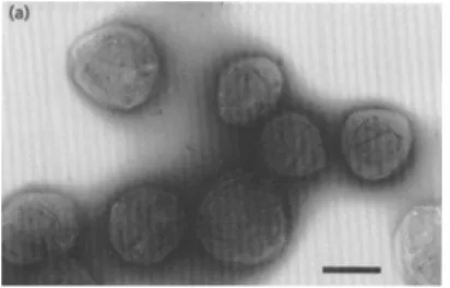
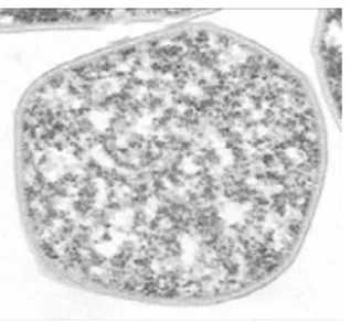
Fig. 1.2 Sulfurisphaera sp. Electron micrographs of
strain TA‐lT. (a) Negative staining; bar, 1 prn. (b)
Thin section. Cell membrane (CM) and envelope (E)
are indicated; bar, 0.5 pm. (8)
Sulfolobus solfataricus and Sulfurisphaera sp.
Scheme 1.1. Taxonomic hierarchy of Order Sulfolobales
(Hhttp://www.taxonomy.nl/taxonomicon/H)
Fig. 1.3. Microscopy image of S.
solfataricus. Image D.Janckovik
and W.Zillig
Sulfolobus solfataricus was discovered by Wolfram Zillig and Karl Stetter in Pisciarelli near Naples, Italy (Fig. 1.3). Up to date it is the most widely studied organism of the crenarchaeal branch of the Archaea, a model for research on archaeal mechanisms and cellular processes like DNA replication, the cell cycle, chromosomal integration, transcription, RNA processing, and translation. It is shaped as highly irregular lobed cocci which usually occur singly, has no flagella, but pilus‐like and pseudopodium‐like structures are often found. Strictly aerobic, its optimal growth temperature is at 87°C and pH 2 to 4, metabolizing sulfur. It produces sulfuric acid. S. solfataricus has been isolated worldwide from continental solfatara fields including Yellowstone National Park, Mount St. Helens, Iceland, Italy, and Russia ‐ almost wherever there is volcanic activity. The genome of the S. solfataricus P2 contains 2,992,245 bp on a single chromosome and encodes 2,977 proteins and many RNAs. One third of the encoded proteins have no detectable homologs in other sequenced genomes. Moreover, 40% appear to be archaeal‐specific, and only 12% and 2.3% are shared exclusively with Bacteria and Eukarya, respectively (9).
1.2.2.
44BMesophilic
model
organism:
Escherichia
coli
A mesophilic bacterium Escherichia coli was discovered in 1885 by the
ithe most widely used prokaryotic system for the synthesis of heterologous proteins and model in scientific research. Many decades of research have resulted in a wealth of genetic, biochemical, and structural information that together is unparalleled in other systems (10). Taxonomic hierarchy of Escherichia coli is presented in Scheme 1.2.
E. coli is the member of large bacterial familyEnterobacteriaceae that are facultatively anaerobic Gram‐negative rods that live in the intestinal tracts of humans and animals in health and disease, physiologically
versatile and well‐adapted to variations of its characteristic habitats.The genome of E. coli consists of 4,639,221 bp of circular duplex DNA (11) that was sequenced in 1997.
Characterization and comparison of E. coli paralogous proteins and protein groups and comparison to other species allows examination of the evolutionary events surrounding protein diversification. Therefore, this organism was our obvious choice for the well known model system for mesophilic organism to compare the results originating from our study regarding archaeal proeome.
Scheme 1.2. Taxonomic hierarchy of
Escherichia coli
(HUhttp://www.taxonomy.nl/taxonomicon/)U
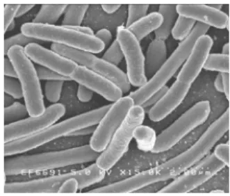
Fig. 1.4. Scanning electron
micrograph of Escherichia coli,
grown in culture and adhered to
a cover slip. Credit: Rocky
Mountain Laboratories, NIAID,
National Institute of Health.
1.3.
7BConvergent
Evolution
Theory
of
Thermal
Adaptation
Dealing with the highly thermostable proteins originating from phylogeneticaly distinct organisms to start with, required the insight into possible background of origins of thermostability. Data from the literature indicate that the choice of a particular strategy depends on the evolutionary history of an organism (12):
Convergent evolution represents a phenomenon when two distinct species with differing ancestries evolve to display similar physical features (13). Environmental circumstances that require similar developmental or structural alterations for the purposes of adaptation can lead to convergent evolution even though the species have different origin. As a consequence of convergent evolution, biological structures or species that exhibit similar functions or/and appearance may appear, even though they evolved through widely divergent evolutionary pathways and had different ancestors. These similarities are typically explained as the result of common adaptive solutions to similar environmental pressures on the level of the organism. On the protein level, these adaptation similarities that arise as a result of the same selective pressures and unfortunately can be misleading to understanding the natural evolution. Therefore, identification of specific residues or fragments which may be more relevant to protein thermostability is influenced by the possibility that some of the differences among the thermophiles and mesophiles rely on phylogenetic differences instead of thermal adaptation or vice versa (14).
Thermostability properties of proteins gave origin to many studies so far. However, up to date no general physical mechanism was found that can be named as the most important factor for increased thermostability. Hyperthermophiles belong to two phylogenetically very different domains of life, the Bacteria and Archaea. Therefore, the strategies of molecular mechanisms including heat adaptation may be rather dissimilar depending on the phylogenetic position of the corresponding organism.
1.4.
8BClusters
of
Orhologous
Groups
proteins (COGs) database has been designed as an attempt to classify proteins from completely sequenced genomes on the basis of the orthology concept (15). Many proteins are members of paralogous gene families and have significant matches in other species. The genes in all genomes are derived from a set of unique ancestral genes present in a progenitor of all organisms. The COGs database relies on phylogenetic classification of the all the proteins encoded in complete sequenced genomes of Bacteria, Archaea and Eukarya, available at the web (HUhttp://www.ncbi.nlm.nih.gov/COGUH). The COG were constructed by applying the criterion of consistency of best hits specific to particular genome to the results of an exhaustive comparison of all protein sequences from those genomes (16). But, as the level of divergence between orthologous genes approaches the level of divergence among paralogs within a species, it is difficult to determine the relation between similar genes in different species. In most of the cases orthologous proteins have the same domain architecture and the same function, but there are also significant exceptions particularly among multicellular eukaryotic organisms.
classification of genes that is conserved across different organisms has provided new information about how these functions are maintained and modified across phylogenetic groups during evolution. However, in overpopulated COGs, the orthologous relationships between members are difficult to delineate precisely. Such COGs might contain proteins that evolved new functions with respect to the original ancestor, and even though these proteins still have significant sequence similarity, at the entire sequence or the domain level, they may be part of different cellular processes. Therefore, proteins involved in biological processes characteristic of eukaryotes may not have the counterparts in bacterial and archaeal genomes.
Fig.1.4. COG functional classification categories
1.5.
9BDesignability
and
evolvability
of
protein
structure
designed and robust against mutations, and that such a selection simultaneously leads to thermodynamic stability (19, 20).
Protein structures are classified into different folds. Proteins that have the same fold also have the same major secondary structures in the same arrangement with the same topological connections, with some variations typically in the loop region. Those that are evolutionary closely related often have high sequence similarity and share a common fold, but the common fold is possible even for proteins with distinct evolutionary origins and different biological functions. Therefore, the number of folds is much lower than the number of proteins (20). The usage of protein folds in nature is known to be non‐uniform: a few folds are used often, while most others are used relatively rarely.
within useful phenotypes. Most of the substitutions destabilize the native structure of a protein, therefore modest raise in thermodynamic stability increases the number and type of substitutions that a protein can tolerate before misfolding (23). Necessity for increased stability in highly expressed proteins would restrict the set of evolutionarily viable sequences and as a consequence slow sequence evolution.
Knowledge of biophysical causes of rate differences in comparing evolvability of various proteins is still scarce. A dominant factor in reducing the evolvability rate is high protein expression level that leads to increased transcription and translation (24). This increase also increases the probability of translational missense errors that may have misfolding as a consequence, and further loss of biological function. Therefore, slow evolvability is a property of highly expressed proteins in nature (25). Chaperoning cellular systems that assist in folding of other proteins and buffer various effects of mutations, therefore are also found to influence evolvability of protein structures (26) by enabling destabilizing mutations to accumulate.
1.6.
10BCellular
environment
Various factors present in the intracellular environment influence protein folding as well as stability of mature protein. As we are dealing with properties of the proteins with highly stable behavior, introduction to an intracellular environment and it’s (de)stabilizing properties gives a better insight into protein “living conditions”. We should keep in mind that the subject of this thesis and research are proteins with elevated stability properties, investigated out of their natural cellular environment, but we can only assume that properties that lead to increased stability within the cell are the same ones that were proven to increase the stability in laboratory controlled environment.
47B
Molecular
crowding
influence
on
proteins´
folding
and
stability
significant part of the total volume of about 40% of the medium. Therefore, the accessible volume in the cell is reduced and a significant fraction of the water is involved in solvation and does not behave as bulk water. This medium is referred to as a solution with molecular crowding. The structure and dynamics of macromolecules and supramolecular assemblies is the result of a large number of small forces such as electrostatic interactions and the hydrophobic effect. Both forces are strongly dependent on the properties of water. In this medium, a significant fraction of the water is involved in solvation and does not behave as bulk water. Therefore, various types of intermolecular forces are strongly affected by the reduced availability of water due to molecular crowding.
macromolecular species present in solution. Association reactions are therefore highly favored under crowded conditions, and association constants under crowded conditions could be several orders of magnitude larger than those measured in dilute solutions (31). This implies that aggregation of refolding protein molecules is a much greater problem under crowded cellular conditions than it is in dilute solutions (32).
Thermophilic and hyperthermophilic organisms generally accumulate compatible solutes as a mechanism of osmotic adjustment and protection of cell components against thermal denaturation. Newly discovered solutes from thermophilic and hyperthermophilic organisms include cyclic‐2,3‐bisphosphoglycerate two isomers of di‐myo‐inositol phosphate, mannosylglycerate and mannosylglyceramide di‐mannosyl‐ di‐myo‐inositol phosphate, diglycerol phosphate and galactosyl‐5‐ hydroxylysine (33) and may constitute an adaptive feature of these organisms to high temperatures. Thermophiles and hyperthermophiles accumulate compatible solutes that have not been found, or are rarely encountered in mesophilic organisms. Therefore, the compatible solutes of (hyper)thermophiles are specifically associated with life at high temperatures. Archaeal compatible solutes are generally negatively charged, while other microorganisms generally accumulate neutral or zwitterionic compatible solutes. Nature of interactions between solutes and exposed groups in the protein structure and its stabilizing effect is attributed mainly to a large contribution from interactions with exposed backbone groups in a partially unfolded state, with side‐chain interactions modulating the specificity of the effect. The interactions should cause a contraction of the protein structure with a concomitant decrease in internal mobility (34) which is in agreement with correlation of higher thermal stability of hyperthermophilic proteins structure rigidification upon in vitro addition of compatible solute (35).
48B
Molecular
chaperones
(18), the presence of molecular chaperones was found to be essential (27). In a living cell self‐folding is inefficient and slow, with danger of misfolding and aggregation due to crowding conditions.
Fig.1.5. Competing reactions of protein folding and aggregation.
Scheme of the funnel‐shaped free‐energy surface that proteins explore
as they move towards the native state by forming intramolecular
contacts. The ruggedness of the free‐energy landscape results in the
accumulation of kinetically trapped conformations that need to
traverse free‐energy barriers to reach a favorable downhill path. In vivo, these steps may be accelerated by chaperones. When several
molecules fold simultaneously in the same compartment, the free‐
energy surface of folding may overlap with that of intermolecular
aggregation, resulting in the formation of amorphous aggregates, toxic
oligomers or ordered amyloid fibrils. Adapted from Hartl et al (30).
acclimation are ubiquitous mechanisms in the extremely thermophilic microorganisms. Molecular chaperones can be classified into three functional groups based on their action mechanism: i) Folding modulators are chaperones that assist and mediate folding processes (DnaK and GroEL) performing their function on conformational changes in the presence of ATP; ii) Holding chaperones that stabilize partially folded protein structure in a case of severe stress situation (Hsp33, Hsp31 and IbpB) awaiting folding chaperones to become available; iii) Chaperone (ClpB) that promotes the solubilization of aggregated proteins as a result of stress (36).
The chaperones that are shared by Archaea and Bacteria include the chaperone machine composed of Hsp70(DnaK), Hsp40(DnaJ), and GrpE (37). In Archaea, proteins coded by these genes are very similar to bacterial homologs, as if the genes had been received via lateral transfer from Bacteria. The chaperonin system in Archaea studied to the present, including those that possess a bacterial‐like chaperone machine, is similar to that of the eukaryotic‐cell cytosol. Hyperthermophilic Archaea like Pyrococcus spp, Sulfolobus spp, Pyrobaculum aerophilum, Methanocaldococcus jannaschii, Metahopyrus kandleri, Archeoglobus fulgidus do not have Hsp90, DnaK, DnaJ, GrpE, Hsp33 and Hsp10 homologs (38).
1.7.
11BDiversity
of
Protein
Conformational
states
Protein stability is defined by imprecise cancellation of two large effects, namely, the hydrophobic effect, favoring folding, and chain entropy, disfavoring it. Because these two effects are similar in magnitude the net stability of proteins is marginal. Indeed, the average Gibbs free energies of denaturation (ΔGD) ranges from 20 to 60 kJ/mol (at 250C), a value
is almost as compact as the native form and have a loosely packed core, while retaining some of its native secondary structure (40). Consequently, each protein has a particular energetic landscape of conformations that it can adopt under physiological conditions. Ultimately this may correspond to a protein in which the most populated ensemble of structural conformations is disordered or that contains highly disordered regions (39, 41). This possibility impacts on the structure‐function paradigm, as disordered proteins are biologically active in functions related to regulation of transcription and translation, protein phosphorylation, storage of small molecules, and regulation of the self assembly of multiprotein complexes (42). Disordered proteins often fold into an ordered structure upon binding to a protein partner. That is, for example, the case of thyroid hormone and retinoid receptors, which occur as unstructured ensembles, and fold upon interacting with a nuclear receptor binding domain. The same occurs upon interaction with a ligand, such as a metal ion or a nucleic acid, as in the zinc‐finger containing transcription factor TFIIIA or the translation initiation factor elF4E (42, 43). Interestingly, disordered domains may adopt distinct ordered conformations depending on the interacting partner, thus reflecting a significant functional flexibility, in agreement with the fact that disordered proteins are mainly involved in signaling and regulatory pathways. This has led to the proposal of generically coining disordered proteins as ‘pliable’ (43). Overall, some proteins comprising intrinsically disordered domains or segments have a plethora of accessible ordered conformations that they can adopt, upon protein‐ligand interaction(s). In thermodynamic terms, coupling folding to binding results in a strategy to use the binding enthalpy to pay the entropic cost to fold a disordered protein.
covalent interactions, metal ion and disulfide cross‐linking interactions assume a particularly relevant role in small proteins comprising irregular domains. Although proteins frequently lack a large hydrophobic core and have minimal secondary structure; covalent interactions allow attaching and stabilizing different parts of the protein. This is what happens, e.g., with scorpion toxin, allergy factor Ra5, and several protease inhibitors (52).
1.9.
13BProteins
with
enhanced
conformational
stability
Some proteins need to be particularly stable as a result of molecular adaptation to a particular physiological condition or to a harsh environmental factor, such as high salinity, extreme pH and high temperatures. Among thermophiles, enhanced protein stability encompasses both thermodynamic and kinetic stability. While the kinetic stability depends on the energy barrier to unfolding, i.e., on the activation energy of unfolding, the thermodynamic stability is reflected in the conformational stabilities (i.e., ΔGD), which may be up to 100 kJ/mol
larger than those from mesophilic proteins (56), and in the midpoint transition temperatures for unfolding (i.e., Tm), which are typically
between 20‐300C above those of mesophiles (57, 58). A recent study
theoretical analyses, diverse stabilizing strategies have been suggested, as putative intrinsic drivers for the enhanced stability exhibited by thermophilic proteins (7). Structural properties like better core (and secondary structure) packing (59‐61), deletion or shortening of loops and increased helical content have been typically ascribed to thermophilic proteins (62). On the physicochemical side, it is generally claimed that thermophilic proteins have more hydrophobic residues (63‐66), a larger amount of main‐chain hydrogen bonds (66) and a higher number of proline residues. However, a recent study, which analyzed 18 non‐ redundant families of thermophilic and mesophilic proteins, reported that these factors do not show consistent, substantial variations between mesophiles and thermophiles (67). The higher number of salt bridges among thermophilic proteins suggests that these interactions play a role in stability enhancement (62, 66‐71). Nevertheless, there is not a general physical mechanism able to rationalize the stability enhancement upon increasing the number of salt bridges, because the net electrostatic free energy of salt bridges can be either stabilizing or destabilizing (72). This results from the fact that energetically favorable Coulombic charge– charge interaction forming in the protein core, is opposed by the unfavorable desolvation of interacting charges – the transfer of a salt bridge from water to nonpolar environment costs ~ 42‐67 kJ/mol (73). Therefore, it is mostly the surface, solvent‐exposed, salt bridges that effectively lead to an increase in protein stabilization (74‐77). Moreover, there is also some evidence that extended networks of salt‐bridges (formed by residues that participate in more than one salt‐bridge) between protein subunits are critical for achieving the superior thermostability of hyperthermostable proteins (63, 67, 78).
1.10.
14BFactors
influencing
protein
stability
an attempt to find new prediction method of theoretical analysis for thermostable proteins.
1.10.1.
45BIntrinsic
factors
influencing
protein
stability
Amino acid composition
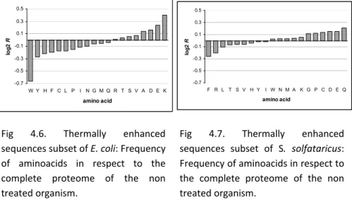
Amino acid composition has long being known to have a certain influence on proteins’ thermostability. Comparing thermophiles and mesophiles, difference of the amino acid composition has been found to be a global trend across a large number of protein families (67). Statistical analyses comparing amino acid compositions in mesophilic and thermophilic proteins indicated trends toward substitutions of certain amino acids with the others that implied in order to increase thermostability. Some of it has been indicated by various studies although the reasons behind the origins of these substitutions are still open for discussion. Many factors might substantially influence the results as well as the conclusions: chosen sample group of thermophilic vs. mesophilic organisms, selection of their representative proteins or availability of data for the comparison. However, the majority of studies would agree to some overall conclusions regarding the frequency of certain amino acids.
Charged residues such as Glu, Asp, Lys, and Arg are frequent and polar residues such as Ser, Thr, Asn, and Gln, are scarce in thermophilic proteins (79, 80). Polar residues Ser, Thr and Asn can hydrogen bond to the backbone peptide groups and this interferes with the hydrogen bonding of the α‐helix; therefore these amino acids are found less frequently in α‐helices, and at the same time less often in thermostable proteins. Substitutions such as Gly Ala and Lys Arg are frequent in thermophiles, where higher alanine content in thermophilic proteins is
Additional aromatic clusters occur in regular secondary structures, implying their location to be in more rigid regions of the protein (84).
Besides from these studies that are focused solely on amino acid composition, some results indicate that frequency of certain combination of amino acids influences thermal stability. Prevalence of either residue combination (IVYWREL)(85) or their ratio ((E+K)/(Q+H))(86) has been noted to lead to thermal stability. It is very important to note on the level of sequence‐encoded increment of thermostability that not just the frequency but the position of certain residues has significant influence.
Intracellular and extracellular proteins have different amino acid compositions as expected, and therefore, their subcellular locations of the protein subpopulation investigated, highly influences the result (87). These differences are consequence almost entirely to residues exposed to the solvent (88). Subcellular location determines the differences in the amino acid composition resulting from the adaptation of the protein surface to the physicochemical environment. Therefore it would be reasonable to expect that the differences in the amino acid compositions between proteins of thermophilic and mesophilic organisms would be much greater on the protein surface than the interior, regarding the differences between the environments in which thermophilic and mesophilic organisms live (79). Therefore, differences in amino acid composition between organisms with different optimal growth temperature (OGT) might often be evolutionarily relevant, rather than an indication of its adaptation to high temperatures ‐ more relevant to thermostability than amino acid composition is the distribution of the residues and their interactions in the protein. With increasing experimental data accumulating, in particular complete genome sequences, it is already obvious that thermophilic adaptation cannot just be defined in relation to significant differences in the amino acid composition (7).
Ion pairs
increased number of salt bridges found in many structures of thermostable and, in particular, hyperthermostable proteins. These results are mostly based on structural comparisons of proteins from hyperthermophiles and mesophiles and have suggested that the ion pairs show a tendency to be increased in number as well as to be organized in large networks, in parallel with increasing melting temperature of the protein (50). As previously mentioned, comparative analysis using complete genome sequences from Bacteria, Eukarya and Archaea, a large difference between the proportions of charged amino acids such as Arg, Asp, His, Glu, and Lys versus polar amino acids was found to be a specific signature of all proteins from hyperthermophilic organisms (60, 80). Proteins from hyperthermophilic organisms are characterized by an increased number of ion pairs with respect to the statistical expectance and/or the number of ion‐pairs in their mesophilic counterparts. This suggests that electrostatic interactions emerge as a factor responsible for the elevation of the melting temperature of proteins from hyperthermophilic organisms. It is important to note that, in addition to the increased number of charged group, their spatial organization influences elevation of the melting temperature of thermostable proteins. In parallel with increasing Tm of the protein, ion pairs tend to be organized in networks that are often found on the protein surface or partially buried at domain or subunit interfaces near to local symmetry axes of protein oligomers. The largest ion‐pair network in any hyperthermostable protein reported has been observed in glutamate dehydrogenase from P. furiosus (63, 67). Statistical appearance of salt bridges is higher for proteins without disulfide bridges than for proteins with disulfide bridges which is explained by the fact that salt bridges often occur between groups distant in the protein sequence, forming cross‐links that therefore stabilize the tertiary structure (80). By contrast, charge burial in the protein environment is energetically unfavorable and can reduce the energy gained by attraction between opposite charges in an ion pair.
electrostatic interactions in both directions: by minimizing repulsive contacts and by increasing the number of ion pairs, so that they gain electrostatic stabilization by minimizing the number of repulsive contacts rather than by creating salt bridges (68).
Hydrophobic interactions
Hydrophobic effect is the main driving force in protein folding (46). Aliphatic amino acids such as Ala, Ile, Leu, and Val in both thermophilic and mesophilic proteins contribute to the hydrophobic interaction, which is main force for maintaining conformational stability in inner part of protein (60). It has been suggested that thermophilic proteins are substantially more hydrophobic and have more surface area buried upon oligomerization as compared with their mesophilic counterparts, even though overall hydrophobicity of thermophilic proteins and their mesophilic homologs are very similar (67). Therefore, it is important to compare the frequency of the residues with the same solvent accessibility. In thermophilic proteins, the amino acid with the short alkyl group tend to interact more closely with neighbouring residues and have better packed form in protein structure. Ala and Val that are surface exposed with lower frequency but high frequent in well‐buried state contribute more to protein thermostability by enhancing conformational stability in the buried part of protein structure (60). With packing, the hydrophobic effect can have consequences at the level of the individual protein chains due to larger and more hydrophobic protein core but also due to the association of the chains (7). Overall numbers of water molecules per protein molecule and the water‐accessible surface area is larger in the mesophilic species proteins than in their thermophilic homologs (89).
Disulfide bridges
significant decrease, together with His. The second reason is that Cys, together with Asn, Gln and Met can be classified as thermolabile due to their tendency to undergo deamidation or oxidation at high temperatures (67). Even though disulfide bonds are not a method to achieve protein thermostability (80) and in general, hyperthermostable proteins contain lower fractions of cysteines and less disulfide bonds than their thermostable and mesostable counterparts, there still are some rare evidence of stabilization due to disulfide bond like in Fd from hyperthermophile Arquiflex aeolicus (91), although not as the only mechanism of stabilization but rather in contribution to electrostatic interactions by enabling dimerization of the subunits (92). Even though the other examples of thermal stabilization of proteins by introduction of disulfide bonds exist, it is obvious that is not the strategy chosen by thermophilic but rather by mesophilic organisms. Fewer cysteines are present in thermophilic proteins due to their property of being the most reactive amino acids in proteins. Their autooxidation, usually catalyzed by metal cations, especially copper, leads to the formation of intramolecular and intermolecular disulfide bridges or to the formation of sulfenic acid. Disulfide bond reshuffling can cause important structural variations. Therefore, after forming incorrect intersubunit disulfide bridges protein often becomes less stable and less thermophilic than the native enzyme (93).
Hydrogen bonds