From the Associação Xavier-Vidal para Direitos Humanos, Ciência, Tecnologia e Desenvolvimento (AXV) and the Biological and Ambient Science Institute,
Santa Úrsula University (USU) – Rio de Janeiro/RJ, Brazil. E-mail: [email protected]
Received for publication on April 12, 2004. Accepted for publication on July 07, 2004.
ORIGINAL RESEARCH
OXYTALAN ELASTIC AND COLLAGEN FIBERS
DURING THE REPAIR PROCESS IN EXPERIMENTAL
NITRIC OXIDE INHIBITION
Ricardo Xavier-Vidal
XAVIER-VIDAL R Oxytalan elastic and collagen fibers during the repair process in experimental nitric oxide inhibition.
CLINICS 60(2):85-92, 2005.
PURPOSE: To evaluate the repair process in rats with experimentally induced arterial hypertension. This study aimed to evaluate lesions in the ventricular myocardium and the repair process during experimental hypertension induced by systemic blockage of nitric oxide using N-omega-nitro-L-arginine methyl ester hydrochloride (L-NAME). Nitric oxide is an endothelial vasorelaxing factor and is necessary for the maintenance of normal arterial pressure, and L-NAME is an analog and antagonist of L-arginine, the substrate of the nitric oxide synthase.
MATERIALS AND METHODS: We used 26 normotensive young male Wistar rats belonging to several litters. Animals were treated with oral administration of L-NAME dissolved in water (75 mg/100 mL) for 43 days . Hearts were weighed and processed by routine methods. Special stains utilized were Gomori’s trichrome (aniline blue), picrosirius red polarization to identify fibrillar collagen, alcian blue technique (pH 0.5 and pH 2.5) to identify glycosaminoglycans, periodic acid-Schiff technique (with and without amylases) to identify proteoglycans, and Weigert’s resorcinol fuchsin solution (with and without oxone) to identify elastic fibers.
RESULTS: The results showed significant elevation of the arterial pressure (P <0.01) and significant increase of
cardiac weight (P <.0001) in the L-NAME (hypertensive) treated group, as compared to an untreated control group. The
histological analysis demonstrated wide infarcted myocardial areas in animals with nitric oxide blockade; several vascular changes such as thickening of the muscular tunica with fibrosis; thickening in the wall of small arteries and arterioles; and fibrinoid necrosis in the wall to nearly complete luminal obliteration. Reparative fibrosis involved mainly oxytalan elastic and collagen fibers.
CONCLUSION: Oxytalan elastic and collagen fibers are of great importance for the postinfarct repair process occurring during experimental nitric oxide inhibition.
KEYWORDS: Oxytalan elastic fibers. Collagen fibers. Nitric oxide. Arterial hypertension. Repair.
Hypertension is a common disorder in modern indus-trial countries.1 In men, arterial hypertension is the most im-portant risk factor in cardiovascular disease, affecting 11% to 20% of the Brazilian population over 20 years of age. In Brazil, cardiovascular diseases are the number-one cause of mortality, representing about 30.8% of the total deaths.2 About 90% to 95% of arterial hypertension is idiopathic,
and the remaining 5% to 10% is largely secondary to renal disease. About 5% of hypertensive patients experience sharp blood pressure elevation, which if untreated, leads to death in 1 or 2 years.3 Arterial hypertension is the most important etiologic factor associated with the appearance of sympto-matic heart failure, and left ventricular hypertrophy is the major risk factor.4 Ventricular hypertrophy also occurs in as-sociation with various forms of arrhythmia and is a risk fac-tor in cardiac sudden death.5
multiple genetic and environmental factors.3 Even if some authors disagree,7 it is now well established that reduced ac-tivity of nitric oxide synthase is, at least in some cases, a pathogenic factor in development of hypertension.8-11
Nitric oxide (NO) is an endothelial vasorelaxing fac-tor.12-17 N-omega-nitro-L-arginine-methyl ester (L-NAME) is an analog and antagonist of L-arginine, the substrate of ni-tric oxide synthase . Nini-tric oxide synthesis is necessary for the maintenance of normal arterial pressure, and its block-age causes arterial hypertension, significant experimental cardiac hypertrophy, and lesions in the myocardium.18-27 The extent of the direct influence of NO and the influence of hypertension produced by NO blockage on hypertrophy and on cardiac lesions is at present the subject of contro-versy and is subject to study using the L-NAME model. Re-duction of arterial hypertension in this model does not sig-nificantly reduce cardiac hypertrophy or the lesions brought about by an NO deficit, these morbid processes are prob-able mainly related to the NO deficit, not the hyperten-sion.25-26,28
The purpose of this work was to study lesions in ven-tricular myocardium mainly concerning oxytalan elastic and collagen fibers of rats at day 43 of arterial hypertension in-duced by NO synthetase blockage (L-NAME model).
MATERIALS AND METHODS
Twenty-nine young normotensive adults Wistar rats from several breeds with initial weights ranging from 179 to 279 grams were used. Arterial pressure was obtained on the 40th day of the experiment using pletismography.20,27,29 To en-sure that animals were normotensive, arterial presen-sure was measured a day before the beginning of the experiment. Af-ter arAf-terial pressures and weights were measured, animals were placed in plastic boxes with no more than 5 to 6 ani-mals per box. The initial arterial pressure was not above 117 mm Hg in any animal (Table 1). Seventeen rats were treated for 43 days with L-NAME (N-omega-nitro-L-ar-ginine methyl ester hydrochloride, Sigma Chemical, St. Louis) via oral administration at a concentration of 75 mg/ 100 mL in drinking water (about 60 mg/kg of body weight).20,27 Nine rats were used as an untreated control group. Six hypertensive animals died during the experi-ment, the majority at days 41 to 43.
At day 43 of the experiment, each animal was anesthetized with ethyl ether, weighed, the thorax opened, and cardiotomy performed. The hearts were weighed, fixed in Bouin’s fixative for 48 hours, processed using routine methods (embedded in Paraplast), and sliced into 6 or 7 mm sections. Sections were stained with Gomori’s trichrome, using aniline blue instead of fast green; picrosirius red, by which fibrillar collagen (probable type I) takes on a red or yellow color and reticular collagen (probable type III) takes on a green color under polarized light; alcian blue tech-nique at pH 0.5 to differentiate sulfated glycosamino-glycans and at pH 2.5 to differentiate carboxylated and phosphated glycosaminoglycans; periodic acid-Schiff tech-nique with and without amylase to differentiate proteo-glycans; and Weigert’s resorcinol fuchsin solution with and without oxone. The periodic acid-Schiff technique with amylase is important for differentiating proteoglycans be-cause it removes glycogen from the tissue.30-31 The treat-ment with Weigert’s resorcinol fuchsin solution using oxone is important because the oxone produces oxidation, which reveals the oxytalan fibers.
RESULTS
Our results demonstrate a sharp elevation of arterial pres-sure in rats treated with L-NAME (hypertensives) compared to the control group (Table 1). The mean tail-cuff pressure in hypertensive rats before cardiotomy (at day 40) was 176 mm Hg (range, 160 to -198 mm Hg). The control group had a mean value of 116 mm Hg before cardiomiotomy (range 100 to -123 mm Hg). Additionally, at day 40, there was a significant difference in mean cardiac weight (P <.001)
be-tween control (0.86 g, standard error = 0.04) and treatment (1.25 g, standard error = .005) groups. The ratio of cardiac weight (mg):body weight (g) was significantly different be-tween groups (P <.001), with values of 3.30 for the control
and 5.38 for the hypertensive group.
Figures 1 to 3 show examples of the histological results of the control and hypertensive groups. The histological analysis revealed that intense reactive and reparative fibro-sis occurred in all layers of myocardium (subepicardial, subendocardial, and inner myocardium) in left and right ventricles of treated animals. The right ventricles were more affected. These results suggested that wide infarcted areas
Table 1 - Tail-cuff arterial pressure measured during the experiments (mean / standard error).
Control (mm Hg) L-NAME (mm Hg)
Initial arterial pressure. 116.87 / 0.84 (n = 9) 115.65 / 0.82 (n = 17)
had developed in the myocardium. We also documented several vascular changes, such as thickening of the muscu-lar tunica with fibrosis replacing part of the cells, narrow-ing of the lumina, and perivascular fibrosis sometimes show-ing dense collagen fibers. We observed systematically a marked thickening in the wall of small arteries and arterioles of the hypertense animals and fibrinoid necrosis in the wall to nearly complete luminal obliteration (Figures 1 and 2).
In the fibrotic areas, we found the fibrillar collagen (prob-able type I) occupying a great proportion of total collagen. Occurrence of fibrillar collagen in reticular disposition (probable type III) was less significant. In spite of the in-tense fibrosis found in the hypertensive animals, the ratio of fibrillar to reticular collagens in these fibrotic areas was similar to that of the myocardial interstitium of the control group (Figure 2). Weigert’s staining for elastic fibers was
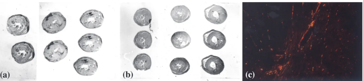
Figure 1 - Panoramic view of heart sections showing hypertrophy and fibrotic scars (dark areas) in the hypertensive hearts (a) compared with
control hearts (b) stained with picro-sirius red; (c) myocardium of a control animal showing fibrillar collagen (in yellow, orange, or red) corresponding to a high proportion of collagen, with a small amount of reticular collagen (in green) (magnification 100X) (picro-sirius red polarization).
(c)
(b)
(a)
Figure 2 - (a) inflammatory infiltrate areas highly suggestive of a myocardial infarct in the subendocardial region of a hypertensive heart (magnification 100X); (b) postinfarct fibrotic areas in the right ventricular myocardium of a hypertensive animal, stained with picrosirius red using polarized light to show fibrillar collagen (in yellow, orange, or red) occupying a great proportion of collagen fibers (magnification 200X);
(c) postinfarct fibrotic areas in the myocardium of a hypertensive animal, showing fibrillar collagen (in yellow, orange, or red) corresponding
to a high proportion of collagen, with a small amount of reticular collagen (in green) (magnification 200X) (picro-sirius-red polarization).
(a)
(b)
(c)
highly positive in the majority of the fibrotic areas, but in perivascular fibrotic areas reactivity was nil for all elastic fibers (Figure 3). Weigert’s staining with oxone was more intense than that without oxone. Alterations in the concen-tration of the sulfated glycosaminoglycans or carboxylated or phosphated glycosaminoglycans in the myocardial mass using the alcian blue technique could not be assessed be-cause there was no reaction.
We did not find fibrotic areas in the myocardium in the control animals. These animals had vessels with character-istic normal appearance.
DISCUSSION
Nitric oxide (NO) is involved in several important sig-nal processes in cardiovascular, nervous, and immunologic systems and also functions as a modulator of extracellular matrix components.15,32-37 Nitric Oxide is produced by car-diac myocytes38 and also acts in the myocardium as a
re-laxing factor.22 Under physiological conditions, NO plays an important role in regulation of tissue perfusion and myo-cardial blood flow.39 The interruption of NO synthesis by L-NAME causes systemic arterial hypertension and signifi-cant cardiac hypertrophy and lesions (reactive and repara-tive fibrosis) in the myocardium.20,21,25-28 This experimental arterial hypertension can be reversed using captopril, losartan, hydralazine, and enalapril.4,20,23,25,28 The adminis-tration of L-arginine (the NO synthase substrate) in rodents treated with L-NAME reverses this experimental arterial hypertension;19 in some cases of human hypertension, the administration of L-arginine reverses the hypertensive dis-ease.9,11
Fibrosis occurs in the myocardium during myocardial hypertrophy due to arterial hypertension. Fibrosis may oc-cur between the cardiac myocytes (reactive or interstitial fibrosis) or secondary to myocyte necrosis (reparative or parenchymal fibrosis, or, scarring). After about 4 weeks of renovascular hypertension, reactive fibrosis occurs, and at 12 weeks, reparative fibrosis appears predominantly in endomyocardium of rats. In diastolic dysfunction (because of a pathological increase of myocardial stiffness), its ma-jor determinant is the tissue component with highest ten-sile strength, which is the collagen matrix of the myocar-dium. Type I collagen, which is predominant in the myocardium¯in rats and monkeys about 70% and 82%, re-spectively, followed by type III at about 20% and 11%¯has great tensile strength, and its concentration determines the stiffness of myocardial tissue. Thus, the origin of heart fail-ure in arterial hypertension with diastolic dysfunction of the left ventricle appears to be rooted in the structural remodeling of the myocardium, particularly in the devel-opment of myocardial fibrosis.4 As the infarcted area heals, the late and persistent arrhythmogenic potential that emerges is not primarily dependent on the effects of ischemia on cellular electrophysiology but rather on the al-teration of the tissue structure stability originating in the postinfarct reparative fibrosis.40
The ideal repair mechanism is not found in adult tis-sue, but rather in embryonic tissue. Because of its relative easy and ideal healing ¯ rapid, efficient, and perfect with-out leaving a scar ¯ embryonic tissue is studied as a favorite paradigm of efficient tissue repair. Identifying how embry-onic tissue heals will lead to new therapeutic strategies for improving adult wound healing.41
Ribeiro et al.,20 observed that rats treated with L-NAME presented a progressive significant increase of the tail-cuff arterial pressure (168 mm Hg at day 30). They also observed systematically a marked thickening in the wall of small ar-teries and arterioles. They found fibrinoid necrosis in the wall of the renal arterioles and nearly complete luminal
ob-Figure 3 - (a) Myocardium with intense reaction by oxytalan fibers (dark dots) of the elastic system in reparative fibrotic areas (Weigert’s resorcinol-fuchsin method using an oxone reaction; magnification 100X); (b) perivascular fibrosis characteristic of the hypertensive cardiac processes using the L-NAME model. Note the degeneration of the inner elastic lamina and lack of reaction for oxytalan fibers of the elastic system (Weigert’s resorcinol-fuchsin solution with oxone; magnification 200X).
(a)
literation in 27% of the cases. Fibrinoid necrosis, which may result in small infarct areas, is the primary histopathologi-cal indication of malignant arterial hypertension.42
In infarcted areas after experimental coronary occlusion in rabbits, there was a 10-fold increase in the inducible form nitric oxide synthase activity.43
In the present work with the L-NAME rat model, we con-firm previously reported data regarding a sharp and signifi-cant increase of the arterial pressure. We found both reac-tive and reparareac-tive fibrosis (including perivascular fibro-sis). We also verified several vascular changes, such as thick-ening of the muscular tunica with fibrosis replacing part of the cells, narrowing of the lumina, and perivascular fibro-sis, sometimes with dense collagen fibers. Weigert’s stain-ing both with and without oxone oxidation was highly posi-tive in the majority of the fibrotic areas. Nevertheless, in perivascular fibrotic areas, the reactivity for Weigert’s fibers was low and concerning oxytalan fibers was nil, probably involved with a lack of major stiffness of the vessels.
The oxone reaction has a high reactivity for oxytalan fibers. Weigert’s resorcinol fuchsin method using oxone oxidation is used to identify oxytalan fibers, staining them violet. The Weigert’s resorcinol fuchsin method without oxone oxidation is used to identify the other fibers of the elastic system.44-45 The occurrence of oxytalan fibers serves to explain at least in part the stiffness of the tissue during diastolic function, because oxytalan fibers are less disten-sible than the other fibers of the elastic system.46
We systematically observed a marked thickening in the wall of small arteries and arterioles of the L-NAME treated hypertense animals and fibrinoid necrosis in the wall nearly to complete luminal obliteration. We also suggest that myo-cardial lesions occurred by vasoconstriction (mainly of arterioles) and by the hypertrophic condition of myocytes leading to local hypoperfusion and ischemia as well as pos-sibly by a degenerative processes of segments of the vas-cular bed in affected regions because of the obstructions. Concerning myocardial lesions that occur because of va-soconstriction, it is well known that in some cases, infarc-tion occurs after vascular spasm.3 Reactive fibrosis (perivas-cular and perimyocyte) possibly occurs by simple interrup-tion of NO producinterrup-tion that would induce collagen prolif-eration.34-35, 38
In this work, we identified in the heart of hypertensive animals intense perivascular fibrosis and scarring of the vas-cular wall in the affected vasvas-cular bed. In the development of cardiac hypertrophy (along the cardiac interstitial remodeling) an abnormal accumulation of fibrillar collagen with thickening of adventitia in the intramyocardial coro-naries also occurs.47 The analysis of Fischer et al.,1 follow-ing the inducement of arterial hypertension usfollow-ing stenosis
of the left renal artery, an interstitial and perivascular focal fibrosis in the heart of hypertensive animals was found. In our material, reparative fibrosis was predominant, and we found a discreet cellular inflammatory infiltration in the le-sions. This occurred because we studied animals at day 43 of administration of L-NAME, and therefore old reparative areas were present. Lukic et al.,48 studying diabetes mellitus in a mouse model, found mild inflammatory infiltration, mainly concerning some cellular types and indicated that NO played an important role in these processes. Consider-ing all factors implicit in the origin of hypertensive dis-ease in men, at least in some cases, the reduction of NO levels is significantly involved.11,49-51 Our present results are also similar to the abnormalities and lesions occurring, at least in some cases, in hypertension, cardiomyopathies, and in arteriosclerosis.
L-NAME. In our laboratory we have used hydralazine (Sigma Chemical, St. Louis) at a concentration of 120 mg/ L25 as well as the commercial preparation (tablet, Ciba-Geigy) at a concentration of 36 mg/L and 120 mg/L. The dose of 120 mg/L reverted arterial pressure and postponed the abnormalities that occur during systemic nitric oxide blockage by L-NAME.50 During renovascular hypertension, pretreatment using captopril has been suggested to prevent hypertension, fibrosis, and hypertrophy of the left ventri-cle.4 An important question is whether the regression of myocardial hypertrophy is linked to the reduction of car-diovascular risk induced by hypertrophy.55 Nevertheless, the cardiovascular risk may also increase in some cases, for in-stance, when the hypertrophy is produced by a myocardial infarct.6 Vasodilators and diuretics reduce arterial pressure,
however, they are not believed to reduce the cardiac weight significantly. It also has been proposed that regression of hypertrophy is only reached when sympathetic activity is reduced, because the sympathetic system is one of the cru-cial factors for the development of the hypertrophy.55
ACKNOWLEDGMENTS
The author acknowledges Professors Doctor Maria de Fátima Maron Ramos, Doctor Doyle Maia, and Doctor José Vanderli Andreata. This work is part of the M.Sc. Thesis from the author, HUAP – UFF (Brazil). During this work, the author had a M.Sc. fellowship from CAPES - Brasília / Brazil. This work was supported by CNPq, CAPES, USU, and AXV.
RESUMO
XAVIER-VIDAL R Fibras elásticas oxitalânicas e fibras colágenas durante o processo de reparo na inibição ex-perimental do óxido nítrico. CLINICS 60(2):85-93, 2005.
OBJETIVO: Avaliar o processo de reparo em ratos sub-metidos à hipertensão arterial experimental. Avaliamos le-sões miocárdicas ventriculares e processos de reparo durante a hipertensão arterial experimental induzida pelo bloqueio da síntese do óxido nítrico (ON) utilizando o Hidrocloreto de L-NAME. ON é um fator de relaxamento endotelial e é necessário para a manutenção da pressão arterial normal. L-NAME é um analogo e antagonista da L-Arginine que é o substrato da enzima óxido nítrico sintase.
MATERIAL E MÉTODOS: Utilizamos 26 ratos Wistar normotensos jovens oriundos de diversas ninhadas. Os ani-mais foram submetidos ao L-NAME (Hidrocloreto de Nw-nitro-L-arginina metil ester) por administração via oral em água durante 43 dias (concentração: 75 mg / 100 ml). Os corações foram pesados e processados por métodos de roti-na. Colorações Especiais utilizadas foram Tricrômico de Gomori (azul de anilina), Picro-Sirious Red sob luz polari-zada para identificar as fibras colágenas, Técnica do Alcian Blue (pH 0,5 e pH 2,5) para identificar as
glicosamino-glicanas, Técnica do Ácido Periódico Schiff (com e sem amilase) para identificar as proteoglicanas e Resorcina Fucsina de Weigert’s (com e sem oxona) para identificar as fibras elásticas.
RESULTADOS: Os resultados demonstram elevação significativa da pressão arterial (p<0,01) e significativo au-mento do peso cardíaco (p<0,001) no grupo tratado com L-NAME (hipertenso), comparado com um grupo de con-troles não tratados. As análises histológicas demonstraram nos animais submetidos ao bloqueio da síntese do óxido nítrico amplas áreas de infarto no miocárdio, várias anor-malidades vasculares como espessamento da túnica mus-cular com fibrose, espessamento da parede de pequenas ar-térias e arteríolas e, ainda, necrose fibrinóide levando à completa obliteração luminal. A fibrose reparativa teve prin-cipal presença de fibras elásticas oxitalânicas e fibras colágenas.
CONCLUSÃO: As fibras elásticas oxitalânicas e fibras colágenas são de grande importância para o processo de re-paro pós-infarto que ocorre na inibição experimental do óxido nítrico.
UNITERMOS: Fíbras Elásticas Oxitalânicas. Fibras Colágenas. Óxido Nítrico. Hipertensão Arterial. Reparo.
REFERENCES
1. Fischer M, Wiest G, Tekesin I, Amann K, Mann J, Hasslacher C, et al. Effects of combined renovascular hypertension and diabetes mellitus on myocardial cells, non-vascular interstitium and capillaries: a stereological study on rat hearts. Virhows Archiv A 1992;420:499-506.
2. Sousa ALL, Jardim PCBV, Monego ET et al. Uma experiência multiproficional na abordagem ao paciente hipertenso. Arq Bras Cardiol 1992;59(1):31-5.
4. Brilla CG, Maisch B, Weber KT. Renin-angiotensin system and myocardial collagen matrix remodeling in hypertensive heart disease: in vivo and in vitro studies on collagen matrix regulation. Clin Investig 1993;71:135-41.
5. Moalic JM, Charlemagne D, Mansier P, Chevalier B, Swynghedauw B. Cardiac hypertrophy and failure - disease of adaptation: modifications in membrane proteins provide a molecular basis for arrhythmogenicity. Circul 1993;87(5 Suppl):IV21-6. 6. Froblich ED - Pathophysiology of systemic arterial hypertension.
In: HURT J.W. and Schlant - The Heart. New York: McGraw-Hill, 1994.
7. Cockcroft JR, Chowienczyk PJ, Benjamin N, Ritter JM. Preserved endothelium-dependent vasodilatation in patients with essential hypertension. N Engl J Med 1994;330:1036-40.
8. Brush JE Jr, Faxon DP, Salmon S, Jacobs AK, Ryan TJ. Abnormal endothelium-dependent coronary vasomotion in hypertensive patients. J Am Coll Cardiol 1992;19:809-15.
9. França M de F. Efeitos da L-arginina por via endovenosa sobre a pressão arterial sistema renina-angiotensina, hemodinâmica renal e excreção de sódio em hipertensos essenciais, 1995. (Tese - Rio de Janeiro, Universidade do Estado do Rio de Janeiro).
10. Panza JA, Garcia CE, Kilcoyne CM, Quyyumi AA, Cannon RO 3rd. Impaired endothelium-dependent vasodilation in patients with essential hypertension. Evidence that nitric oxide abnormality is not localized to a single signal transduction pathway. Circulation 1995;91:1732-8.
11. Sander M, Chavoshan B, Ronald GV. A large blood pressure-raising effect of nitric oxide synthase inhibition in humans. Hypertension 1999;33:937-42.
12. Gruetter CA, Barry BK, McNamara DB, Gruetter DY, Kadowitz PJ, Ignarro L. Relaxation of bovine coronary artery and activation of coronary arterial guanylate cyclase by nitric oxide, nitroprusside and a carcinogenic nitrosoamine. J Cyclic Nucleotide Res 1979;5:211-24.
13. Palmer RMJ, Ferrige AG, Moncada S. Nitric oxide accounts for the biological activity of endothelium-derived relaxing factor. Nature 1987;327:524-6.
14. Furchgott RF. Studies on relaxation of rabbit aorta by sodium nitrite: the basis for the proposal that the acid-activated inhibitory factor from bovine retractor penis is inorganic nitrite and the endothelium-derived relaxing factor is nitric oxide. In: Vanhoutte PM (ed) Mechanisms of Vasodilatation. New York, Raven Press, 1988, v. IV, p. 401-14.
15. Ignarro LJ. Role of nitric oxide in the pathophysiology of hypertension; physiology and pathophysiology of nitric oxide. Kidney Intern 1996;49(S55):S2-S5.
16. Ignarro LJ, Byms RE, Wood KS. Biochemical and pharmacological properties of EDRF and its similarity to nitric oxide radical. In: Vanhoutte PM (ed) Mechanisms of vasodilatation. New York: Raven Press, 1988, vol. IV, p. 427-37.
17. Palmer RMJ, Ashton DS, Moncanda S. Vascular endothelial cells synthesize nitric oxide from L-arginine. Nature 1988;333:664-6.
18. Aisaka K, Gross SS, Griffith OW, Levi R. NG-methylarginine, an inhibitor of endothelium-derived nitric oxide synthesis, is a potent pressor agent in the guinea pig: does nitric oxide regulate blood pressure in vivo? Biochem Biophys Res Comm 1989;160:881-9.
19. Rees DD, Palmer RM, Moncada S. Role of the endothelium-derived nitric oxide in the regulation of blood pressure. Proc Natl Acad Sci 1989;86:3375.
20. Ribeiro MO, Antunes E, de Nucci G, Lovisolo SM, Zatz R. Chronic inhibition of nitric oxide synthesis. A new model of arterial hypertension. Hypertension 1992;20:298.
21. Arnal JF, el Amrani AI, Chatellier G, Menard J, Michel JB. Cardiac weight in hypertension induced by nitric oxide synthase blockade. Hypertension 1993; 22(3):380-7.
22. Sellke FW, Dai HB. Responses of porcine epicardial venules to neurohumoral substances. Cardiovasc Res 1993;27(7):1326-32.
23. Leão MC. Efeito do captopril na hipertensão arterial e na resposta pressora induzida pelo L-NAME (inibidor da síntese do oxido nítrico), 1993 (Dissertação - Universidade do Estado do Rio de Janeiro).
24. Murray RK, Granner DK, Mayes PA, et al. Harper’s biochemistry. London, Prentice-Hall International Inc., 1993.
25. Numaguchi K, Egashira K, Takemoto M, Kadokami T, Shimokawa H, Sueishi K, et al. Chronic inhibition of nitric oxide synthesis causes coronary microvascular remodeling in rats. Hypertension 1995;26(1):957-62.
26. Moreno H Jr, Nathan LP, Metze K, Costa SK, Antunes E, Hyslop S, et al. Non-specific inhibitors of nitric oxide synthase cause myocardial necrosis in the rat. Clin Exper Pharmacol Physiol 1997;24:349-52.
27. Xavier-Vidal R, Madi K, Lima RJ et al. Estudo anatomo-patológico experimental em miocárdio de animais submetidos à hipertensão arterial via bloqueador da síntese do óxido nítrico. Rev Port Cardiol 1999;17:381-91.
28. Moreno H Jr, Piovesan Nathan L, Pereira Costa SK, Metze K, et al. Enalapril does not prevent the myocardial ischemia caused by the chronic inhibition of nitric oxide. Eur J Pharmacol 1995;287:93-6.
29. Zatz RA. Low-cost tail-cuff method for estimation of mean arterial pressure in conscious rats. Lab Anim Sci 1990;42:198-201. 30. Bancroft JD, Cook HC. Manual of histological techniques and
their diagnostic application. London, Churchill Livingstone, 1994.
31. Junqueira LC, Carneiro J. Histologia Básica. Rio de Janeiro, Guanabara Koogan, 1995.
32. Culotta E, Koshland DE. NO news is good news. Science 1992;258:1862-5.
34. Kato H, Hou J, Chobanian AV, Brecher P. Effects of angiotensin II infusion and inhibition of nitric oxide synthase on the rat aorta. J Clin Invest 1996;96:2469-77.
35. Hou J, Kato H, Cohen RA, Chobanian AV, Brecher P. Angiotensin II - induced cardiac fibrosis in the rat is increased by chronic inhibition of nitric oxide synthase. J Clin Invest 1995;96:2469-77.
36. Ramires FJ, Sun Y, Weber KT. Myocardial fibrosis associated with aldosterone or angiotensin II administration: attenuation by calcium channel blockage. J Mol Cell Cardiol 1998; 30:475-83.
37. Hocher B. Endothelin system-dependent cardiac remodeling in renovascular hypertension. Hypertension 1999;33:816-22. 38. Brady AJ, Poole-Wilson PA, Harding SE, Warren JB.. Nitric oxide
production with cardiac myocytes reduces their contractility in endotoxemia. Am J Physiol 1992;263(32):1963-6.
39. Avontuur JA, Bruining HÁ, Ince C. Inhibition of nitric oxide synthesis causes myocardial ischemia in endotoxemic rats. Circ Res 1995;76(3):418-25.
40. Saffitz JE. Myocyte interconnection at gap junctions and the development of anatomic substrates of ventricular arrhythmias. Cardiovasc Pathol 1994;3:87-91.
41. Nodder S, Martin P. Wound healing in embryos: a review. Anat Embryol 1997;195:215-28.
42. McGee JD, PG Isaancson, AW Nicholas. Oxford Textbook of Pathology; Pathology of Systems. Oxford, Oxford University Press, 1992.
43. Akiyama K, Suzuki H, Grant P, Bing RJ. Oxidation products of nitric oxide, NO2 an NO3, in plasma after experimental myocardial infarction. J Mol Cardiol 1997;29:1-9.
44. Fullme HM, Sheetz JH, Markates AJ. Oxytalan connective tissue fibers: a review. J Oral Pathol 1974;3:291.
45. Corrêa EM. Estudo histoquímico e ultra-estrutural da matriz extracelular na córnea de embriões de Gallus gallus dom.. Estudo experimental em aves, 1986 (Dissertação - Universidade do Estado do Rio de Janeiro).
46. Ghadially FN. Ultrastructural Pathology of the Cell and Matrix. London, William Clowes, 1982.
47. Weber KT, Brilla CG .Pathological hypertrophy and cardiac interstitium; fibrosis and renin-angiotensin-aldosterone system. Circul 1991;83(6):1849-50.
48. Lukic ML, Stosic-Grujicic S, Ostojic N, Chan WL, Liew FY. Inhibition of nitric oxide generation affects the induction of diabetes by streptozocin in mice. Biochem Biophys Res Communic 1991;178(3):913-20.
49. Xavier-Vidal R. Hipertrofia cardíaca durante a inibição sistêmica experimental da síntese do óxido nítrico. Rev Cient Cent Univ Barra Mansa 2001;3(6):54-63.
50. Xavier-Vidal R, Madi K, Reis A de A, et al. Hydralazine reduces myocardial tissue damage in rats submitted to chronic inhibition of systemic nitric oxide synthesis during days 4, 14 and 28. J Bras Patol Med Lab 2003;39:249-56.
51. Xavier-Vidal R, Carvajal SS, Cunha SB, et al. Multivariate allometry and myocardium abnormalities during experimental systemic nitric oxide blockage. J Bras Patol Med Lab 2004, 40:203-8. 52. Xavier-Vidal R. Alterações morfológicas do miocárdio devidas a hipertensão arterial induzida por inibidor da síntese do óxido nítrico. Estudo experimental em ratos. Niterói, RJ: Universidade Federal Fluminense (Dissertação de Mestrado em Patologia), 1995.
53. Xavier-Vidal RR, MADI K. A hipertensão arterial e a hipertrofia cardíaca com ênfase ao uso experimental de bloqueadores da síntese do óxido nítrico: uma breve revisão. UNIMAR 1997, 19 (2):593-610.
54. Xavier-Vidal R. Avaliação morfológica, à microscopia óptica convencional, do miocárdio ventricular de ratos Wistar submetidos ao bloqueio sistêmico da síntese do óxido nítrico. Rio de Janeiro, RJ: Universidade Federal do Rio de Janeiro (Tese de Doutorado em Ciências Morfológicas), 2000. 55. Simko F. Pathophysiological principles of the relation between