Roberto M. A. Lima Filho*, Carlos Jorge Vogel**, Estélio Zen***,
Ana Maria Bolognese****, José Nelson Mucha*****, Telma Martins de Araújo******
Brazilian Board of Orthodontics and Facial
Orthopedics: Certifying excellence
The Brazilian Board of Orthodontics and Facial Orthopedics (BBO) is the institution that certifies the standards of clinical excellence in the practice of this specialty. This article describes the his-tory of BBO’s creation and the examination structure and phases to obtain the BBO Certification. It also presents a detailed report of the first exam applied in Brazil. Its purpose is to expand the knowledge, among professionals in the area, about the importance of BBO Certification as assur-ance of the highest level of quality in orthodontic treatments.
Abstract
Keywords: Examination. Certiication. Orthodontics.
* Post Graduate Degree in Orthodontics, University of Illinois at Chicago. MSc and PhD in Orthodontics, Federal University of Rio de Janeiro, Rio de Janeiro, Brazil (UFRJ). Diplomate of the American Board of Orthodontics. Former President of the Brazilian Board of Orthodontics and Facial Orthopedics (BBO). ** MSc, University of Illinois, Chicago, USA. PhD in Orthodontics, University of São Paulo (USP), São Paulo, Brazil. Member of the Angle Society of Ortho
-dontics. Former President of the Brazilian Board of Orthodontics and Facial Orthopedics (BBO).
*** Post Graduate Degree in Orthodontics, UFRJ. MSc in Orthodontics, UFRJ. Former President of the Brazilian Board of Orthodontics and Facial Orthopedics (BBO). **** MSc and PhD in Orthodontics, UFRJ. Specialist Degree in Radiology, UFRJ. Full Professor, Orthodontics, UFRJ. Former President of the Brazilian Board
of Orthodontics and Facial Orthopedics (BBO).
***** MSc and PhD in Dentistry, UFRJ. Specialist Degree in Radiology, UFRJ. Full Professor, Orthodontics, Fluminense Federal University (UFF), Rio de Janeiro, Brazil. Former President of the Brazilian Board of Orthodontics and Facial Orthopedics (BBO).
****** MSc and PhD in Orthodontics, UFRJ. Full Professor and Head of the Orthodontic Center “Professor José Édimo Soares Martins”, Federal University of Bahia, Salvador, Brazil. Specialist Degree in Radiology, UFRJ. Former President of the Brazilian Board of Orthodontics and Facial Orthopedics (BBO).
The advances in medical sciences in the be-ginning of the 20th century positively affected the practice of specialties. Although such ad-vances promoted improvements in service qual-ity, there was no system to ensure, for the pa-tient, that the professional that advertised as a specialist was actually qualified. Therefore, in 1908, Derrick T. Vail, then President of the American Academy of Ophthalmology and Otolaryngology, came up with the concept of a Board for specialties in health care.1 Essentially, a Board evaluates the knowledge and clinical skills of professionals in a certain specialty. In May 1916, the pioneering American Board of Ophthalmic Examination was founded.
Since then, this new concept extended to other specialties. In dentistry, orthodontics was the first to establish its Board. In July 1929, dur-ing the 28th Conference of the American Soci-ety of Orthodontics in the USA, the American Board of Orthodontics (ABO) was founded.2 In 1950, the Council on Dental Education of the American Dental Association (ADA) recog-nized the ABO as the official certifying agency for excellence in orthodontics.3
In Brazil, the idea of creating a Board was also born from the need to promote the achievement of clinical excellence standards in the practice of orthodontics. In 1998, the Brazilian Association of Orthodontics and Facial Orthopedics (ABOR),
» The authors report no commercial, proprietary, or inancial interest in the products or companies described in this article.
How to cite this article: Lima Filho RMA, Vogel CJ, Zen E, Bolognese AM,
presided by Eros Petrelli, established a Special Committee, whose members were Kurt Faltin Jr., Roberto Mario Amaral Lima Filho and Airton O. Arruda. In 1999, during the 2nd ABOR Meeting, a project to create the Brazilian Board was dis-cussed and evaluated during the ABOR Council Meeting, and its principles were approved by all council members.
In May 2000, members of the ABOR Special Committee participated in a meeting of the ABO in Chicago, USA, to learn about the operations of the American Board. The event was directed to countries interested in the implementation of a certification system. The essential resources to operate a Board were available and provided by the ABO Directors. The Brazilian Committee es-tablished contacts to learn about the mechanisms necessary to establish the Brazilian Board and re-ceived full support and promises of effective as-sistance. The material resulting from this meeting was presented in an extraordinary meeting of the ABOR during the Orto Rio Premium Conference in Rio de Janeiro in July 2000.
The professionals appointed to participate in the first Brazilian Board were: Roberto Mário Amaral Lima Filho, Carlos Jorge Vogel, Fran-cisco Damico, Estélio Zen, Anna Letícia Lima, Ana Maria Bolognese, José Nelson Mucha and Telma Martins de Araújo. The legitimacy to hold those positions was obtained in examina-tions applied during the 101st Meeting of the American Association of Orthodontics (AAO) held in Toronto, Canada, on May 7, 2001. On that occasion, the members of the group were examined by Dr. Jack Dale and Dr. Eldon Bills, former ABO presidents.
The Brazilian Board of Orthodontics and Facial Orthopedics (BBO) was founded on September 2, 2002, in São Paulo. The founding members were Roberto Mário Amaral Lima Filho, Carlos Jorge Vogel, Estélio Zen, Ana Maria Bolognese, José Nelson Mucha and Telma Martins de Araújo, who also participated on the first Board of Directors.
Similarly to what occurred in the United States, the BBO had a pioneering role in health care in Brazil and acted as an exemplary model for other specialties in dentistry and medicine.
The BBO Board of Directors has eight members: President; President-elect; Secretary; Treasurer; 1st Director; 2nd Director; 3rd Di-rector; and 4th Director. The Directors serve one-year terms. After that, the President leaves his position, becomes a member of the group of former presidents and retains membership. The President-elect then becomes President and, sequentially, the other members are ap-pointed to the immediately higher position. The 4th Director position becomes vacant and, on the same date, a new member for that posi-tion is elected by the General Assembly. This model gives the members the chance to be-come familiar with all the institutional struc-tures and prepares and motivates the Directors acting in the different positions.
The candidates to obtain the certification as “Diplomate of the Brazilian Board of Orthodon-tics and Facial Orthopedics” are evaluated in the areas of diagnosis, treatment planning and knowl-edge about different aspects of orthodontic treat-ments. The examinations provide a unique op-portunity for candidates to review their practices, reflect about the importance of carefully main-taining quality records, of mechanical control in performing the treatment and of the attention to the final treatment phase.
To ensure the continuous professional qual-ification and recycle his or her clinical skills and scientific knowledge, the BBO diplomate must undergo periodic revalidation of the Cer-tificate of Excellence.
Board Brasileiro
de Ortodontia
e Ortopedia Facial
BBO
A B C
Board does not grant any professional license or academic degree. It is a certificate of excellence and, therefore, does not confer any privileges in the practice of orthodontics. The best definition of the feelings of professionals that seek certi-fications came from the American orthodon-tist George Ewans: “The title conferred by the Board will not make you better than others, but it will definitely make you better than before.”
Symbols
The BBO logo was developed using a classi-cal lettering style, which conferred a tradition-al character to this symbol, compatible with the status of an agency that certifies profes-sional excellence. The figure that accompanies the lettering suggests smoothness and stands for the concept of non-traumatic correction: a plant shoot being guided to grow up. As an analogy, this image refers to the aim of our fession (orthodontic correction), to the pro-fessional practice per se and the educational guidelines in the area. The colors are referenc-es to the Brazilian flag. The seal has the tradi-tional shape of a stamp, and keeps the logo in an outstanding position. This logo is also print-ed on the lapel pin that all Diplomates receive when certification is granted (Fig 1).
FIGURE 1 - BBO symbols: A) Logo; B) Seal and C) Lapel pin.
Examination
The BBO certification examination has two phases. Phase 1 is the evaluation of the diagno-sis and planning of cases presented by the BBO; phase 2, the presentation of ten cases treated by the candidate.4 The cases presented in phase 2 should meet the following criteria: 1) Angle Class II or III malocclusion treated without extractions and with growth control; 2) Angle Class I maloc-clusion treated with extractions of permanent teeth; 3) Angle Class II malocclusion treated with extractions of permanent teeth; 4) Malocclusion with marked anteroposterior discrepancy: Angle Class III relationship and ANB angle equal to or smaller than -2 degrees; Angle Class II relationship and ANB angle equal to or greater than 5 degrees; 5) Malocclusion with transverse discrepancy and at least one quadrant with crossbite; 6) Malocclusion and marked overbite; 7 to 10) free choice.
Orthodontic records
the end of treatment (B) may be obtained up to one year after the appliance is removed.
To ensure that evaluations are uniform and balanced, records should be standardized. The cases submitted should include dental casts, radio-graphs and photoradio-graphs. The requisites for dental cast trimming and the cephalometric evaluation (tracing, angles, linear measures and superimposi-tions) follow international norms for case presen-tations and are available in the BBO website.
Dental casts
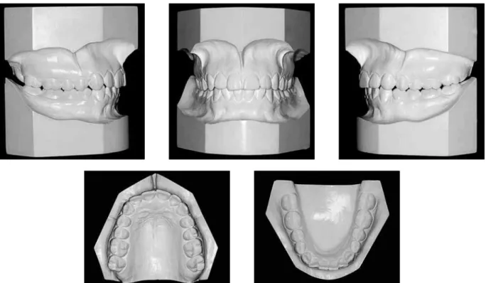
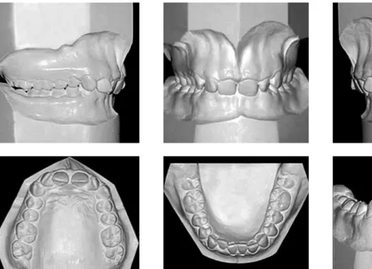
Casts should accurately reproduce dental arches and the buccal area to serve as accurate models of the malocclusion. The casts should be trimmed to maximum intercuspation, as shown in Figure 2.5
Adjustments or trimming in the anatomic por-tion (teeth and buccal area) of the casts should
be limited to removing bubbles and small flaws. Changes in tooth anatomy are considered adul-terations, which will lead to the automatic rejec-tion of the case. Dental casts must be polished so that all anatomic details are preserved (Fig 3). When preparing casts for cases in which it is not possible to use the recommended heights and an-gles, symmetry, proportion and esthetics should be taken into consideration.
Radiographs
Panoramic, periapical and supplemental ra-diographs should be of good quality. The films should be accurately oriented, and the right and left sides should be marked. Panoramic ra-diographs without a satisfactory definition in the incisor areas (maxillary and mandibular) should be accompanied by periapical radio-graphs of these areas (Fig 4).
A B FIGURE 3 - Polished casts with preserved details and accurate reproduction of malocclusion.
FIGURE 4 - Panoramic radiograph (A) and periapical radiograph of maxillary and mandibular incisors (B).
Lateral cephalometric radiographs should be properly standardized, and bone and soft tissue profiles should be clearly visible. In cases of evi-dent facial asymmetry posteroanterior cephalo-grams should be properly examined and submit-ted in addition to profile cephalograms (Fig 5). To preserve anonymity, the names of the radiology service and of the dentist should be blacked out.
The patient’s name and the radiograph date should be visible.
A B
SNA ANB
SN-GoGn
SNB
FMA Facial Convex.
Ls-S 1NA mm 1NB mm Li-S IMPA
1 -APog 1:1 1NA ang
1NB ang
FIGURE 5 - Lateral (A) and posteroanterior (B) cephalometric radiographs.
FIGURE 6 - Cephalometric tracing with reference lines and indication of places where measurements should be included.
not accepted. Templates may be used to trace tooth outlines. Cephalometric landmarks should be carefully identified to ensure reliability of the reference lines drawn.
The examinee should be familiar with all as-pects of cephalograms, tracings and measurements, as well as their meanings. Tracings should be sepa-rated from the lateral radiographs and placed in the plastic envelopes found in the folders.
At least three tracing superimpositions are re-quired: Total or craniofacial, to evaluate general changes during growth and/or treatment; and par-tial, maxillary and mandibular, to demonstrate den-tal changes in the maxilla and mandible and their
respective supporting bones. Total superimposi-tions may be prepared using one of two methods (Fig. 7): (a) Plane of the sphenoid bone and eth-moid cribriform plate, registered on the midpoint between the wings of the sphenoid bone; and (b) the sella-nasion plane, registered on sella. Partial superimpositions should be prepared as follows (Fig 8): Maxilla – best fit of the maxillary bony complex, registered on the palatal curve; mandible – best superimposition in the lower limit of the cortical bone of the mandibular body, registered on the internal cortical outline of the symphysis.
The three superimpositions should be hand-traced by the examinee using pen or pencil. In cases of treatments with intermediate tracings, superimpositions should be presented as follows: A-A1 (beginning–intermediate), A1-B (inter-mediate–final) and A-B (beginning–final). Cases with post-treatment records should include A-B-C (beginning–final–post-treatment). Superimposi-tions should be arranged on white paper, but not fixed to it, and placed into separate envelopes. In cases treated with orthognathic surgery, presurgi-cal intermediate tracings should be included.
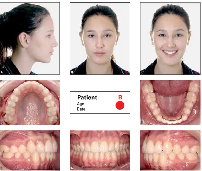
Photographs
Patient records should include the following face photographs: (a) Frontal; (b) Right lateral profile;
A B
A B
FIGURE 7 - Total superimpositions: A) Plane of the sphenoid bone and ethmoid cribriform plate, registered on the midpoint between the wings of the sphenoid bone; B) the Sella-Nasion line, registered on Sella.
FIGURE 8 - Partial superimpositions: A) Maxilla – best fit of the maxillary bony complex, registered on the palatal curve; B) Mandible – best superimposition in the lower limit of the cortical bone of the mandibular body, registered on the internal cortical outline of the symphysis.
and (c) whenever possible, a frontal smile pho-tograph. These photographs should be oriented to Frankfort horizontal, and the line between the pupils should be parallel to the ground. They should be taken with relaxed lips and depict the patient’s actual labial relationship.
The background should be neutral, preferably white; good-quality lighting should reveal facial con-tours without shadows; the ears should be visible for
purposes of orientation; the eyes should be open and looking straight ahead; glasses and other ac-cessories should be removed.
A B C
F G H
D E
such as occlusal views of the maxillary and mandib-ular dental arches. Photographs should be as close as possible to a 1:1 ratio with the patient´s teeth. If mirror images are used, they should be printed vertically fliped. Attention should be paid to a few other aspects: clean teeth, free of bacterial biofilm, bleeding or saliva; cheek retractors; adequate light-ing to show anatomic contours, completely free of shadows; standardized colors; no visual distractions (cheek retractors, labels, fingers).
If the facial and intraoral images are computer generated, their resolution should be high, and
they should accurately demonstrate soft and hard tissues. Photographs may be printed in color to achieve the best possible framing, using the land-scape layout and printing them on glossy photo paper. The examinees should keep in mind that records are legal documents and must not be al-tered. For malocclusions with marked skeletal discrepancies and indication of orthodontic treat-ment associated with orthognathic surgery, im-mediate preoperative records must be submitted. Below an example of a photo mount with three facial and five intraoral photographs (Fig 9).
FIGURE 9 - Photograph layout: A, B, C) facial - right-side profile, frontal and frontal smiling photographs; D, E, F, G,H) intraoral - upper occlusal, lower occlusal, right lateral, frontal and left lateral photographs.
Age Date
First examination
BBO conducted its first examination from March 19 to 21 in 2004, in the city of São Pau-lo, Brazil. Interestingly, in that same year, the American Board celebrated its 75th anniversary. The examination had the special participation of Jack Dale, renowned Canadian orthodontist, former ABO president and Professor Emeritus of the University of Toronto. In May of the same year, during the 104th AAO Annual Session in Orlando, Florida, Jack Dale was honored for his services to the American Board. At that time, he mentioned the work of the BBO Board of Directors and highlighted the effort and hard work that were landmarks of the beginning of the journey into BBO’s mission.6 In special ref-erence to it, he delivered a speech, freely repro-duced below, which translated his view of the integrity of the Board efforts in Brazil:
The California redwoods, as magnificent as they are, do not grow alone; they need each other. They grow strong together by intertwining and en-tangling their roots, thus supporting one another. Without this mutual support they could not be nearly as robust and magnificent. With mutual sup-port, we can remain strong and effective in our ser-vice to society. Maintaining our standard of care is a vital part of our strength… all over the world.
It was my honor and privilege to be invited as an external consultant for the first BBO examina-tion. I found the treatment to be superb and the organization by the board of directors outstand-ing. There were problems, but that was expected. I am sure that these problems will be dealt with and solved in the future, because I am aware of the integrity, dedication, competence and con-cern of the BBO Directors. The American Board of Orthodontics has also had to solve problems along its 75 years of existence. In the future, these problems will certainly remain challenges.
In Brazil, records were standardized, uniform and beautifully done. You could examine any of the case reports on display and find that the
presentation was identical to the others. How I wish that this standard of excellence existed all over the world.
The exam was divided into two parts: (a) a written exam about case reports presented by the BBO; and (b) case report displays by each examinee.
a) Written examination: examinees from eight Brazilian states had four hours to examine two cases presented by BBO. For that, they were allowed to make cephalo-metric tracings and carry out any proce-dures that they used in their practices. I sat in the room for the four hours allotted for the examination, and observed men and women working hard at their tasks. The more I observed them, the more my admiration and respect grew.
B) Case report displays: the ten cases submit-ted by each examinee included six with specific malocclusions and four optional. The cases were on display in the room to be examined for two days. After that, there was a round table with the participation of all the examinees. The discussion was most valuable and constructive for the BBO. The motto on the Brazilian flag means “Or-der and Progress”. BBO exemplified this motto to perfection. They certainly achieved progress and did it step by step in an orderly way.
FINAL CONSIDERATIONS
As Jack Dale said, the motto on the Brazil-ian flag was put into practice by BBO. Accord-ing to that prominent professional, the level of excellence was achieved in the organization of the examination structure, which makes Brazil stand out as a model for the countries aspiring to become members of the World Board of Or-thodontics (14 countries already have a Board of Orthodontics). Brazilian professionals should
believe in such effort so that the seed sown by the words of the Canadian professor germinates and bears good fruit as more specialists apply for excellence certification by the Brazilian Board of Orthodontics and Facial Orthopedics. The BBO certification system has been constantly updating. Therefore, orthodontists interested in taking the Certification Examination should regularly check the website www.bbo.org.br.
REFERENCES
Contact address
Roberto M. A. Lima Filho Avenida Alberto Andaló 4.025
CEP: 15.015-000 – São José do Rio Preto/SP, Brazil E-mail: [email protected]
Submitted: June 13, 2011 Revised and accepted: July 3, 2011
1. Little DM. The founding of the specialty boards. Anesthesiology. 1981;55:317-21.
2. Cangialosi TJ, Riolo ML, Owens S Jr, Dykhouse VJ, Mofitt AH, Grubb JE, et al. The American Board of Orthodontics and specialty certiication: the irst 50 years. Am J Orthod Dentofacial Orthop. 2004;126(1):3-6.
3. The American Board of Orthodontics. [Cited 2010 Jan 11]. Available from: www.americanboardortho.com.
4. Board Brasileiro de Ortodontia e Ortopedia Facial. [Acesso 2010 Jan 11]. Available from: www.bbo.org.br.
5. Habib F, Fleischmann LA, Gama SLC, Araújo TM. Obtenção de modelos ortodônticos. Rev Dental Press Ortod Orthop Facial. 2007;12(3):146-56.