Marinho Del Santo Jr*, Luciano Del Santo**
Decodify
®
System: Cephalometrics as a risk
manager applicative and administrative tool for
the orthodontic clinic
Introduction: Cephalometrics may have limited use in orthodontics because of its sub-jective interpretation. An Artificial Intelligence (AI) system, the Decodify® System, was
developed to allow the customized quantitative assessment of contextualized cephalo-metric data. In this article, the system is tested as an administrative tool in orthodon-tic offices. Methods: The development of algorithms includes the norms and standard deviations modeling of Brazilians’ cephalometric data, measured in lateral radiographs. In order to test the system, initial cephalograms of 60 orthodontic patients of two ferent orthodontic offices (30 cases each) were processed and re-processed by three dif-ferent technicians. The intra-observer and inter-observer reproducibility and reliability indices were checked by paired comparisons. The risk in each orthodontic case, assessed by the electronic analysis, was compared by covariance matrices and agreement coef-ficients. Results: Levels of paired agreement inter-observers (versus golden-pattern) for 23 pairs of variables ranged from 0.68 (S-Go distance) to 0.98 (Na-Me distance) in an orthodontic clinic (JU) and from 0.66 (L1.APg angle) to 0.98 (S-Go distance) in the other (SP). All the correlations were significant at the p<0.001 level. The average of the agreement coefficients was 0.78 for one clinic (JU) and 0.75 for the other (SP). The agreement coefficients were significant at the p<0.001 level. Conclusions: The results of such research support that the analyses provided by the Decodify® System are
re-producible and reliable. Therefore, the system can be applied in order to contextualize conventional cephalometric measurements and to generate individualized risk indices. The system may be used by orthodontists as an administrative tool in the daily profes-sional evaluations.
Abstract
Keywords: Orthodontics. Diagnosis. Artiicial Intelligence.
* Master of Orthodontics, Baylor College of Dentistry, Dallas, Texas.
** Master of Oral and Maxillofacial Surgery at the Hospital Heliopolis, São Paulo/SP.
» The authors declare to be developers of Decodify® System. How to cite this article: Del Santo Jr M, Del Santo L. Decodify® System:
LIteRAtuRe RevIew
Although cephalometrics presents known limitations, it is an important diagnostic tool for the orthodontist.1-4 One of its limitations
is the dependence of personal opinion, since each specialist “interprets” cephalometric data according to the biases built up by his/her aca-demic education, clinical experience and type of clinical service. Technically, cephalometrics presents a limited internal validity due to the identification of cephalometric landmarks5,6,7
and other methodological8-11 or geometrical
problems.12 Naturally, new solutions in
orth-odontic diagnosis has been presented.13-18
An important update in cephalometrics would be the customization of cephalometric values measured in each case, what each orthodontist already subjectively makes in daily assessments. Such kind of improvement would not eliminate the need of different sources of information, as cast models and photos. However, in regard to cephalometrics, would be close to the ideal.
Up to date, cephalometric values were not considered in a contextualized model, that means, in the particular scenario of each patient. However, such constraint is more mathematical than biological. Such contextualization would be possible if an artificial intelligence system could provide decisions, imitating what the human being thinking already provides. Such software would need to take into account the degree of uncertainty and inconsistency associated to each cephalometric number, increasing or decreasing the importance of its contribution for the “final degree” of skeletal and dental compromise which each case of malocclusion presents.
Mathematicians and computer engineers have worked in diverse models of “intelligent” algo-rithms, based upon different types of logic and applied in diverse fields of science as logistics, ro-botics, defense, economics and medicine.19,20
When artificial intelligence systems make deci-sions in the medical field, they are called specialist
systems, programmed to support physicians and other professional personnel of the health area, which; however, provide the final diagnosis or the hypotheses of diagnosis, at their own.
The model of logic applied in this proj-ect21-24 allowed that diverse cephalometric
variables were contextualized in each specific craniofacial scenario.
The fuzzy logic has been applied in medi-cine25,26,27 and orthodontics28,29 in order to
pre-vent inadequate rigid allocations in pre-defined categories. However, fuzzy logic considers only certainty and it is not a sufficient mathematical tool in decision making processes. In other hand, paraconsistent logic22,24 also works with the
un-certainty, inconsistency and insufficiency of data, common features in cephalometric data bases, and because of that, was applied in the decision making processes here described.
In another article,30 the Decodify® System
was tested against the opinions of three special-ists in orthodontics and, showing an expected variance, behaved as a specialist system.
In the current paper, the results of the De-codify® System were obtained by two trained
technicians and, in a paired matter, were com-pared with the results of an experienced tech-nician in the processing, source called “golden-pattern”. Therefore, the article has two main goals: 1) To test the reproducibility and reli-ability of the results obtained by the Decodi-fy® System and; 2) If the Decodify® System is
reproducible and reliable to be introduced to the orthodontic community as an administra-tive tool used by the orthodontist to measure the degree of risk involved in each proposed orthodontic treatment.
Implementation
The Decodify® System was written in
2
3
4
1
9
10 11 14 13 18
15 8
17 12 6 16
7 5 integrated, considering the uncertainty,
inconsis-tency and insufficiency carried by each variable. The inferences of the system are based upon the degrees of evidence of abnormality (DEA) in specific units: Skeletal (anteroposterior and ver-tical) and dental (upper and lower teeth).
The components of the Decodify® System
(soft-wares Decodify® e DecodeCAD®) are registered in
Brasil in the INPI (Instituto Nacional da Proprie-dade Industrial) under the licences 00070981 and 00075342. In the USA, the softwares are registered in the Copyright Office – Library of Congress/USA under the protocols TXu1-326-513 (7/31/06) and TXu1-326-514 (7/31/06).
MAteRIAL And Methods samples
The samples, retrospectively collected, in-cluded 60 initial cephalograms, from patients of both genders, who seek for orthodontic treatment in two clinics, in Jundiaí (JU) and São Paulo (SP). In Jundiaí, 13 male and 17 fe-male individuals, from 12 to 29 year-old, were included in the sample. In São Paulo, 10 male individuals and 20 female individuals, from ages of 19 to 55 year-old, were included in the sample. Patients who presented compromised lateral cephalograms or craniofacial deformi-ties were not included in the sample. There was not discriminate rule in regard to the mal-occlusion initially presented by the patient, neither in regard to its severity.
data collection
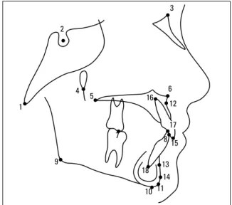
Eighteen cephalometric landmarks (Fig 1) were identified, traced, re-identified and re-traced in ace-tate paper, with mechanical pencil 0.3 mm, by three trained technicians (golden-pattern, Jundiai clinic and São Paulo clinic). All the tracing were digitalized in the tablet Trust TB 7.300 Wide Screen Design Ta-ble (PO Box 8043, 3301 CA Dordrecht, The Neth-erlands) and the data analyzed by the Excel software (Windows 7, Redmond, Washington, USA).
Landmarks and cephalometric measurements The following landmarks and cephalomet-ric measurements were identified and digi-talized (Fig 1):
1) Basion (Ba): The most postero-inferior point on the posterior margin of the fora-men magnum.
2) Sella (S): The center of the pituitary fossa of the sphenoid bone.
3) Nasion (N): The junction of the frontal and nasal bones, at the fronto-nasal suture. 4) Pterygo-maxillary fissure (PtgI): the most
in-ferior point of the pterygo-maxillary fissure. 5) Posterior nasal spine (PNS): The most
pos-terior point on the bony hard palate.
6) Anterior nasal spine (ANS): The tip of the median anterior bony process of the maxilla. 7) Upper molar: The most inferior point of the mesial cuspid tip of the first upper molar, posterior reference for the occlusal plane. 8) Anterior reference of the occlusal plane:
Es-tablished by bisecting the overbite or open bite of the incisors, considering the incisal edges of the upper and lower incisors. 9) Gonion (Go): The most postero-inferior
5 1
13
12 11
8
10
9 6
7
4 2
15 14 3 10) Menton (Me): The most antero-inferior
point on the mandibular symphysis. 11) Gnathion (Gn): The most antero-inferior
point on the contour of the symphysis. Determined by bisecting the angle formed by the mandibular plane (Go-Me) and the Nasion-Pogonion line.
12) A Point: The most posterior point on the anterior curvature of the maxilla.
13) B Point: The most posterior point on the an-terior curvature of the mandibular symphysis. 14) Pogonion (Pg): The most anterior point on
the contour of the bony chin.
15) Upper incisor edge: The incisal tip of the maxillary central incisor.
16) Upper incisor apex: The root tip of the maxillary central incisor.
17) Lower incisor edge: The incisal tip of the mandibular central incisor.
18) Lower incisor apex: The root tip of the mandibular central incisor.
The following cephalometric measurements were considered (Fig 2):
1) S-N: Plane that represents the anterior cranial base.
2) Palatine plane: Angle between the anterior cranial base (S-N) and the palatine plane, considering the landmarks ANS and PNS. 3) Occlusal plane: Angle between the anterior
cranial base (S-N) and the occlusal plane, considering the landmarks molar and incisor. 4) Mandibular plane: Angle between the
anterior cranial base (S-N) and the man-dibular plane (Go-Me).
5) Ba-Na: Plane that represents the cranial base.
6) Y Axis: Smaller angle between the cranial base (Ba-N) and the facial axis (Ptg-Gn). 7) S-Go: Distance between Sella and Gonion,
representing the posterior facial height. 8) N-ENA: Distance between Nasion and
ANS, representing the upper part of the an-terior facial height.
9) ANS-Me: Distance between the ANS and Menton, representing the lower part of the anterior facial height.
10) N-Me: distance (mm) between Nasion and Menton, representing the antero-pos-terior facial height.
11) SNA: angle between the anterior cranial base (S-N) and the A Point, representing the antero-posterior positioning of the maxilla. 12) SNB: angle between the antero-posterior
cranial base (S-N) and the B Point, repre-senting the antero-posterior positioning of the mandible.
13) Long axis of the upper incisor. 14) Long axis of the lower incisor.
15) A Point-Pg plane: Plane representing the maxilla-mandible skeletal profile.
Research model and statistical method The system was developed in 3 units: 1) An-tero-posterior, 2) Vertical and 3) Dental. The cen-tral tendency measurements (average and stan-dard deviation) for each age (6 to 18 year-old) from both genders were obtained for the Cranio-facial Growth Atlas from Bauru.31
The chosen golden-pattern was the digitaliza-tion and processing operated by the a technician (BioLogique S/S Ltda Company, São Paulo-SP, Brazil) and hosted in the central server Hostloca-tion (HostLocaHostloca-tion S/C Ltda. Company, R. Mae-stro Cardim 7081, 01323-001, São Paulo-SP). Each one of the two offices provided 30 lateral radiographs and a technician (examiner) to digi-talize and process the data.
After the golden-pattern was established, the intra-examiners reproducibility was tested for each one of the examiners. The results of each examiner were independently compared with the golden-pattern (it was called inter-examiner comparison). The results of each sub-sample of 30 cases were also self-compared (re-digitalized 4 weeks after the first trial). The intra-examiners correlations targeted to measure the systematic error and the inter-examiner correlations tar-geted to measure the method error. Epistemo-logically, the null hypothesis of no difference intra-examiners and the null hypothesis of no difference inter-examiner (examiner against the golden pattern) were tested.
Coefficients of correlation compared similar cephalometric variables in a paired manner, isolat-ing as dependent variable the examiner. The risk involved in each case, result of the electronic pro-cessing by the Decodify® System and presented as
a quantitative ranking, was measured in an ordi-nal mode. The risks were matched by matrices of covariance and such comparisons were expressed by agreement indices, again testing the null hy-potheses of no difference intra-examiners and the null hypotheses of no difference inter-examiner (examiner against the golden pattern).
Logic of the artiicial intelligence system Decodify® is an artificial intelligence system
that can calculate the degrees of cephalometric severity, skeletal and/or dental, after the mathe-matical contextualization of the selected variables. The “neural” network is build with paraconsistent logic,22,24 capable of making non-trivial decisions,
based in its sensibility to uncertainty, inconsisten-cy and insufficieninconsisten-cy of the treated data.
ResuLts
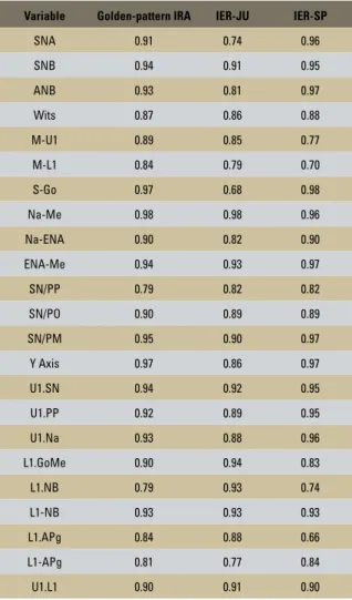
The results of reprodicibility and reliability of the selected cephalometric variables are described
Variable Golden-pattern IRA IER-JU IER-SP
SNA 0.91 0.74 0.96
SNB 0.94 0.91 0.95
ANB 0.93 0.81 0.97
Wits 0.87 0.86 0.88
M-U1 0.89 0.85 0.77
M-L1 0.84 0.79 0.70
S-Go 0.97 0.68 0.98
Na-Me 0.98 0.98 0.96
Na-ENA 0.90 0.82 0.90
ENA-Me 0.94 0.93 0.97
SN/PP 0.79 0.82 0.82
SN/PO 0.90 0.89 0.89
SN/PM 0.95 0.90 0.97
Y Axis 0.97 0.86 0.97
U1.SN 0.94 0.92 0.95
U1.PP 0.92 0.89 0.95
U1.Na 0.93 0.88 0.96
L1.GoMe 0.90 0.94 0.83
L1.NB 0.79 0.93 0.74
L1-NB 0.93 0.93 0.93
L1.APg 0.84 0.88 0.66
L1-APg 0.81 0.77 0.84
U1.L1 0.90 0.91 0.90
TABLE 1 - Matched correlation, golden-pattern intra-examiners (IRA) and inter-examiners (IER). Significance level: [p< 0.001].
in the Tables 1 and 2. Table 3 describes the rank-ing of assessed risk and the Table 4 shows the agreement indices between the risks presented in the two examiners assessments compared with the pre-defined golden-pattern.
dIsCussIon
Cephalometrics is a worldwide accepted orth-odontic diagnostic tool and considered essential information to offer a reliable treatment plan to the patient. It is based upon measurements on head lateral radiographs and it describes skeletal and dental discrepancies with considerable preci-sion. An experienced clinician can well interpret, although subjectively, cephalometric numbers and apply such information in his/her daily prac-tice. Because only numbers cannot be directly applied in the clinic, such subjectivity has been referred as a drawback in the potential value of cephalometrics for routinely use.
The point to be discussed is not if cephalo-metrics should be or should not be used, but how cephalometric numbers might be interpreted be-fore its application, since that interpretation holds significant variance. This occurs because of two main reasons: first, because the degree of clini-cal abnormality is not quantitatively measured and; second, because there is no way to establish a golden-pattern. Golden-pattern is the pattern reference established in order to have other refer-ences compared to it and, according to this com-parison, become acceptable or not.
TABLE 2 - Matched correlation, between intra-examiner and golden-pattern. Significance level (p< 0.001).
TABLE 4 - Degrees of agreement measured by matrices of covariance, comparing the measured risk by the two examiners against the golden-pattern. Significance level: [p< 0.0001].
TABLE 3 - Definition of ranking of risk. Ordinal classification of the quan-titative results.
*As a reference, up to each clinician.
Variable IER-JU IER-SP
SNA 0.86 0.73
SNB 0.94 0.64
ANB 0.69 0.85
Wits 0.81 0.92
M-U1 0.88 0.60
M-L1 0.83 0.56
S-Go 0.98 0.83
Na-Me 0.98 0.62
Na-ENA 0.86 0.77
ENA-Me 0.96 0.69
SN/PP 0.83 0.73
SN/PO 0.90 0.67
SN/PM 0.97 0.75
Y Axis 0.94 0.61
U1.SN 0.86 0.62
U1.PP 0.85 0.61
U1.Na 0.81 0.55
L1.GoMe 0.91 0.81
L1.NB 0.78 0.73
L1-NB 0.91 0.77
L1.APg 0.52 0.62
L1-APg 0.67 0.83
U1.L1 0.75 0.69
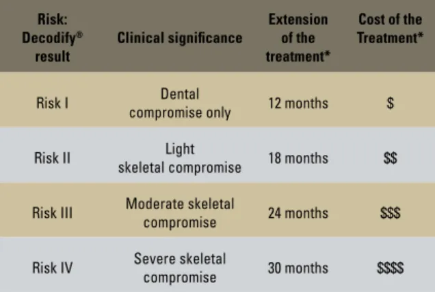
Risk: Decodify®
result
Clinical signiicance Extension of the treatment*
Cost of the Treatment*
Risk I Dental
compromise only 12 months $
Risk II Light
skeletal compromise 18 months $$
Risk III Moderate skeletal
compromise 24 months $$$
Risk IV Severe skeletal
compromise 30 months $$$$
Risk-Result JU SP
Minor Correlation 0.54 0.47
Major Correlation 0.90 0.88
Average Correlation 0.78 0.75
Degree of
Agreement 0.78 0.75
In our research project, we established a golden-pattern (Table 1), contextualizing ceph-alometric measurements of wide application. The result of such contextualization is called risk. It is important to highlight that such risk was based upon expected norms for each one of the elected measurements, individualized by gender and age, measured in the same popula-tion of the evaluated cases (Brazilians, Cauca-sians, with average degree of ethnic miscegena-tion). The contextualization (or risk) can be de-fined as: “What we should expect as the degree of severity of malocclusion, skeletal or dental, in that particular patient.”
From our results, it was observed that the inter-examiners comparison (against the gold-en-pattern) varies according to the cephalo-metric measurement. Such result is what we expected from the human evaluation of cepha-lometric landmarks with different degrees of identification and reproduction. Notice that such degree of variation involves the degree of accuracy of the examiner and of the golden-pattern as well. As examples of landmarks vul-nerability in regard to the identification and reproduction the point A (due to the thickness of the maxillary bone), the inclination of the lower incisor (due to the lower incisors images superimposition) and the geometric location of the Gonion point (constructed bisect).
Such variations also account the intra-exam-iner variation, isolated in the Table 2. The varia-tion in reproducibility of the technicians implies in the fact that there are examiners with better knowledge and/or expertise to trace a cephalo-gram, what is reasonably expected.
The quantitative results provided by the De-codify® System as risk are presented in an ordinal
ranking in the Table 3. Then, parameters that we consider useful for the daily orthodontic prac-tice are suggested. For instance, the greater the skeletal compromise of a case, greater its risk and consequently greater the treatment time
required and the cost involved.
The contribution of our work is evident when the degrees of agreement are presented. When we have a calibrated system which pres-ents degrees of severity, throughout algorithms that contextualize individual cephalomet-ric variables, we have the level of risk in each evaluated case. The degrees of agreement show that such level of risk is reproducible and trust-able and, therefore, evaluations are minimally based in personal opinions. The “cephalometric guessing” is exchanged by the “evidence” of the cephalometric risk. In few words, the Decodify®
System works as a “ruler”, to measure the degree of difficulty to treat a specific orthodontic case.
With such instrument, therefore, the orthodon-tist can measure, with high level of precision, how much “energy” the office must dedicate to that particular case. And the practical consequences of such measurement are many: The orthodontist might estimate the extension of the treatment, the approximated number of appointments, and the need of his/her attention as the chief clinician (and consequently the possibility to delegate less important functions to his /her assistants), the po-tential problems, the fee to be charged etc.
ConCLusIon
Our results support that the risk measured by the presented system is reproducible and re-liable. Therefore, we accept the null hypotheses of no difference intra-examiner and inter-exam-iners evaluations, in all the matched compari-sons performed.
As a direct consequence of the acceptance of these null hypotheses, we suggest that the De-codify® System is an important cephalometric
tool for the orthodontist to establish clear pa-rameters about the service that will be provided to his/her clients. Then, the patient can have a reliable estimation on the degree of severity
of his malocclusion, the difficulties to treat it and the necessary time to accomplish such goal. Consequently, the patient will pay the fair fee for the contracted service, according to the mar-ket in which it is inserted.
ACKnowLedgeMents
1. Wylie WL. Present beliefs in the practicability of cephalometric studies in individual case analysis, prognosis and treatment. Am J Orthod. 1946;32:836-42.
2. Moorrees CFA. Normal variation and its bearing on the use of cephalometric radiographs in orthodontic diagnosis. Am J Orthod. 1953; 39:942-50,
3. Sved A. A critical review of cephalometrics. Am J Orthod. 1954; 40:567-90,
4. Dreyer CJ, Joffe BM. A concept of cephalometric interpretation. Angle Orthod. 1963;33:123-6.
5. Baumrind S, Frantz RC. The reliability of head ilm measurements. 1. Landmark identiication. Am J Orthod. 1971;60:111-27. 6. Major PW, Johnson DE, Hesse KL, Glover KE. Landmark
identiication error in posterior anterior cephalometrics. Angle Orthod. 1994;64(6):447-54.
7. Trpkova B, Major P, Prasad N, Nebbe B. Cephalometric landmarks identiication and reproducibility: a meta-analysis. Am J Orthod Dentofacial Orthop. 1997;112(2):165-70,
8. Björk A, Solow B. Measurements on radiographs. J Dent Res. 1962;41:672-83.
9. Savara BS, Tracey WE, Miller PA. Analysis of errors in
cephalometric measurements of three dimensional distances on the human mandible. Arch Oral Biol. 1966;11(2):209-17. 10, Buschang PH, LaPalme L, Tanguay R, Demirjian A. The technical
reliability of superimposition on cranial base and mandibular structures. Eur J Orthod. 1986;8:152-6.
11. Buschang PH, Tanguay R, Demirjian A. Cephalometric reliability. A full ANOVA model for the estimation of true and error variance. Angle Orthod. 1987;57(2):168-75.
12. Del Santo M Jr. Inluence of the occlusal plane inclination on ANB and Wits assessments of anteroposterior relationship of the jaws. Am J Orthod Dentofacial Orthop. 2006;129(5):641-8.
13. Baumrind S, Muller DM. Computer-aided head ilm analysis: the University of California San Francisco method. Am J Orthod. 1980;78(1):41-65.
14. Eriksen E, Solow B. Linearity of cephalometric digitizers. Eur J Orthod. 1991;13(5):337-42.
15. Fine MB, Lavelle CLB. Diagnosis of skeletal form on the lateral cephalogram with a inite element-based expert system. Am J Orthod Dentofacial Orthop. 1992;101(4):318-29.
16. Ferraro VF, Sforza C, Dallorca LL, De Franco DJ. Assessment of facial form modiications in Orthodontics: proposal of a modiied computerized mesh diagram analysis. Am J Orthod Dentofacial Orthop. 1996;109(3):263-70,
RefeRenCes
17. Forsyth DB, Shaw WC, Richmond S. Digital imaging of cephalometric radiography, part 1: advantages and limitations of digital imaging. Angle Orthod. 1996;66(1):37-42.
18. Forsyth DB, Shaw WC, Richmond S, Roberts CT. Digital imaging of cephalometric radiography, part 2: image quality. Angle Orthod. 1996;66(1):43-50,
19. Aminzadeh F, Jamshidi M. Soft computing: Fuzzy logic, neural networks and distributed artiicial intelligence. Upper Saddle River: Prentice Hall; 1994.
20, Russell S, Norvig P. Artiicial Intelligence. 2nd ed. Upper Saddle River: Prentice Hall; 2002.
21. Da Costa NCA. On the theory of inconsistent formal systems. Notre Dame J Formal Logic. 1974;15(4):497-510,
22. Da Costa NCA, Subrahmanian VC, Vago C. The paraconsistent logic. Zeitschrift für Math Logik und Grundlagen der Math. 1991;37:137-48.
23. Kifer M, Subrahmanian VS. Theory of generalized annotated logic programming and its applications. J Logic Program. 1992;12(4):335-67.
24. Abe JM. Paraconsistent artiicial networks: an introduction. Lect Notes Artiic Intellig. 2004; 3214:942-8.
25. Roy MK, Biswas R. I-V Fuzzy relations and Sanchez’s approach for medical diagnosis. Fuzzy Sets Syst. 1992;47(1):35-8.
26. Sanchez E. Truth-qualiication and fuzzy relations in natural languages, application to medical diagnosis. Fuzzy Sets Syst. 1996;84(2):75-86.
27. Kuncheva LI, Steimann F. Fuzzy diagnosis. Artiicial intelligence in medicine. 1999;16:121-8.
28. Takada K, Sorihashi Y, Stephens CD, Itoh S. An inference modeling of human visual judgment of sagittal jaw-base relationships based on cephalometry: Part I. Am J Orthod Dentofacial Orthop. 2000;117(2):140-7.
29. Sorihashi Y, Stephens CD, Takada K. An inference modeling of human visual judgment of sagittal jaw-base relationships based on cephalometry. Part II. Am J Orthod Dentofacial Orthop. 2000;117(3):303-11.
30, Del Santo M Jr, Del Santo LM. Diagnóstico cefalométrico eletrônico: contextualização de variáveis cefalométricas. Dental Press J Orthod. 2011;16(2):75-84.
31. Martins DR, Janson GRP, Almeida RR, Pinzan A, Henriques JFC, Freitas MR. Atlas de crescimento craniofacial. São Paulo: Ed. Santos; 1998.
Contact address
Marinho Del Santo Jr.
Rua Mal. Hastimphilo de Moura 277, Casa 1 CEP: 05.641-000 - Morumbi - São Paulo / SP, Brazil E-mail: [email protected]