ISSN 1517-7076 artigo 11760, pp.987-995, 2016
Autor Responsável: Lilian Ferreira de Senna Data de envio: 19/01/2016 Data de aceite: 01/06/2016
10.1590/S1517-707620160004.0091
Carbon steel corrosion induced by sulphate-reducing
bacteria in artificial seawater: electrochemical
and morphological characterizations
Mariana Silva de Paula1, Márcia Monteiro Machado Gonçalves1, Monick Alves da Cruz Rola1, Diana José Maciel 1, Lilian Ferreira de Senna1, Dalva Cristina Baptista do Lago1
1
Instituto de Química - UERJ, Rua São Francisco Xavier, 524 PHLC, CEP: 20550-900 Rio de Janeiro, RJ
e-mail: [email protected], [email protected], [email protected], [email protected], [email protected], [email protected]
ABSTRACT
In this work, the corrosion behavior of carbon steel AISI 1020 was evaluated in artificial seawater in the presence of mixed sulfate-reducing bacteria (SRB) culture isolated from the rust of a pipeline. The corrosion evaluation was performed by electrochemical techniques (open circuit potential (Eocp), polarization curves and electrochemical impedance spectroscopy (EIS)), while the formation of a biofilm and corrosion products were observed by scanning electron microscopy (SEM) and X-ray energy dispersive spectroscopy (EDS). The presence of SRB in the medium shifted the open circuit potential to more positive values and increased the corrosion rate of the steel. Electrochemical and morphological techniques confirmed the presence of a biofilm on the steel surface. EDS spectra data showed the presence of sulfur in the corrosion products. After removing the biofilm, localized corrosion was observed on the surface, confirming that localized corrosion had occurred. The biogenic sulfide may lead to the formation of galvanic cells and contributes to cathodic depolarization.
Keywords: sulphate-reducing bacteria, biofilm formation; carbon steel, electrochemical impedance spectros-copy, morphological characterization.
1. INTRODUCTION
Corrosion, in its various forms, can cause damages to bridges, ships, platforms and pipelines. It has been es-timated that approximately 20 % of corrosion cost is due to microbiologically influenced corrosion (MIC), and a significant part of this biocorrosion process can be induced by sulphate-reducing bacteria (SRB) [1, 2]. Biocorrosion is an electrochemical process of metal dissolution initiated or accelerated by bacteria and other microorganisms through their metabolic activities [3]. The interaction of bacteria with the metal surface may also result in the formation of biofilms, which can severely affect the kinetics of cathodic and/or anodic reac-tions in an electrochemical process [4]. Moreover, a synergistic interaction of microorganisms may occur, resulting in biofilms and metabolic products that enhance corrosion processes [5].
The main characteristic of SRB is the use of a sulphate ion as the final electron acceptor in their bio-energetic process. In their metabolic process, sulphate ions are reduced to sulphide ions, which can be present in three forms: H2S (soluble), HS- and S2-, depending on the pH of the environment [6]. SRB are anaerobic microorganisms generally found in anoxic environments, such as soil sediment, oil fields and the anaerobic reactors used in wastewater treatment [7]. In the oil, gas and shipping industries, SRB are particularly aggres-sive, generally causing pitting corrosion in metal equipment, which results in high corrosion costs [8].
988 with ferrous ions act as sites for the reduction of H+ ions to molecular hydrogen, enhancing the corrosion [10,
11].
Currently, researchers are investigating the formation of biofilms on the corrosion of carbon steels us-ing various species of SRB and microbial consortia [12, 13, 14, 15]. In these cases, artificial seawater is the main incubation medium for these bacteria [16]. Therefore, in this work, the corrosion of carbon steel AISI 1020 in artificial seawater was evaluated under anaerobic conditions in the presence of a mixed culture of SRB using an experimental exposure time of 35 days. The corrosion process was investigated by open circuit potential (Eocp), polarization curves, electrochemical impedance spectroscopy (EIS), scanning electron mi-croscopy (SEM) and X-ray energy dispersive spectroscopy (EDS). The aim of this study was to investigate the electrochemical and morphological impacts of a mixed culture of SRB, obtained directly from a real ma-rine environment, in the MIC process on carbon steel in an artificial saline medium using a longer experi-mental time.
2. EXPERIMENTAL
2.1 Metal coupon preparation
Rectangular carbon steel AISI 1020 coupons (1.5 cm x 3.5 cm x 0.1 cm) were used for cellular quantification and morphologic evaluation. The composition of the carbon steel was (% mass): C 0.18, Mn 0.63, P 0.035 max, S 0.035 max [17].
Coupons of the same type of steel were used in the electrochemical tests. First, they were attached to a copper conductor wire and then embedded in epoxy resin, so that only one face (with an area of 2 cm2)would be exposed to the solution.
In all cases, the coupons were sequentially polished with a series of sandpaper (grades 100, 200, 300, 400, 500 and 600), washed with distilled water and ethanol, and dried in a current of air. The coupons were polished on both sides for the cellular quantification and morphologic evaluation.
2.2 SRB cultures
The mixed culture of SRB used for the study was isolated from the surface of a rusty pipeline that was sub-merged in Guanabara Bay, Rio de Janeiro, Brazil. The layer of rust was removed and then inoculated into flasks containing 50 mL of culture medium (0.5 g KH2PO4, 1.0 g NH4Cl, 4.5 g Na2SO4, 0.04 g CaCl2.2H2O, 0.06 g MgSO4.6H2O, 9.4 mL sodium lactate (50 % m/v), 1.0 g yeast extract, 0.3 g sodium citrate, 0.004 g FeSO4.7H2O, 1.9 g agar, 4.0 mL resazurin) in 1 L of artificial seawater (0.003 g NaF, 4.0 g Na2SO4, 0.02 g SrCl2.6H2O, 10.78 g MgCl2.6H2O, 0.03 g H3BO3, 23.5 g NaCl, 0.1 g KBr, 0.02 g NaSiO3.9H2O, 0.7 g KCl, 0.001 g Na2EDTA, 1.113 g CaCl2, 0.2 g NaHCO3) [18].
The medium was prepared anaerobically under nitrogen purge due to the reductive metabolism of the SRB. The pH was adjusted to 7.6 using 1 mol L-1 NaOH, and the medium was then autoclaved at 121 °C for 15 min. The samples were incubated at 30 °C for 7 days.
2.3 Test conditions
Biocorrosion tests were conducted in 100 mL bottles containing 80 mL of the culture medium previously described in the absence or presence of the mixed culture of SRB (10 % v/v) isolated in this study. Nitrogen gas was used to purge the medium and remove the dissolved oxygen (O2) at the exact moment that the spec-imens were immersed in the reactors. Although the medium composition did not perfectly resemble the natu-ral conditions of the MIC process, it may be interesting for inducing biofilm production and accelerating the MIC testing as performed by PÉREZ et al. [19].
All of the tests were performed for 35 days, and the data were collected at intervals of 7 days, consid-ering the time zero after inoculation of the medium.
2.4 Enumeration of SRB
extrac-989 tion of the biofilm from the steel coupon surface was carried out by transferring the coupon to a flask con-taining a dilution solution (0.124 g sodium thioglycollate, 0.100 g ascorbic acid, 4.0 mL resazurin in 1 L of artificial seawater), shaking it on vortex, and then sonicating it under ultrasound. After the removal of the biofilm, aliquots of the suspension were taken for quantification. The inoculated tubes were incubated at 30 °C for 28 days, and the growth of the SRB was indicated by the formation of a black FeSx precipitate.
2.5 Surface analysis
The surface morphologies of carbon steel 1020 specimens after 35 days of exposure to the culture medium were examined with a scanning electron microscope (SEM; LEO 1450VP SEM) coupled to an energy disper-sive X-ray spectroscopy (EDS) system (IXRF-EDS 2000 Microanalysis System). The specimens were visual-ised in variable pressure mode (low vacuum), making fixation and dehydration steps unnecessary. The metal coupons were washed carefully with distilled water and ethanol and then conditioned in a desiccator for 3 hours prior to coating with gold. Only one side of the coupon was covered with gold for morphological eval-uation; EDS analysis was performed on the reverse side of the gold-coated specimen due to the overlap of the gold and sulphur spectral lines [20].
After 35 days-biofilm evaluation, the corrosion products of the specimens were pickled in Clark solu-tion for 1 minute, and the naked corroded surfaces were also observed by SEM.
2.6 Electrochemical Techniques
The electrochemical experiments, consisting of open circuit potential measurements (EOC), polarization curves and electrochemical impedance spectroscopy (EIS), were performed in a three-electrode cell using a computer-controlled potentiostat/galvanostat (Autolab PGSTAT302N). The three electrodes consisted of a carbon steel AISI 1020 coupon as the working electrode, a saturated calomel electrode as the reference elec-trode, and a platinum wire as the counter electrode.
All the experiments were conducted in the culture medium earlier described, either with or without SRB, in triplicate. The open circuit potential of the steel was measured after 15 minutes of stabilization. Po-larization curves were then performed and the Tafel curves were recorded by scanning the potential from -1.2 V to 0.0 V (SCE) with a rate of 20 mV s-1. A rate above 10 mV s-1 was chosen based on the literature [11,
21]. The corrosion current density (Icorr) and the corrosion rate were obtained by extrapolation of the Tafel curves.
EIS measurements were carried out in duplicate using a frequency range of 100 kHz to 0.001 Hz with an AC wave of ±10 mV after 30 minutes of stabilization in the open-circuit potential. The experiments were performed in the same electrolytic media earlier used for the Eocp measurements and polarization tests.
3. RESULTS AND DISCUSSION
3.1 SRB Growth
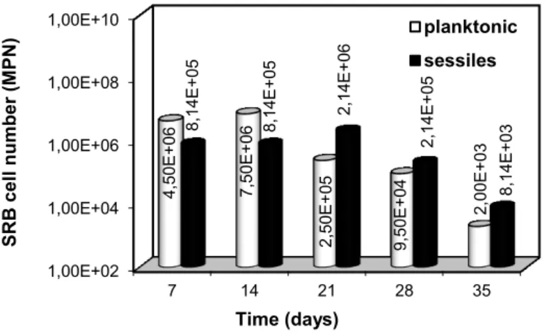
The results for the enumeration of planktonic and sessile bacteria using the MPN technique are shown in Figure 1.
Figure 1: Quantification of planktonic (mL-1) and sessile (cm-2) SRB cells by the most probable number (MPN) method.
1,00E+02 1,00E+04 1,00E+06 1,00E+08 1,00E+10
7 14 21 28 35
990 The initial concentration cell in the inoculum was 105 cells mL-1. The maximum planktonic cell densi-ty (7.50 x 10⁶ cells mL-1) was reached at 14 days of exposure, when an intense blackening of the medium was observed, most likely due to the precipitation of iron sulphide. From the 21st day onward, the number of planktonic cells decreased by an order of magnitude every 7 days, maintaining this trend until the end of the test. During this same interval, visual inspection of the biofilm on the metal surface revealed that it had be-come thicker.
According to GAYOSSO et al. [8], the kinetics growth is different for planktonic microorganisms and for those established at the metal surface (i.e., sessile microorganisms). Thus, it is also important to quantify the number of sessile cells, and this result is also presented in Figure 1. The sessile cell quantification showed maximum and minimum cell densities of 2.14 x 10⁶ cells cm-2 and 8.14 x 10³ cells cm-2, respectively. It is important to note that the highest concentration of sessile bacteria occurred on the 21st day of immersion, when the reduction in the number of planktonic cells began. From the 28th day onward, both the number of sessile bacteria cells attached to the biofilm and the number of planktonic cells decreased. This behaviour may be attributed to a lack of nutrients and/or to the generation of biogenic hydrogen sulphide as the meta-bolic product of SRB, which is considered a growth inhibitor [22].
3.2 Surface analysis
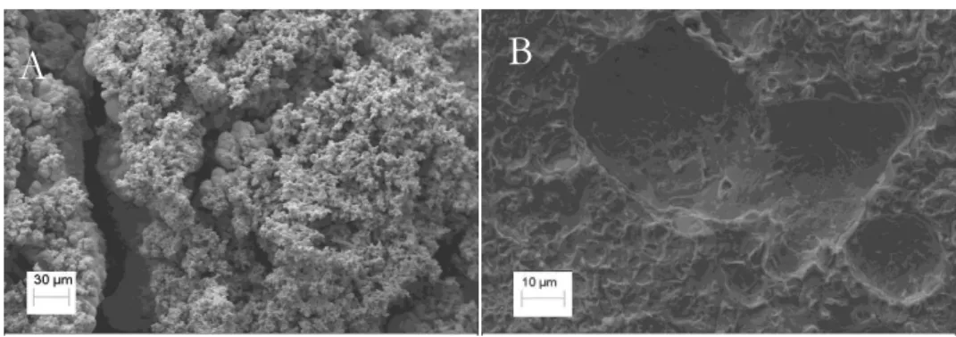
Morphological analysis of the steel coupons in the presence of SRB is shown in Figure 2.
Figure 2: SEM micrograph of the steel coupon surface exposed to culture medium with SRB (993x) after 35 days of exposure (A). The same coupon after pickling the biofilm and corrosion products (4000x) (B).
As expected, the micrograph in Figure 2A shows a nonhomogeneous, porous biofilm on the steel sur-face, confirming that the biofilm was not a protective layer and that it most likely accelerated the metal cor-rosion. After removal of the biofilm (Figure 2B), localized corrosion can be observed. This result confirms that inoculating the medium with SRB can result in harmful corrosive processes.
The EDS spectrum of the steel surface covered with a biofilm (Figure 3) shows the presence of sul-phur and iron, indicating the formation of FeSx in the corrosion product, as observed by MIRANDA et al. [3]. The formation of FeSx may be the result of a reaction between the sulphides generated by SRB metabolism and the Fe2+ ions produced by the oxidation of the substrate [7]. Additionally, the spectrum in Figure 3 shows an intense carbon line, which is most likely due to extracellular products (EPS) and colonies of bacteria at-tached to the substrate. Therefore, this result suggests that the film observed on the carbon steel in Figure 2A is mainly formed by both organic and inorganic matter, the last one originated from the corrosion process of steel in a SRB medium.
A
991 Figure 3: EDS spectrum from the carbon steel surface after 35 days of exposure in the presence of SRB.
3.3 Electrochemical Analyses
3.3.1 Open circuit potential (Eocp)
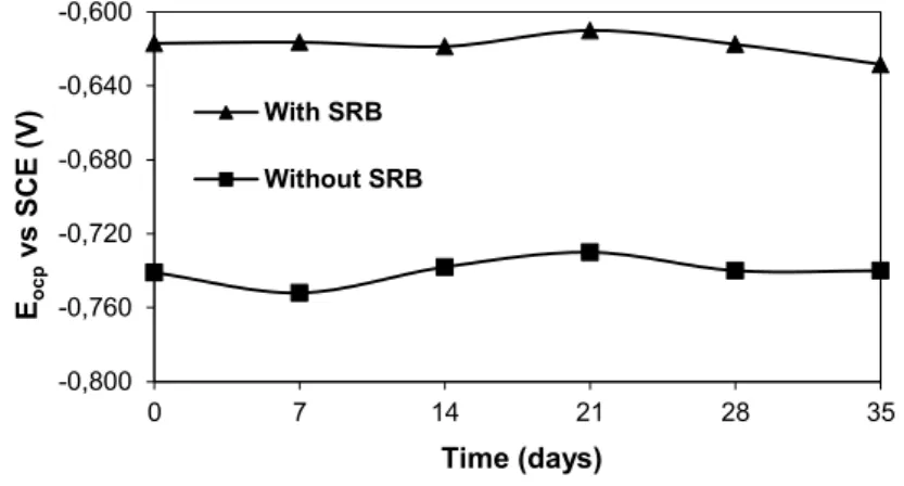
Figure 4 shows the variation of the measured Eocp with the exposure time of the steel sample immersed in the culture medium. It can be observed that there were no significant changes in the Eocp values with the expo-sure time, for both the media with or without SRB. The presence of SRB in the medium shifted the Eocp val-ues in the positive direction by approximately 120 mV, causing the so-called ennoblement of the substrate due to the biofilm formation [5]. Therefore, the Eocp was affected by the microbial activity produced by the SRB, which were attached to the coupon surface to form a biofilm. However, as already shown in Figure 2A, the steel surface was covered by a porous biofilm, which means that this layer may not be considered as pro-tective.
Figure 4: Variation of the Eocp with the duration of immersion of the steel sample in the culture media with and without SRB.
3.4 Polarization curves
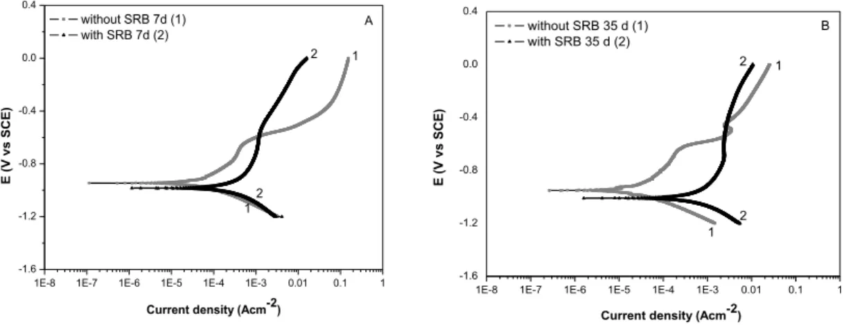
The Tafel plots of the polarization curves of the steel electrodes immersed in the culture medium either in the absence or in presence of SRB at 7 and 35 days are shown in Figures 5A and 5B, respectively.
-0,800 -0,760 -0,720 -0,680 -0,640 -0,600
0 7 14 21 28 35
Eo
c
p
v
s
SC
E (V
)
Time (days)
With SRB
992
1E-8 1E-7 1E-6 1E-5 1E-4 1E-3 0.01 0.1 1 -1.6
-1.2 -0.8 -0.4 0.0 0.4
2 1
E
(V
v
s
SC
E)
Current density (Acm-2)
without SRB 7d (1) with SRB 7d (2)
1 2
A
Figure 5: Polarization curves of carbon steel in the culture medium in the absence and presence of SRB after 7 days (A) and 35 days (B) of exposure.
Both polarization curves were obtained by scanning the potential from -1.2 V to 0.0 V (SCE). The curves of carbon steel exposed to the culture medium containing SRB were shifted to more negative poten-tials and to higher current density values independent of the exposure time. These results indicate that SRB accelerated the corrosion process, as observed by ANANDKUMAR et al. [21], acting mainly by enhancing the anodic branch. This behaviour could be attributed to the fact that the SRB activity can produce H2S as a secondary metabolite, increasing the corrosive ability of the solution [23]. Even though, it is important to mention a small cathodic depolarization observed for the experiments performed after 35 days of exposure. This result suggests that the corrosion process in SRB medium may have changed with the exposure time. It is known that the iron sulphides (FeSx) formed by the precipitation of biogenic sulphide with ferrous ions can act as a cathode in the galvanic couple [11]. Therefore, the presence of biogenic sulphide and the FeSx pre-cipitated on the steel surface could likely contributed to the depolarization observed for the cathodic branch of the polarization curve after 35 days of exposure, enhancing the corrosion process.
The highest differences between biotic and abiotic experiments were observed for 35 days of exposure (Figure 5B), where the Ecorr and the corrosion current density (Icorr) in the absence and presence of SRB, var-ied from - 0.951 V to -1.010 V and from 3.16 x 10⁻⁶ A cm-2 to 2.87 x 10⁻⁴ A cm-2, respectively. Comparing the results obtained for the substrate exposed to a medium containing SRB (Figures 5A and 5B), there was an increase in the corrosion process with the exposure time (Icorr = 1.26 x 10
-4
A cm-2 and Icorr = 2.87 x 10 -4
A cm-2, for 7 days and 35 days, respectively). However, the anodic branches observed in the SRB curves pre-sent a narrow potential range (from -0.7 V to -0.5 V). It may indicate passivity behaviour, probably due to the formation of the corrosion products (mainly FeSx) and biofilm on the steel surface, as shown in Figure 3. ZHAO et al. [24] suggested that FeS2 and other non-stoichiometric polysulphide layers, which are initially protective, could become loose, porous and easily desquamated as the time goes on. Therefore, the integrity of the total biofilm and corrosion products layer could have been affected, exposing the steel substrate to the electrolytic medium and accelerating the corrosion process [25]. Moreover, the SRB activity under this film continued, which can lead to the acceleration of the corrosion rate [23].
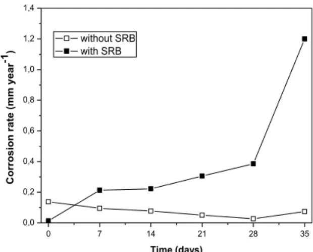
The corrosion rates as a function of exposure time, obtained from the polarization curves, are shown in Figure 6.
1E-8 1E-7 1E-6 1E-5 1E-4 1E-3 0.01 0.1 1 -1.6
-1.2 -0.8 -0.4 0.0 0.4
Current density (Acm-2)
E
(V
v
s
SC
E)
without SRB 35 d (1) with SRB 35 d (2)
1
1 2 2
993
0 7 14 21 28 35
0,0 0,2 0,4 0,6 0,8 1,0 1,2 1,4
C
o
rr
o
s
io
n
r
a
te
(
m
m
y
e
a
r
-1)
Time (days)
without SRB with SRB
Figure 6: Corrosion rates as a function of exposure time obtained from the polarization curves
The presence of SRB in medium largely increased the corrosion rate of the carbon steel relative to the medium without this microorganism, reaching 1.2 mm year-1 after 35 days of exposure. In general, when the corrosion rate increases, it can be related to the breakdown of the passivity film, composed by the corrosion products and biofilm [23]. ALABBAS et al. [5] also found a high corrosion rate (approximately 1.8 mm year-1) for high-strength steel after 30 days of exposure in modified Baar’s medium.
3.5 EIS analysis
The EIS data were also analysed to verify the influence of SRB on the corrosion of carbon steel, and the re-sults are shown in Nyquist plots for 7 and 35 days of exposure (Figure 7). It is evident that the polarization resistance of carbon steel was reduced in the presence of SRB, independent of the exposure time. This result confirms that the metabolic products of the SRB and extracellular polymeric substances (EPS) caused a change in the electrochemical properties of the systems [26].
0 2 4 6 8 10 12 14
0 1 2 3 4 5 6 7
Z
imag
(
k
.cm
²
)
Zreal(k.cm²)
without SRB 7d with SRB 7d without SRB 35 d with SRB 35 d
Figure 7: Nyquist plots of the carbon steel in the media in the absence and presence of SRB cells for 7 and 35 days of exposure
All of the curves in the diagrams present only one capacitive loop. The diameter of the capacitive semicircle increased with the immersion time of carbon steel in the sterile medium. This behaviour could be attributed to the formation of a surface layer due to the oxidation of steel and the precipitation of corrosion products on steel surface. DUAN et al. [1] and HEITZ et al. [27] suggested that this corrosion product be-comes more compact on the surface of the substrate with the exposure time.
994 behaviour could be induced by changes in the microenvironment and in the biofilm, as well as by the corro-sion of the steel substrate, that resulted from the decay of the SRB number.
4. CONCLUSIONS
A non-homogeneous and porous film, probably composed by a biofilm and corrosion products (mainly FeSx), was observed on the steel surface in the biotic medium, as a result from the SBR action. The Eocp measurements, the polarization curves and the EIS analysis indicated the presence of this film on the surface of the steel exposed to the biotic medium. However, the polarization and EIS results showed that it was not stable enough to act as a protective layer. Localized corrosion on carbon steel surface was observed when this film was removed, corroborating that the corrosion process continued under the biofilm/corrosion products layer.
It is important to note that although we used a mixed culture found in a natural marine environment in Brazil, the MIC processes observed in this study were not different from those described in works using isolated SRB cultures or any other natural marine consortium from anywhere in the world.
5. ACKNOWLEDGMENTS
The authors are grateful to CAPES, CNPq and FAPERJ for their generous financial support.
6. BIBLIOGRAPHY
[1] DUAN, J., WU, S., ZHANG, X., et al. “Corrosion of carbon steel influenced by anaerobic biofilm in nat-ural seawater”,Electrochimica Acta, v. 54, n. 1, pp. 22-28, Dec. 2008.
[2] JAVAHERDASHTI, R. “A review of some characteristics of MIC caused by sulfate-reducing bacteria: past, present and future”,Anti-Corrosion Methods and Materials, v. 46, n. 3 pp. 173-180, Apr. 1999. [3] MIRANDA, E., BETHENCOURT, M., BOTANA, J.F., et al., “Biocorrosion of carbon steel alloys by an hydrogenotrophic sulfate-reducing bacterium Desulfovibrio capillatus isolated from a Mexican oil field sepa-rator”, Corrosion Science, v. 48, n. 9, pp. 2417-2431, Sep. 2006.
[4] XU, C., ZHANG, Y., CHENG, G., et al., “Localized corrosion behavior of 316L stainless steel in the presence of sulfate-reducing and iron-oxidizing bacteria”, Materials Science and Engineering, v. 443, n. 1-2, pp. 235 – 241, Jan. 2007.
[5] ALABBAS, F.M, WILLIAMSON, C., BHOLA, S.M, et al., “Influence of sulfate reducing bacterial bio-film on corrosion behavior of low-alloy, high-strength steel (API-5L X80)”, International Biodeterioration &
Biodegradation, v. 78, n. 1, pp. 34-42, Mar. 2013.
[6] AL-ZUHAIR, S., EL-NAAS, M. H., AL-HASSANI, H. “Sulfate inhibition effect on sulfate reducing bac-teria”, Journal of Biochemistry Technology, v. 1, n. 2, pp. 39-44, Sep. 2008.
[7] CETIN, D. AKSU, M.L. “Corrosion behavior of low-alloy steel in the presence of Desulfotomaculum sp.”
Corrosion Science, v. 51, n. 8 pp.1584-1588, Aug. 2009.
[8] GAYOSSO, M.J.H., OLIVARES, J.Z., ORDAZ, N.R., RAMIREZ, C.J., ESQUIVEL, R.G., VIVEROS, A.P. “Microbial consortium influence upon steel corrosion rate, using polarisation resistance and el ectro-chemical noise techniques”,Electrochimica Acta, v. 49, n. 25, pp. 4295-4301, Oct. 2004.
[9] JAVAHERDASHTI, R. “Impact of sulphate-reducing bacteria on the performance of engineering materi-als”,Applied Microbiology and Biotechnology, v. 91, n. 6, pp. 1507–1517, Sep. 2011.
[10] THIERRY, D., SAND, W. “Microbially Influenced Corrosion”, In: Marcus, P. (ed), Corrosion
Mecha-nisms in Theory and Practice, 3 ed., chapter 17, Pennsylvania, USA, CRC Press, 2011.
[11] ANTONY, P.J., CHONGDAR, S., KUMAR, P., et al., “Corrosion of 2205 duplex stainless steel in chlo-ride medium containing sulfate-reducting bacteria”,Electrochimica Acta, v. 52, n. 10, pp. 3985-3994, Mar. 2007.
[12] CANTERO, E. V., CABRIALES, J.J. P. J. “Effects of iron-reducing bacteria on carbon steel corrosion induced by thermophilic sulfate-reducing consortia”,Journal of Microbiology and Biotechnology, v. 24, n. 2, pp. 280-6, Fev. 2014.
995 [14] XU, J., WANG, K., SUN, C., et al., “The effects of sulfate reducing bacteria on corrosion of carbon steel Q235 under simulated disbanded coating by using electrochemical impedance spectroscopy”, Corrosion
Science, v. 53, n. 4, pp. 1554–1562, Apr. 2011.
[15] WAN, Y., ZHANG, D., LIU, H., et al., “Influence of sulphate-reducing bacteria on environmental pa-rameters and marine corrosion behavior of Q235 steel in aerobic conditions”,Electrochimica Acta, v. 55, n. 5, pp. 1528-1534, Fev. 2010.
[16] CASTANEDA, H., BENETTON, X.D. “SRB-biofilm influence in active corrosion sites formed at the steel-electrolyte interface when exposed to artificial seawater conditions”,Corrosion Science, v. 50, n. 4, pp.1169-1183, Apr. 2008.
[17] ASTM. Standard Specification for Steel Tubes, Carbon and Carbon Manganese, Fusion Welded, for Boiler, Superheater, Heat Exchanger and Condenser Applications. 2012.
[18] ASTM. Standard Specification for Substitute Ocean Water. ASTM D1141-98. 2008.
[19] PÉREZ, E.J., CABRERA-SIERRA, R., GONZÁLEZ, I., et al. “Influence of Desulfovibrio sp. biofilm on SAE 1018 carbon steel corrosion in synthetic marine medium”,Corrosion Science, v. 49, n. 9, pp. 3580-3597, Sep. 2007.
[20] GOLDSTEIN, J. I., NEWBURY, D. E., ECHLIN, P., et al., Scanning Electron Microscopy and X-Ray
Microanalysis, 1 ed. New York and London, Plenum Press, 1992.
[21] ANANDKUMAR, B., GEORGE, R.P., MARUTHAMUTHU, S., et al., “Corrosion behavior of SRB Desulfobulbus propionicus isolated from an Indian petroleum refinery on mild steel”,Materials and Corro-sion, v. 62, n. 4, pp. 1-8, Jan. 2011.
[22] OUDE ELFERINK, S.J.W.H., VISSER, A., POL, L.W.H., et al., “Sulfate reduction in methanogenic bioreactors”,FEMS Microbiology Reviews, v. 15, n, 2-3, pp. 119-136, Oct.1994.
[23] RODRIGUEZ-HERNANDEZ, M., GALVAN-MARTINEZ, R., OROZCO-CRUZ, R., et al., “Influence of the sulfate reducting bacteria on API-X70 steel corrosion”,Materials and Corrosion, v. 60, n. 12, pp. 982-986, May, 2009.
[24] ZHAO, X., DUAN, J., HOU, B., et al., “Effect of Sulfate-Reducing Bacteria on Corrosion Behavior of mild steel in Sea Mud”,Materials Science Technology, v. 23, n. 3, pp. 323-328, Oct. 2007.
[25] LIU, F., ZHANG, F., LI, W., et al., “Effect of sulphate reducting bacteria on corrosion of Al-Zn-In-Sn sacrificial anodes in marine sediment”,Materials and Corrosion, v. 63, n. 5, pp. 431-437, May. 2012. [26] NGUYEN, T. M.P., SHENG, X., TING, Y., et al., “Biocorrosion of AISI 304 stainless steel by Desul-fovibrio desulfuricans in seawater”,Industrial & Engineering Chemistry Research, v, 47, n. 14, pp, 4703-4711, Jun. 2008.