*e-mail: [email protected], [email protected], [email protected]@metalmat.ufrj.br
Development and Characterization of 5% mol Zn Bioceramic in Granular Form
Ingrid Russoni de Limaa, Andrea Machado Costab, Ivan Napoleão Bastosc*, José Mauro Granjeirod*, Gloria de Almeida Soaresa*
a
Metallurgical and Materials Engineering Department, Federal University of Rio de Janeiro,
PEMM/COPPE/UFRJ, PO Box 68505, 21941-972 Rio de Janeiro, RJ, Brazil
b
Department of Materials Science (III), Biomaterials, University of Erlangen-Nuernberg,
Henkestr 91, 91052, Erlangen, Germany
c
State University of Rio de Janeiro – UERJ, PO Box 97282, 28610-974 Nova Friburgo, RJ, Brazil
dMolecular and Cellular Biology Department, UFF, Niteroi, RJ, Brazil
Received: May 4, 2006; Revised: September 11, 2006
Hydroxyapatite (HA) is capable of accepting substitute ions within its lattice, including zinc ions. Zinc is a trace element that activates the osteogenesis of osteoblastic cells and therefore plays an important role in the activity of alkaline phosphatase enzyme. The purpose of this work was to produce and characterize 5% mol Zn bioceramic in granular form (Zn-granules) for clinical applications and compare it with granules made from HA by using the same production route. Granules with addition of porogen agents were produced from powders of HA and zinc-containing HA by uniaxial pressing and heat treatment. The granules were subsequently ground and sieved. The results indicated that zinc contributed to the reduction of sample crystallinity and formed a biphasic structure after calcination at 1200 °C. Additionally, zinc release from granular material may have clinical applications as bone graft.
Keywords: hydroxyapatite, zinc, granules, bone graft
1. Introduction
The need to substitute lost body structures sparked interest in materials capable of integrating with the human body. Such materials should be able of mimetizing lost structures, to be biocompatible and should not be antigenic nor carcinogenic1. Hydroxyapatite (HA) is
one of the most popular biomaterials as it fulfills these conditions. The use of synthetic HA [Ca10(PO4)6(OH)2] in granular form has found increasing applications in dentistry and medicine in the last few decades2,3.
The mineral part of bone is composed of carbonated and non-stoichiometric HA structure containing substitutions of Na+, K+, Mg2+,
Sr2+, Cl-, F-, HPO 4
2- ions originated in the extracellular
matrix-medi-ated process of mineralization found in in vivo body fluid medium. Moreover, c-axes of HA crystals are parallel to the long axis of Type I collagen fibrils, which reveals the interplay of biomaterial and tis-sue. On the other hand, in vitro experiments using simulated body fluid revealed the presence of Na+ and Mg2+ ions on the biomimetic
coatings of titanium after a two-week soaking exposure, rather similar to the biological apatites of human bones2,4. These HA substitutions
offer the possibility of using hydroxyapatite as a means to transfer essential ions directly to where is needed.
Zinc may activate the osteogenesis of osteoblastic cells, and it participates in the activity of more than 300 types of enzymes, in-cluding alkaline phosphatase5,6. These enzymes are essential because
they take part in bone metabolism. It would therefore be desirable to obtain a hydroxyapatite with a certain amount of zinc so as to enhance several biochemical processes such as osteogenesis and increase the production of hundreds of enzymes.
HA powders can be partially decomposed into tricalcium phosphate (TCP) by heat treatment7. In addition, the calcination
temperature and stoichiometry of hydroxyapatite affect HA→TCP
transformation8. This transformation can be useful for the
develop-ment of materials with biphasic structure and a degradation rate between HA and TCP9. Zinc seems to decrease the thermal stability
of powder HA and, indeed, zinc-containing a TCP phase was identi-fied in ZnHA in powder form calcined at temperatures higher than 800 °C10. Regarding the solubility of these phases, the literature shows
that TCP is more soluble than HA when immersed in Tris-buffer (tris(hydroxymethyl)aminomethane)/HCl medium11, and ZnTCP
powder is more soluble than ZnHA in milliqui water9.
Zinc plays an important role in proliferative effects on osteoblastic cells apart from inhibiting osteoclastic resorption. Zinc-substituted calcium phosphates has thus received considerable attention. In addi-tion, hydroxyapatite solubilizes high fractions of Zn, approximately 15%mol10, which is a necessary condition for delivering efficacy. The
possibilities of increasing Zn releasing rate by controling the surface area and/or phase transformation are a promising means to achieve such purposes. However, clinical applications demand materials in blocks or granular form. Thus, the aim of this paper is to produce a Zn-containing bioceramic in granular form (Zn-granules) and compare its physicochemical properties and in vitro degradation with those of granular material produced from stoichiometric hydroxyapatite (HA-granules).
2. Materials and Methods
Hydroxyapatite (HA) and HA with 5% mol of zinc (ZnHA) powders were prepared by the precipitation method, according to a procedure described elsewhere9. In order to produce porous tablets,
for 2 hours at this temperature (heating rate was 1 °C min-1) followed
by furnace cooling. Part of the samples calcined at 400 °C were maintained for 1 hour at 1200 °C (heating rate from 400 to 1200 °C was 20 °C min-1) and also furnace cooled.
The porogen agent was pearl-shaped stearic acid of analytical grade (Vetec) with the size of powder ranging from 0.25 to 1.50 mm. The calcined tablets were ground and sieved to obtain HA-granules and Zn-granules within the 250–1,000 μm range. The porous structure facilitates the grinding and sieving step and the pores remaining in the granules can increase the surface area.
The physicochemical characterization of the calcined samples (HA-granules and Zn-granules) included X ray diffractometry (XRD), Fourier-transform infrared spectrometry (FT-IR), scanning electron microscopy (SEM) and transmission electron microscopy (TEM). For XRD, TEM and FT-IR characterizations, the granular calcined material was ground and sieved in order to obtain a homogeneous powder.
The crystalline structure of the samples was investigated by conventional XRD (Rigaku Miniflex) operating at 30 KV and 15 mA with CuKα radiation, and the data were acquired using the double angle range 10-100°. A diffusion reflectance FT-IR spectrometer (ABB Bomem Inc.) was used to identify the vibration modes of spe-cies such as phosphate, carbonate and O-H bonds of granules. The spectra were collected at room temperature at a nominal resolution of 4.00 cm-1 with number of scans equal to 100. The spectra were
recorded from 400 to 4000 cm-1. A scanning electron microscope
(ZEISS DSM 940A) was employed to image the topography and porosity of granules.
The samples calcined at 1200 °C were further examined by using a transmission electron microscope (TEM, JEOL 2000 FX), operating at 200 kV. The samples analyzed by TEM were prepared by crush-ing small quantities of granular material, dispersed in ethanol and subsequently dropped on a carbon-coated copper grid.
A degradation test of the calcined samples at 1200 °C was car-ried out in accordance to the ISO 10993-9 standard. The samples of 1.000 g of HA-granules and Zn-granules were immersed in 20 mL of Tris-buffer (tris(hydroxymethyl)aminomethane)/HCl solution in an isothermal bath at 30 °C for 120 hours, under gentle shaking. According to the ISO standard, the pH of degradation medium was maintained at 7.4 ± 0.1. The experiments were run in triplicate and the concentrations of calcium and zinc in solution were measured by atomic absorption spectrometry (AA). This technique shows a limit of detection (approximately μg/L) suitable for these experiments18. The
differences in the results from both types of samples were analyzed by the statistic ANOVA method.
3. Results and Discussion
Figures 1a and 1b show the diffraction patterns of the granules produced from HA and Zn-HA powders before and after calcination at 400 and 1200 °C, respectively. The XRD pattern of non-calcined HA-granules showed good correlation with stoichiometric hydroxya-patite (JCPDS 09-0432 card), with a small shift to higher angles, probably due to zinc substitution. Apart from hydroxyapatite, no other phases were identified, even after calcination at 1200 °C. In fact, the calcination temperature had little effect on crystallinity. On the basis of the great similarity between the standard pattern and the experimental results, we suggest a Ca/P ratio close to 1.67 and high chemical purity for these samples.
The non-calcined Zn-granules were characterized as low-crys-talline hydroxyapatite since the diffraction peaks have low intensity and are relatively broad. In this case, zinc affects the structure when it partially replaces calcium in the hydroxyapatite crystal sites. Zinc
substitution reduces markedly the crystallite size9,10 and causes,
there-fore, a reduction of cristallinity. As expected, heating at 400 °C did not change significantly the structure of the crystals nor altered XRD pattern. Therefore, a second phase was not observed. However, with calcination at 1200 °C, two phases were identified: a Zn-containing HA (JCPDS 09-0432 card) and a Zn-containing β-TCP (tricalcium phosphate, JCPDS 09-0169 card). Comparing the divalent ion substi-tutions of Mg2+ and Ca2+ in apatite we believed that the heat-treatment
above 700 °C produces the same biphasic mixtures23. In the present
study they are ZnHA and ZnTCp, as shown in Figure 1b. The pres-ence of zinc in the hydroxyapatite lattice affects its stability, inducing HA thermal decomposition. Indeed, this phenomenon is observed for hydroxyapatite with carbonate substitution or for those calcium deficient when calcined above approximately 1000 °C8. According
to Miyaji et al.10, the diffraction peaks of β-TCP shifted to higher
angles compared with those present in JCPDS data.
As for infrared spectroscopy, the spectra of HA-granules and Zn-granules (Figure 2a and 2b, respectively) present similar bands,
Intensity (u.a.)
non-calcined calcined at 400 °C calcined at 1200 °C
25.97 31.96
33.31
39.96
46.75 49.60
20 25 30 35 40 45 50 55 60
2Q (a)
Intensity (u.a.)
20 25 30 35 40 45 50 55 60
2Q
non-calcined calcined at 400 °C calcined at 1200 °C
25.99
28.10 32.01
31.24 33.16
34.71 40.01 46.91 49.55
ZnHA ZnTCP
(b)
though, with different relative intensities. These results can be at-tributed to small variations in the sample surface compositions. The bands of OH- absorption characteristics of HA9 at 3570 and 633 cm-1
and bands at 109322, 103420, 96021, 60220, 56520 and 47520 cm-1
cor-responding to PO43- groups were identified. Small peaks related to
C-O vibration bands of CO32- groups20 at 1414-1450 cm-1 suggest
that some carbonate was incorporated during the low temperature HA processing. When the samples were calcined at 1200 °C, these vibration bands disappeared. For the Zn-granules treated at 1200 °C, we did not clearly identify β-tricalcium phosphate (β-TCP) bands, albeit they were revealed by XRD. A possible reason for that could be the sample region covered by each technique, i.e., XRD is a bulk technique whereas diffusion reflectance FT-IR obtains information especially from the surface of samples.
Figure 3 shows SEM images of HA-granules and Zn-granules for the two different temperatures of calcination. The presence of
4000 3600 3200 2800 2400 2000 1600 1200 800 400
Wavenumber (cm-1)
Intensity
3570
3570
3570
non-calcined calcined at 400 °C calcined at 1200 °C
1093 1032 14481415
1093 1034
960 602
565 960
602 565
14531425 1034
602 565 1093
(a)
Intensity
non-calcined calcined at 400 °C
calcined at 1200 °C 3570
3570 1425
1095
14531424
1093 1032
962 602
595 565 602 963 1450 1090
602
1030
560
1034
4000 3500 3000 2500 2000 1500 1000 500
Wavenumber (cm-1)
(b)
Figure 2. Infrared spectroscopy spectra: a) HA-granules; and b) Zn-gran-ules.
Figure 3. SEM images for two temperatures of calcination: a) HA-granules, 400 °C; b) HA-granules, 1200 °C; c) Zn-granules, 400 °C; and d) Zn-gran-ules, 1200 °C.
15 kV x100 100 Mm COPPE/UFRJ
a
15 kV x100 100 Mm COPPE/UFRJ
b
15 kV x100 100 Mm COPPE/UFRJ c
15 kV x100 100 Mm COPPE/UFRJ
Table 1. Chemical analysis of solution after the degradation test. Three samples were used for each condition.
Ca (mg/L) Zn (mg/L)
HA-granules 17.1 ± 0.6
-Zn-granules 12.7 ± 1.1 40.1 ± 7.6
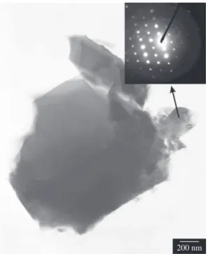
Figure 4. TEM micrography of Zn-granules calcined at 1200 °C. Inset: electron diffraction pattern.
200 nm
superficial concavities due to the porogen agent can be observed. This morphology can be attributed to the size of the granules, which was close to that of porous. Moreover, no significant morphological differences between both granules were observed from SEM images. However, it is worth noting that the material calcined at 400 °C exhibited low mechanical integrity when evaluated by simple finger touch and was, therefore, discarded before the degradation test. This high brittleness precludes it from being used for in vivo application. Therefore, it was not tested in the degradation solution.
Figure 4 shows TEM micrography for Zn-granules calcined at 1200 °C and the correspondent electron diffraction pattern. The TCP particles exhibit a massive morphology whose particles are bigger than the usual needle-like HA crystals12. The diffraction lattices
depicted in the inset of Figure 4 highligths the crystallinity of TCP phase, as already detected by XRD analysis. In an unpublished work we have identified zinc in both hydroxyapatite and TCP crystals.
The solution composition of calcium and zinc content after the 120 h degradation test is shown in Table 1. The average Ca content for HA-granules is 17.1 mg/L and for Zn-granules is 12.7 mg/L. Significant differences (p < 0.05) among calcium concentrations were observed for both set of samples. The reduction of calcium content after dissolution of Zn-granules can be associated with the competi-tion between calcium and zinc to be dissolved.
Zinc replaces calcium in the hydroxyapatite crystal lattice and affects a- and c-axis unit cell parameters of hydroxyapatite due to difference of ionic radii10. The radius of Ca is 0.099 nm and for Zn,
0.074 nm, and zinc presence steadily reduces c-axis. For a-axis, there is a decrease up to 5% mol Zn, and above this fraction the a-axis size
increases. In the present study, the choice of 5% mol was made to obtain a bioceramic material of small crystal size and consequently large surface area9, which accelerates surface reactions, such as those
occurring in the degradation test.
The conjoint effect of double dissolution (Ca and Zn) results in bigger averaged dissolution of Zn-granules than of HA-granules. This result is consistent with data available in the literature11,13. This
behavior encourages the use of synthesized Zn-granules as an effective zinc deliver agent. On the other hand, the types of dissolution media play an important role because they can promote a selective dissolu-tion of atoms. In this sense, two standardized media19, pure water9
or Tris (present work), have different effects on apatite dissolution. The Tris is present in high concentration (1 molL-1), approximately
20 times its content in simulated body fluid17. Therefore it is relevant
to know the values of the stability constant of M(Tris)2+ in aqueous
media. Fisher et al.14 reported logK ML
M as 1.94 for Zn(Tris)2+ and less
than 0.7 for Ca(Tris)2+. In this equation M stands for the metal and L
for the ligand (Tris). Hence, Zn complex is more stable than that of Ca, and this difference could explain the high Zn content depicted in Table 1 for Zn-granules. The AA technique quantifies the element concentration in solution regardless of its presence as a metallic ion or complex, i.e, Ca2+ or Ca(Tris)2+. These complexations do not occur
when milliqui water is used as degradation medium.
The recommended tests15 of calcium salts encompass elemental
analyses, dissolution/solubility and biocompatibility for characteriza-tion of the candidate biomaterials3. However, the link between in vitro
and in vivo responses is not straightforward16,17, as the in vitro tests
are an inert closed environment. Consequently, before carrying out
in vivo tests, at least cytotoxicity of Zn-granules must be evaluated, even for bioceramics that show good solubility, such as the Zn-gran-ules studied in the present paper.
4. Conclusions
Zn-granules can be easily produced by cold pressing zinc-con-taining HA powders followed by heat-treatment and ground-sieving steps. The temperature of calcination was found to increase the crystallinity of the granules especially above 400 °C. The presence of ZnTCP phase in Zn-granules calcined at 1200 °C can contribute to its higher solubility. Moreover, zinc, rather than calcium, dissolves in Tris medium, resulting in zinc release when in vitro tested. Thus, the route herein employed seems to be effective to produce biphasic granules with zinc release for clinical applications.
Acknowledgments
The authors acknowledge the financial support of CNPq (CT-504.808/2004-4), CAPES and FAPERJ Brazilian agencies. They also thank CBPF for powder synthesis.
References
1. Webster T, Massa-Schlueter E, Smith J, Slamovich E. Osteoblast response to hydroxyapatite doped with divalent and trivalent cations. Biomaterials. 2004; 2(11):2111-2121.
2. LeGeros R. Properties of osteoconductive biomaterials: Calcium phos-phate. Clinical Orthopaedics and Research. 2002; 395:81-98.
3. Conz MB. Physicochemical characterization of six commercial hydroxya-patites for medical-dental applications as bone graft. Journal of Applied Oral Science. 2005; 13(2):136-140.
4. Vallet-Regí M, González-Calbet JM. Calcium phosphate as substitution of bone tissues. Progress in Solid State Chemistry. 2004; 32(1-2):1-31.
6. Ito A, Otsuka M, Kawamura H, Ikeuchi M, Ohgushi H, Sogo Y et al. Zinc-containing tricalcium phosphate and related materials for promoting bone formation. Current Applied Physics. 2005; 5(5):402-406.
7. Bhattacharjee S, Swain SK, Sengupta DK, Singh BP. Effect of heat treatment of hydroxyapatite on its dispersibility in aqueous medium. Colloids and Surfaces A: Physicochemical Engineering Aspects, 2006; 277(1-3):164-170.
8. Oliveira JF, Sena LA, Pérez CAC, Aguiar PF, Rossi AM, Soares GA. Influence of processing parameters on structural characteristics of po-rous calcium phosphate samples: A study using an experimental design method. Materials Research. 2005; 8(1):71-76.
9. Costa AM, Soares GA, Calixto R, Rossi AM. Preparation and properties of zinc containing biphasic calcium phosphate bioceramics. Key Engineering Materials. 2004; 254-256:119-122.
10. Miyaji F, Kono Y, Suyama Y. Formation and structure of zinc sub-stituted calcium hydroxyapatite. Materials Research Bulletin. 2005; 40(2):209-220.
11. Daculsi G. Biphasic calcium phosphate concept applied to artificial bone, implant coating and injectable bone substitute. Biomaterials. 1998; 19(16):1473-1478.
12. Mavropoulos E, Rossi AM, Rocha NCC, Soares GA, Moreira MJ, Moure GT. Dissolution of calcium-deficient hydroxyapatite synthesized at dif-ferent conditions. Materials Characterization. 2003; 50(2-3):203-207.
13. Fulmer MT, Ison IC, Hankermayer CR, Constantz BR, Ross J. Measure-ments of the solubilities and dissolution rates of several hydroxyapatites. Biomaterials. 2002; 23(3):751-755.
14. Fisher BE, Häring UK, Tribolet R, Sigel H. Metal ion/buffer interac-tions, stability of binary and ternary complexes containing 2-amino-2(hydroxymethyl)-1,3-propanediol (Tris) and adenosine 5´-triphosphate (ATP). European Journal of Biochemistry. 1979; 94:523-530.
15. Class II Special Controls Guidance Document: Resorbable calcium salt bone void filler device; Guidance for Industry and FDA, June, 2003. US Department of Health and Human Services, Food and Drug Administra-tion, Center for Devices and Radiological Health.
16. Xin R, Leng Y, Chen J, Zhang Q. A comparative study of calcium phos-phate formation on bioceramics in vitro and in vivo. Biomaterials. 2005; 26(33):6477-6486.
17. Kokubo T, Takadama H. How useful is SBF in predicting in vivo bone bioactivity? Biomaterials. 2006; 27(15):2907-2915.
18. Ribeiro AS, Arruda MAZ, Cadore S. Electrothermal atomic absorption spectrometry with tungsten coil: a critical re-view. Química Nova. May 2002; 25(3):396-405. (in portuguese).
19. Standard test method for Evaluation of the environmental stability of cal-cium phosphate calcal-cium coatings, ASTM F 1926-99, West Conshohocker PA, USA.
20. Andrade MC, Sader MS, Filgueiras MRT, Ogasawara T. Microstructure of ceramic coating on titanium surface as a result of hydrothermal treatment, Journal of Materials Science: Materials in Medicine. 2000; 11:751-755.
21. Kumar M, Xie J, Chittur K, Riley C. Transformation of modified brushite to hydroxyapatite in aqueous solution: effects of potassium substitution. Biomaterials. 1999; 20(15):1389-1399.
22. Stoch A, Jastrzebski W, Brozek A, Stoch J, Szaraniec J, Trybalska B et al. FTIR absorption-reflection study of biomimetic growth of phos-phates on titanium implants. Journal of Molecular Structure. 2000; 555(1-3):375-382.