In-Frame Amber Stop Codon Replacement
Mutagenesis for the Directed Evolution of
Proteins Containing Non-Canonical Amino
Acids: Identification of Residues Open to
Bio-Orthogonal Modification
James A. J. Arpino1¤‡, Amy J. Baldwin1‡, Adam R. McGarrity1, Eric M. Tippmann2, D. Dafydd Jones1*
1School of Biosciences, Main Building, Park Place, Cardiff University, Cardiff, CF10 3AT, United Kingdom, 2Department of Chemistry, Indiana–Purdue University Fort Wayne, Fort Wayne, Indiana, 46815, United States of America
¤ Current address: Centre for Synthetic Biology and Innovation, Imperial College London, London, United Kingdom
‡These authors are joint first authors on this work.
Abstract
Expanded genetic code approaches are a powerful means to add new and useful chemistry to proteins at defined residues positions. One such use is the introduction of non-biological reactive chemical handles for site-specific biocompatible orthogonal conjugation of pro-teins. Due to our currently limited information on the impact of non-canonical amino acids (nAAs) on the protein structure-function relationship, rational protein engineering is a“hit and miss”approach to selecting suitable sites. Furthermore, dogma suggests surface ex-posed native residues should be the primary focus for introducing new conjugation chemis-try. Here we describe a directed evolution approach to introduce and select for in-frame codon replacement to facilitate engineering proteins with nAAs. To demonstrate the ap-proach, the commonly reprogrammed amber stop codon (TAG) was randomly introduced in-frame in two different proteins: the bionanotechnologically important cytb562and
thera-peutic protein KGF. The target protein is linked at the gene level to sfGFP via a TEV prote-ase site. In absence of a nAA, an in-frame TAG will terminate translation resulting in a non-fluorescent cell phenotype. In the presence of a nAA, TAG will encode for nAA incorporation so instilling a green fluorescence phenotype onE.coli. The presence of endogenously ex-pressed TEV proteases separatesin vivotarget protein from its fusion to sfGFP if expressed as a soluble fusion product. Using this approach, we incorporated an azide reactive handle and identified residue positions amenable to conjugation with a fluorescence dye via strain-promoted azide-alkyne cycloaddition (SPAAC). Interestingly, best positions for efficient conjugation via SPAAC were residues whose native side chain were buried through analy-sis of their determined 3D structures and thus may not have been chosen through rational
OPEN ACCESS
Citation:Arpino JAJ, Baldwin AJ, McGarrity AR, Tippmann EM, Jones DD (2015) In-Frame Amber Stop Codon Replacement Mutagenesis for the Directed Evolution of Proteins Containing Non-Canonical Amino Acids: Identification of Residues Open to Bio-Orthogonal Modification. PLoS ONE 10 (5): e0127504. doi:10.1371/journal.pone.0127504
Academic Editor:Nediljko Budisa, Berlin Institute of Technology, GERMANY
Received:February 2, 2015
Accepted:April 13, 2015
Published:May 26, 2015
Copyright:© 2015 Arpino et al. This is an open access article distributed under the terms of the Creative Commons Attribution License, which permits unrestricted use, distribution, and reproduction in any medium, provided the original author and source are credited.
Data Availability Statement:All relevant data are within the paper and its Supporting Information files.
protein engineering. Molecular modeling suggests these buried native residues could be-come partially exposed on substitution to the azide containing nAA.
Introduction
The advent of expanded genetic code approaches has allowed proteins to be engineered to con-tain new chemistry not normally present in the natural amino acid repertoire (see [1–3] for re-cent reviews). Through the use of reprogrammed codons or entirely new codon systems in conjunction with engineered translation machinery, upwards of 100 non-canonical amino acids (nAAs) can be incorporated into a wide range of cell types (from bacteria to mammalian) and even whole organisms (e.g.Caenorhabditis elegans[4],Drosophila melanogaster[5] and
Arabidopsis thaliana[6]). Reprogrammed genetic code systems essentially makes any member of the proteome, whether native or recombinant, accessible through defined and targeted nAA incorporation during cellular protein synthesis. Furthermore, as proteins containing nAA be-come more valuable in a commercial context, recombinant expression can generate high yields of protein product for downstream applications [7,8].
The most common expanded genetic code approach is to reprogramme the low usage amber (TAG) stop codon [9,10]. Reprogramming is implemented using engineered tRNA-amino-acyl/tRNA synthase pairs that incorporate a desired nAA in response to the UAG codon during cellular protein synthesis [2]. This approach has recently been further enhanced through the development of newE.colistrains with all native TAG stop codons removed from the genome along with it associated ribosomal release factor RF1 [11].
While protein engineering using nAAs is becoming more established it still suffers from the drawback of traditional rational site-directed mutagenesis: limited understanding of the effect of a nAA on protein structure and function leading to the use of a fairly rudimentary design processes. As has been repeatedly observed during rational protein engineering (see reference [12,13] for two excellent recent examples), the impact of nAA incorporation at a particular site may not always be obvious, and while our understanding of the effect of a particular nAA on the protein folding-structure-function relationship is increasing [14–16] it is still very limited. Directed evolution (see [17–19] for a sample of available reviews) was developed as a concept to address the shortcomings in rational protein engineering by taking a Darwinian approach through the generation of molecular diversity followed by library screening and selection. By sampling the whole protein backbone variants with new or enhanced properties containing non-intuitive mutations are routinely isolated.
Directed evolution approaches to widen nAA sampling were until recently restricted to sampling defined regions [20]. The advent of transposon-based approaches that allowed ran-dom trinucleotide/codon (TriNEx) mutations events across the length of a gene [21,22] opened up the opportunity for whole gene TAG sampling [23,24]. This has proved useful in identifying residues in proteins such as GFP not previously sampled by traditional mutagenesis by which nAAs can modulate function [15]. The main problem with the basic approach was the quality of the libraries generated due to the inherent nature of the method. Trinucleotide exchange makes no allowance for codon positioning thus replacement may occur across two adjacent codons generating an out of frame TAG replacement (Fig 1A). Additionally, the cas-sette donating the TAG codon may insert in the wrong direction resulting in CTA codon inser-tion. In some instances CTA codon sample may be useful in expanding sampling by rescuing some cross codon replacement events where the last nucleotide of a codon is G (e.g. aaC TAg).
One important area that would benefit from combining nAA incorporation and broad pro-tein sampling is defined and bioorthogonal propro-tein conjugation [1,26]. Attachment of chemi-cally useful adducts to proteins is important for applications ranging from bioimaging to therapeutics. Traditionally, attachment has relied on utilising the inherent chemistry in a pro-tein, normally amine, carboxyl or thiol groups. However, these groups are ubiquitous both within an individual protein and the proteome as whole leading to non-specific, non-optimal and in the case of complex biological mixtures targetless labelling. Thus the ability incorporate a reactive handle into a desired position in protein without any native biological reactivity is of great utility. Click chemistry [27], especially strain-promoted azide-alkyne cycloaddition (SPAAC) [28,29] is becoming of particular interest due to it speed and compatibility with bio-logical systems. Incorporation of the phenyl azide Click handle into a protein via the nAAp -azido-L-phenylalanine (azF) [30] makes Click chemistry a reality bothin vivoandin situ[31]. However, little is known about the influence of residue microenvironment on azide reactivity in SPAAC in terms of efficiency and kinetics. Recent work suggests that the link between sur-face accessibility of the azide group and SPAAC is not straight forward and counter intuitive [32].
The challenge of trinucleotide exchange is to quickly identify desired TAG mutations with-out extensive sequencing and functional screening. Here we present a general approach that al-lows the generation of in-frame TAG codons across a gene of interest. The method utilises read-through of the target gene to superfolder GFP (sfGFP) [33] in the presence of a nAA. In the absence of nAA, the TAG reverts to a stop codon so prematurely stopping translation in the target gene before reaching the sfGFP segment. A TEV protease cleavage site is present be-tween the target and sfGFP so that the activity of the two proteins can be decoupled soon after
Fig 1. In-frame TAG codon replacement.(A)Step 1involves trinucleotide deletion followed by TAG donation using a combination of the engineered transposon MuDel [25] and the DNA cassette SubSeq [21] essentially as described previously [23] and outlined in B. The TAG substitution library is then cloned in front of the TEV-sfGFP cassette in plasmid pIFtag (S1 Fig).Step 2outlines the selection for in-frame TAG substitutions. Initially, cells are grown in the absence of a nAA and non-fluorescent colonies selected; fluorescent colonies are removed at this stage as they are deemed not to have an in-frame TAG due to the generation of a full translation product. The second selection involves plating the selected colonies in the presence of nAA. Those cells that regain fluorescence suppress TAG termination due to nAA incorporation and thus produce sfGFP. (B) Alternate versions of the new SubSeq DNA cassette for donating TAG. The two alternatives are shown in the red boxes at the far left. (C) The two nAAs used in this study, p-azido-L-phenylalanine (azF) andp-iodo-L-phenylalanine (iodoF).
production through endogenously expressed TEV. We demonstrate the approach using two proteins, cytochromeb562(cytb562) and keratinocyte growth factor (KGF). Modification by SPAAC with a fluorescent dye was achievable for only certain cytb562residues that were buried in the native, non-mutagenized structure.
Materials and Methods
Materials
AzF and iodoF were purchased from Bachem and dissolved in 0.25 M NaOH prior to use. DBCO-585 was purchased from Click Chemistry Tools and dissolved in DMSO to a stock con-centration of 2.5 mM.
Fusion plasmid construction
The pET-22b(+) vector (Novagen/Merck) formed the basis for constructing pIFtag, with the gene of interest expressed as a fusion with sfGFP under control of the T7 promoter. The two fused proteins are separated via a TEV protease sequence (Fig 2BandS1 Fig). In this study ei-ther cytb562or KGF were used as the N-terminal fusions to sfGFP. This plasmid was initially constructed by splicing PCR amplified fragments containing either the mature portion of the
cybCgene (primers 109/112) orsfGFP(primers 104/105) via a linker encoding a new XhoI rec-ognition site and the TEV cleavage site. NdeI and SalI sites were added to the 5’and 3’ends of
the fragment, respectively. The resulting fragment was ligated into the NdeI/XhoI sites in pET-22b(+), thereby destroying the XhoI site within pET-pET-22b(+), but adding an additional XhoI site 5’of the TEV cleavage site. Alternative genes of interest can be easily sub-cloned into this vector upstream of the TEV site and sfGFP gene using NdeI/XhoI.
The 3 mutants, cg-51TAG/51Naa,cg-51TAG+1/CG-51Ileandcg-51TAG+2/CG-51Asn, whereby a
TAG codon was either added in-frame or +1 or +2 nucleotides with respect to K51 was achieved by site-directed mutagenesis. Primer pairs 092/093, 092/102 and 092/103 were used
Fig 2. Selection for in-frame TAG replacement.(A) Schematic of the cytochrome (red) and sfGFP (green) fusion with the TEV cleavage site between the two shown. The residue targeted (K51) for replacement and the rational TAG replacements are shown. (B) cellular fluorescence of each variant in the absence (-nAA) and presence (+nAA) of either azF or iodoF. The wt refers to the cytb562-sfGFP fusion without any TAG codon
replacement. (C) SDS-PAGE (top panel) and Western blot (bottom panel) analysis of fusion protein expression in the soluble fraction of the cell lysate. The—v lane represents cells producing CG-wt in the absence of the pAB vector. The + and—lanes refer to the presence or absence of the nAA, respectively. CG and G refer to the cytb562-sfGFP fusion (~40 kDa) and the sfGFP alone (27 kDa) generated through TEV
cleavage. The Western blot detection was performed with an antiGFP primary antibody.
to generate each respective TAG mutation. The mutations were confirmed by DNA sequenc-ing. All primers sequences are listed inS1 Table.
Western blotting
E.coliTuner cells hosting the pAB and pIFtag were grown in LB broth supplemented with 100μg/ml ampicillin and 34μg/ml chloramphenicol. The pAB plasmid harbours the iodoF
aminoacyl-tRNA-synthetase (aaRSiodoF) and two tRNACUArequired for nAA incorporation during cellular protein synthesis (S1 Fig). The aaRSiodoFis promiscuous and can be used for
high efficiency and fidelity incorporation of both iodoF and azF [15]. Protein production was induced with 100μM IPTG, and culture medium supplemented with 5 mM p-iodoPhe. Cells
were then incubated at 37°C with shaking overnight. Cells were collected and resuspended at an OD600of 10 in BugBuster (1 ml), 200μg/ml lysozyme and benzonase (0.5μl/1 ml
BugBus-ter) and incubated at room temperature for 30 min. Following fractionation by centrifugation (20 min, 16000g, 4°C) 10μl of soluble fraction was analysed on a 15% SDS-PAGE alongside
NEB prestained protein marker. Proteins were blotted and detected with anti-GFP antibody (1:10,000) and 2° antibody (1:10,000). Detection was performed using the BioRad Immun-Blot AP Colorimetric Kit.
Library construction: cyt
b
562The initial cytb562library was constructed in the plasmid pNOM [25] plasmid essentially as described previously [21,23]. Introduction of the MuDel transposon throughout the pNOM:
cybCplasmid was performed using MuA and generated ~4.3x10⁴cfu on transformingE.coli.
The colonies were pooled and plasmid DNA isolated. Replacement of MuDel by SubSeqTAG/CTA in the vector was achieved by digestion of the plasmid pool with MlyI to remove MuDel and 3 bp from the host plasmid, followed by dephosphorylation, purification following agarose gel electro-phoresis, and ligation with the SubSeqTAGor SubSeqCTAcassette DNA. Transformation ofE.coli
DH5αresulted in ~2.3 x10⁴CFU, with ~55% incorporating SubSeqTAG(~1.3x10⁴CFU) and 45%
SubSeqCTA(1.0x10⁴CFU), as calculated from individual transformations. The colonies were
pooled and a second digestion with MlyI removed SubSeq. Intramolecular ligation of digested vector gave ~8.7 x10⁴CFU following transformation ofE.coliDH5α.
ThecybCTAGlibrary was sub-cloned into a modified version of pIFtag (S1 Fig) between the
NcoI and XhoI sites upstream of encoded TEV cleavage site and sfGFP. Modified pIFtag has the more amenable NcoI restriction site in place of NdeI but is otherwise identical to that inS1 Fig. It was necessary to use PCR on this occasion to add the relevant 5’NcoI recognition se-quence. The library was amplified using Phusion High-Fidelity DNA Polymerase and primers 131 and 111 in 16 separate tubes for 20 cycles before subcloning into modified pIFtag.
Transformation ofE.coliDH5αgave ~5.0x10³CFU, which were pooled and plamid DNA
isolated. The pooled pIFtag DNA was then used to transform electrocompetentE.coliTuner cells already containing pAB (S1 Fig). Colony fluorescence was determined using a transillumi-nator. Non-fluorescent colonies (192 selected at random) were transferred to 96-well microtitre plates and replica plated onto rectangular agar plates containing chloramphenicol (34μg/ml),
carbenicillin (100μg/ml), IPTG (100μM) and iodoF (2 mM). After 24 hrs the plates were moved
to 4°C. Colonies that became fluorescent in the presence of iodoF were sequenced. All sequenced variants contained in-frame TAG codons and variants covered ~15% of thecybCgene.
Library construction: KGF
Transposition of MuDel into pNOM-XP6:kgfusing MuA for either 4 hours or overnight yielded approx. 2.0x10⁴cfu after transformation intoE.coliBL21 Gold (DE3) electrocompetent cells. Following transformation, cells were plated on LB agar containing chloramphenicol (34μg/ml) and cells scrapped from the plate and pooled. DNA was purified from the pooled
cells and digested with NcoI and XhoI. The band corresponding to MuDel incorporated within thekgfgene (~1.7 kb) was isolated and ligated with pNOM-XP6 also digested with NcoI/XhoI. The resulting sample was used to transformE.coliBL21 Gold and yielded 1.3 x10⁴cfu. MuDel
was removed fromkgfusing MlyI and replaced by SubSeqTAGand SubSeqCTA. Subsequent liga-tion and transformaliga-tion resulted in 2.4 x10⁴variants selected on kanamycin containing LB
agar plates. SubSeq was excised via MlyI digestion and the remaining vector was recircularised via intramolecular ligation. Subsequent transformation ofE.coliDH5αresulted in more than
1.5 x10⁶cfu when selected on LB agar plates supplemented with 100μg/ml carbenicillin. The
resulting library DNA was digested with NcoI/XhoI and subcloned into modified pIFtag.E.
coliBL21 Gold (DE3) cells containing pAB were transformed and plated on LB agar plates con-taining 100μg/ml ampicillin, 34μg/ml chloramphenicol and 200μM IPTG. A total of 77
non-fluorescent colonies were transferred to 96-well format and replica plated onto LB agar con-taining 100μg/ml carbenicillin, 34μg/ml chloramphenicol and 2 uM IPTG plus 2 mM azF,
and incubated at 30°C overnight. The sequence of 16 colonies that were non-fluorescent in the absence of nAA and fluorescent in the presence were determined.
Protein production
E.coliBL21 Gold (DE3) cells were used to produce cytb562or KGF incorporating azF in re-sponse to the amber (TAG) stop codon using a reprogrammed genetic code system similar to that described previously [23,36]. Cells were co-transformed with the relevant pIFtag plasmid housing either the cytb562or KGF gene and pAB. Single colonies grown on ampicillin and chloramphenicol LB agar were used to inoculate 1 mL LB broth cultures supplemented with the same antibiotics. After 6 hours of growth at 37°C, the 1 mL cultures were used to inoculate 10 mL LB supplemented with ampicillin and chloramphenicol. Cultures were grown at 37°C with shaking until an OD600of 0.4–0.8 was achieved then 2.5 mM azF was added and the
cul-tures grown for a further hour. Protein expression was induced by the addition of 500μM
IPTG for cytb562-TAG variants or 100μM IPTG for KGF-TAG variants and incubated at 37°C
for 24 hours. Cells were harvested by centrifugation and lysed in 50 mM Tris-HCl, pH 8.0, 150 mM NaCl containing 0.2 mg/mL lysozyme, 1 mM PMSF and 1 mM EDTA by sonication.
Strain-Promoted Azide-Alkyne Cycloaddition (SPAAC)
SPAAC reactions were performed, as described elsewhere [32], using crude cell lysates (to mimic intracellular conditions more closely) as the source of azF-Cytb562or azF-KGF. SPAAC was performed using cell lysates from 5 mL expression cultures standardised to an A600of 1.
Reactions were performed with 5μM of DBCO-585 at 25°C for 24 hours.
Fluorescent SDS-PAGE Analysis
In silico
modelling of azF containing cyt
b
562variants
Structure files for iron protoporphyrin IX were made using Avogadro [37]. Force field parame-ters were derived based on the electrostatic model approach, which uses electrostatic potential (ESP) to describe the electronic structure of the porphyrin [38]. Geometry optimization and ESP calculations were performed using GAMESS-US [39] at the HF/6-31Glevel in order to be consistent with the AMBER99sb force field [40]. The published crystal structure for holo-cyt
b562(PDB code: 256B [41]) was used as the starting point for simulation of both proteins. For the P45azF cytb562variant the published structure for apo-cytb562(PDB code: 1APC [42]) was used instead. The structures were altered using a python script within MacPyMol to mutate the chosen residue to azF prior to energy minimisation, which was carried out using the GRO-MACS software package [43]. Molecular dynamic simulations were carried out using the AMBER99sb force field, modified with the parameters for iron protoporphyrin IX and azF. The starting structure was placed within a triclinic box with dimension of 6.4x6.1x7.8 nm. This was populated using the SPC water model to solvate the system to a total number of 16986 sol-vent molecules. The system was first energy minimised by performing 500 steps of steepest de-scent method followed by 500 steps of Conjugant Gradient method. The lowest energy state of the system was used as the starting conformation for the molecular dynamics simulation. The simulations were conducted at a constant temperature and pressure of 300 K and 1 atm (NPT). A cutoff of 8Å was chosen for nonbonded interactions and long range electrostatic interactions were characterised using the Particle Mesh Ewald (PME) [44]. The simulations were carried out for a total of 2 ns with a time step of 2 fs and the trajectories were analysed using various GROMACS components and visualised using VMD [45] (www.ks.uiuc.edu/Research/vmd/).
Results and Discussion
In-frame TAG selection process
The general approach for selecting in-frame TAG codons required for nAA incorporation is outlined inFig 1A. TAG replacement in a target gene using the TriNEx process has been de-scribed previously [23]. However, there was no easy way to determine in-frame and correctly orientated TAG codons. To address this problem and to make the method more general in its application, a new plasmid system was constructed (S1 Fig). The vector pIFtag housed the sfGFP gene within the cloning site to allow direct gene fusion downstream of the target protein bridged by a TEV protease sequence. The pAB vector was based on pAA [46] but housed two additional elements. The first is a gene encoding inducible and endogenous production of TEV protease to allow forin situcleavage of the target protein-sfGFP fusion to prevent any potential adverse downstream effects of the fusion event. The second is the promiscuousp -iodo-L-phe-nylalanine (iodoF) aminoacyl tRNA synthetase under the control of the promoter and termina-tor from the endogenously expressed glnS gene (E.coliglutaminyl-tRNA synthetase). This iodoF aminoacyl tRNA synthetase is known to efficiently encode incorporation of various nAAs [15]. To maximise the number of clones with in-frame TAG codons, a new version of the TAG donating DNA cassette (SubSeq [21,23]) was generated which sampled the comple-ment sequence (CTA) in case of reverse cassette insertion (Fig 1B).
generate substitutions leading to translational read through to sfGFP resulting in cellular fluo-rescence. Non-fluorescent colonies are retained for the second selection round in which the presence of the nAA will result in its incorporation at the position dictated by the TAG codon. Translational read through from the target gene to sfGFP will occur resulting in cellular fluo-rescence. Variants that were non-fluorescent in the first selection round due to other undesir-able mutational events will thus also be removed. While we have used colony picking to separate fluorescence and non-fluorescent cells, automated and high throughput systems such as FACS can be used to facilitate the process.
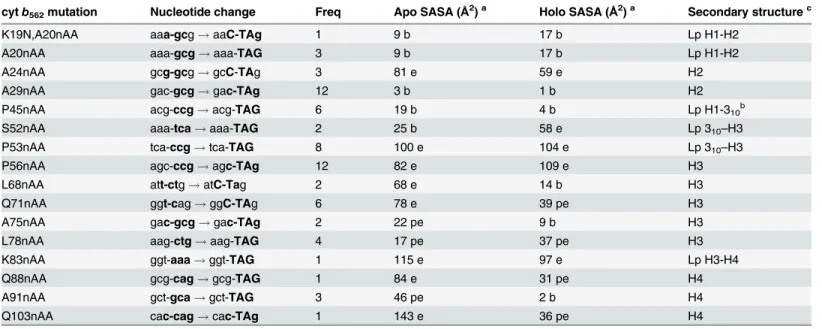
To test that the system can select for in-frame TAG codons, a set of site-directed mutants were generated in the small electron transfer protein, cytochromeb562(cytb562;Fig 2A). Cyto-chromeb562is proving to be an important system for studying metalloprotein electron transfer at the single molecule level, and as a novel molecular electronic [47–49], biosensing [50,51] and controlled assembly scaffold [52–55]. To facilitate its use for both fundamental and appli-cative studies, optimal interfacing with non-biological materials is required, which will be aided by the use of nAAs with suitable coupling chemistry. For example, incorporation ofp -azido-L-phenylalanine (azF;Fig 1D) can introduce new photochemical cross-linking and bioorthogonal conjugation properties onto a target protein [31].
Cytb562placed in-frame with sfGFP and the two linked via the TEV protease cleavage se-quence (Fig 2A) resulted in cells displaying an obvious green fluorescence phenotype (Fig 2B). Replacement of the codon for Lys51 with TAG (cg-51TAG/CG-51nAA) gave a nAA-dependent cell fluorescence phenotype; cellular fluorescence was only observed in the presence of a nAA in the culture medium. Shifting the TAG codon +1 (cg-51TAG+1/CG-51Ile) or +2 (cg-51TAG+2/CG-51Asn) nucleotides with respect to K51 did not instill a nAA-dependent green fluorescence phenotype (Fig 2B); despite the native amino acid substitution mutations in cytb562, read through to sfGFP occurred as predicted. Both SDS-PAGE and Western blot confirmed that protein was only pro-duced in the presence of nAA for the in-frame TAG variant and that cellular production of TEV protease gave rise toin situcleavage of the sfGFP from cytb562(Fig 2C).
Bioconjugation of cyt b
562by SPAAC
The SPAAC reaction is outlined inFig 3. Essentially, the azF acts as the protein embedded azide component that can react orthogonally under biologically compatible conditions with an activated alkyne (cyclooctyne) [29]. As the azide group is prerequisite for SPAAC reactions and not present in any natural biomolecule, modification with a derivative harbouring an al-kyne (CC) will be specific to defined residue position within the target protein amongst the
cellular milieu orin vitro. Several variants identified during TAG codon screening were as-sessed for the reactivity of the introduced phenyl azide group to its alkyne partner (Fig 4A) that sampled a wide variety of cytb562structural features. Dibenzylcyclooctyne (DBCO) housing the strained ring alkyne group linked to the fluorescence rhodamine dye Texas red (here on in termed DBCO-585) was used to test for residue conjugation ability and efficiency [32].
The 11 variants selected for further analysis were successfully expressed in the presence of azF as indicated by the red-pink phenotype due to the production of full length holo cytb562
(Fig 4B); little or no expression was apparent in the absence azF. The colour of cells expressing
Table 1. Sequence and characteristics of observed cytb562TAG replacement variants.
cytb562mutation Nucleotide change Freq Apo SASA (Å2)a Holo SASA (Å2)a Secondary structurec
K19N,A20nAA aaa-gcg!aaC-TAg 1 9 b 17 b Lp H1-H2
A20nAA aaa-gcg!aaa-TAG 3 9 b 17 b Lp H1-H2
A24nAA gcg-gcg!gcC-TAg 3 81 e 59 e H2
A29nAA gac-gcg!gac-TAg 12 3 b 1 b H2
P45nAA acg-ccg!acg-TAG 6 19 b 4 b Lp H1-310b
S52nAA aaa-tca!aaa-TAG 2 25 b 58 e Lp 310–H3
P53nAA tca-ccg!tca-TAG 8 100 e 104 e Lp 310–H3
P56nAA agc-ccg!agc-TAg 12 82 e 109 e H3
L68nAA att-ctg!atC-Tag 2 68 e 14 b H3
Q71nAA ggt-cag!ggC-TAg 6 78 e 39 pe H3
A75nAA gac-gcg!gac-TAg 2 22 pe 9 b H3
L78nAA aag-ctg!aag-TAG 4 17 pe 37 pe H3
K83nAA ggt-aaa!ggt-TAG 1 115 e 97 e Lp H3-H4
Q88nAA gcg-cag!gcg-TAG 1 84 e 31 pe H4
A91nAA gct-gca!gct-TAG 3 46 pe 2 b H4
Q103nAA cac-cag!cac-TAg 1 143 e 36 pe H4
acalculated solvent accessible surface area (SASA) using GETAREA (http://curie.utmb.edu/getarea.html) b, buried; pe, partially exposed; e, exposed. b
residue interacting with heme.
cH1, H2, H3 and H4 refers to helices 1, 2, 3 and 4 running from the N- to C-terminus, with Lp referring to the corresponding linking loops.
doi:10.1371/journal.pone.0127504.t001
Fig 3. SPAAC between genetically encoded azide (red sphere) within a protein (blue spheres) and an activate alkyne (dibenzylcyclooctyne;DBCO).A triazole link is formed between the azide and
alkyne groups.
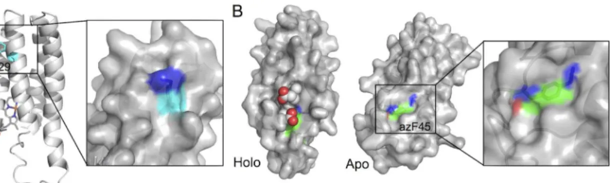
two variants, A29azFand A91azF, were different to the others; the A29AzFcell pellet was more orange and A91azFbrown. Both A29 and A91 lie close to each other in the protein close at the
opposite end of the protein from the haem binding pocket (Fig 4A). SDS-PAGE revealed that TEV protease cleavagein situwas largely complete leaving free cytb562separate from sfGFP (Fig 4C). One variant, A29AzF, appeared to be produced at a higher level than the others, as in-dicated by the darker band equivalent to sfGFP (~27 kDa) and cytb562(~13 kDa).
To assess how placement within the protein affected the reactivity of the phenyl azide group, conjugation with DBCO-585 was performed. Only two variants, A29azFand P45azF,
were modified to any extent with the DBCO-585 derivative in cell lysates (Fig 4C). The native residues at both these positions are buried in the determined structures of apo and holo forms, with P45 being close to the heme co-factor in the holo-form (Table 1). The higher apparent brightness of the P45azFcytb562variant suggests it was modified to a greater extent even
though more A29azFcytb562protein was produced (Fig 4C). The maintenance of a coloured phenotype suggests that both variants still retain the capacity to bind haem on
azF incorporation.
There does not appear to a simple correlation between residue position encoding azF, its rel-ative solvent accessibility of the nrel-ative residue, position in secondary structure and the ability to undergo SPAAC when replaced by azF. Most of the observed azF containing cytb562 vari-ants involve replacement of an exposed or a partially exposed residue (in apo or holo cytb562). Overexpressed engineered cytb562is routinely produced as a mixture of apo and holo protein [58], both of which are produced as soluble proteins, so it is pertinent to take into account both these forms. Three residues replaced with azF have their original residues buried in both apo and holo forms according to the determined structures (Table 1), two of which (A29 and P45) are the only ones open to conjugation via SPAAC. A24 resides in the same helix (H2) as A29 with both residues separated by just over one helical turn and occupying similar positions in
Fig 4. Production and conjugation of cytb562azF-containing variants.(A) Schematic structure of cyt b562showing the residues substituted with azF. (B) Expression of cytb562variants housed in the pIFtag
plasmid in the presence (+azF) or absence (-azF) of azF. Cells successfully expressing functional cytb562
display a red phenotype. (C) SDS-PAGE analysis of SPAAC conjugation. The top panel shows the
Coomassie Blue stained and the bottom is fluorescence imaging of the DBCO-585 moiety. CG, G and C refer to cytb562-sfGFP fusion, sfGFP and cytb562, respectively. The lower band is unreacted DBCO-585. For the
sake of transparency, the samples Q88 and A91 were run on separate gels to the others and the resulting images electronically linked to the other samples.
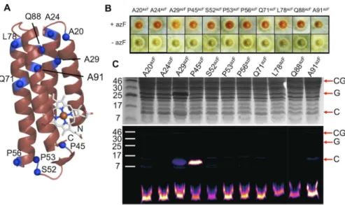
both the apo and holo cytb562structures. However, the A24 residue is exposed to the solvent while A29 is buried: only when azF is incorporated at residue 29 can cytb562be modified. This suggests that on replacement of A29 with the bulkier and longer azF, the side chain must at least become partially accessible to the solvent in either the apo and/or holo form. In an at-tempt to rationalise the effect of A29azFmutation, the variant was modelledin silicousing the
holo-cytb562structure as a template. The putative molecular model of holo-cytb562[41,59] A29azFrevealed that the phenyl azide may potentially exert a significant local structural effect
and the reactive azide component may become surface exposed (Fig 4A). Rather than the phe-nyl azide pointing directly out to the solvent, the azide group may still be closely associated with the protein surface. Thus the modelling suggests replacement of the buried A29 with azF results in the critical reactive azide group becoming surface exposed without full exposure of the side chain as a whole.
A similar modelling approach was taken to rationalise the effect of the P45azF. The putative model of holo cytb562P45azFsuggested that haem may block accessibility for the incoming DBCO group (Fig 4B). The structure of apo-cytb562is known to be less structured around the C-terminal helix H4 [42,60], making accurate modelling more difficult. Nonetheless, given that over-expressed cytb562is known to be a mix of holo and apo forms [58] modelling of the P45azF based on the available NMR structure of apo protein was attempted. The putative apo cytb562
model of the P45azFvariant suggested that the azide group becomes surface accessible and thus
available for modification (Fig 5B). As with the A29azFmodel, the azide group and the phenyl azide side chain as a whole remains closely associated with the protein surface. Such a close sur-face association of the azide group was associated with the residues displaying the highest modifi-cation efficiency by SPAAC in sfGFP suggesting that a fully protruding phenyl azide may not be ideal [32]. Therefore, a key observation, at least in terms of SPAAC modification of protein em-bedded phenyl azides, is that burial of the native residue should not be overlooked as potential useful modification site and that positions likely to retain a degree of surface association on con-version to azF may be the ideal in terms of selecting high efficiency modification sites. Given that Click chemistry approaches like SPAAC are becoming more and more utilised, these observa-tions may have wider implicaobserva-tions in terms of the design of modification sites.
Application of TAG in-frame mutagenesis to KGF
One of area of significant potential is the development of protein-based biopharmaceuticals through the defined and controlled modification with polymers and other useful agents [8,62].
Fig 5.In silicomodelling of azF cytb562variants.(A) Models of the A29azFholo cytb562. The mutated residue, azF29, is coloured in cyan at its carbon
atoms. The right hand panel is a close up surface view of the model. (B) Models of the P45azFapo and holo cytb
562variant. The mutated residue, azF45, is
coloured green at its carbon atoms. Haem is represented as spheres. The models were based on the PDB coordinate files 265B (Holo) and 1APU (apo). The figures where generated using PyMol [61].
Derivatising a protein with large chemical adducts such as PEG shields them from proteolytic degradation and rapid removal from the body resulting in improved pharmacokinetics. How-ever, most of the current approaches rely either on the inherent chemistry of the natural 20 amino acids (e.g. amine, carboxyl and sulfhydryl) or the introduction of terminal“ modifica-tion”tagging sequences. Such lack of specificity in terms of position within the protein can give rise to unwanted heterogeneity of the modified protein sample and the site of modification may not be optimal for maximising protein stability/activity. One way of overcoming such problems is through the defined placement of nAAs with useful bioorthogonal and biocompat-ible reactivity, such as azF [31].
Initially, a range of different non-antibody therapeutic proteins were screened for their suit-ability for use with the sfGFP fusion library selection approach: (1) Keratinocyte growth factor (KGF) fragment (Kepivance); (2) Ethropoietin (EPO); (3) Uricase (URI; Rasburicase); (4) As-paraginase (ASP; Oncaspar); (5) Streptokinase (SK; Streptase). When cloned in-frame with sfGFP all the selected proteins conferred a fluorescence phenotype onE.coli(Fig 6A) suggest-ing that they will be compatible with the library screensuggest-ing approach. As a test for the system, li-brary construction was focused on KGF as it was relatively small (139 amino acids) and conferred the brightest green fluorescent phenotype onE.coliunder our expression conditions. The pharmaceutical version of KGF used here (sequence provided inS3 Fig), also known as Palifermin [63], is a recombinant analogue of human KGF with the first 23 residues at the N-terminal removed to improve stability. It stimulates keratinocyte growth in a variety of tissues and aids patient recovery from chemotherapy.
Using a combination of the two phase negative (non-fluorescent clones in absence of azF) and positive (fluorescent clones in the presence of azF) selection procedure, 13 different vari-ants at the amino acid level were identified (Fig 6B). The requirement of the nAA for cellular gene fluorescence was confirmed by culturing cells expressing variants in the presence and ab-sence of azF, three of which are show inFig 6Cas representatives. Only in the presence of azF
Fig 6. In-frame TAG replacement of KGF.(A) Compatibly of various therapeutic proteins with theE.coli sfGFP fusion screening approach. KGF, keratinocyte growth factor fragment; EPO, ethropoietin; URI, Uricase; ASP+ Asparaginase with terminal signal pepetide sequence; ASP- Asparaginase lacking N-terminal signal peptide sequence; SK, Streptokinase. The‘con’sample is the control cell sample. The proteins shown are all fused to sfGFP via the TEV digestion sequence. (B) Sequence of observed KGF variants. (C) azF-dependent expression of selected KGF-sfGFP variants. (D) Map of observed mutations on the structure of KGF, including an indication of whether the native residue is surface exposed (blue spheres) or buried (grey spheres) based on surface accessibility.
was a fluorescence phenotype observed. Mapping the observed residues onto the known struc-ture of KGF (PDB 1QQK;[64]) revealed that all but 3 (L119azF, A166azF, L167azF) of the vari-ants were equivalent to exchange of surface exposed residues for azF (Fig 6D). While the current dogma suggests that surface exposure is critical as we have seen here and previously [32], this is not a guarantee for efficient modification by SPAAC especially when the mutation may result in a residue converting from buried to partially exposed. Thus, we did not ignore their potential for modification by SPAAC at this stage.
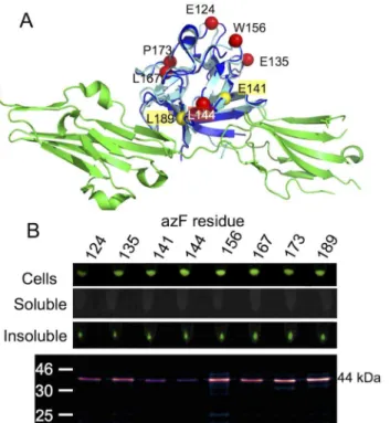
Another important requirement is that both mutation and the subsequent modification will not impede function by, for example, disrupting a key protein-protein interaction. KGF inter-acts with its cognate receptor FGFR2 and triggers various signalling pathways that simulate production of proteins required for cell growth and survival in epithelial cells [65]. There is currently no structure available for KGF bound to its cognate receptor FGFR2 so the KGF was modelled against the structurally analogous FGF10-FGFR complex (Fig 7A; 1EV2[66]). Analy-sis of the model revealed that six variants incorporate azF at positions distant from the protein-protein interface: E124, E135, L144, W156, L167 and P173. These variants together with two others with azF incorporated at E141 and L189 located at the receptor interface were chosen to assess their modification with DBCO-585 by SPAAC (Fig 7).
While attachment of sfGFP is supposed to aid solubility of attached proteins [33], all the KGF fusion variants were expressed as insoluble fusion proteins (Fig 7BandS4 Fig). While whole cells expressing the KGF-sfGFP were fluorescent suggesting that sfGFP matured (and thus folded) correctly, all the fluorescence was observed in the insoluble pellet fraction after cell lysis. The product size (~44 kDa) indicates that the insoluble KGF-sfGFP fusion was not cleaved by TEV suggesting the requirement for a soluble product for successful cleavage. This
Fig 7. Production and SPAAC modification of KGF variants.(A) Superimposition of the KGF structure on the homologous FGF10-FGFR complex. Yellow and red spheres represent residues contributing to or not involved in the receptor interface. (B) Inherent sfGFP fluorescence of different cellular fractions (top two panels) and SPAAC modification with DBCO-585 of each analysed variant.
is in contrast to the cytb562fusions that produced largely soluble protein fusions that were es-sentially fully cleaved (Figs2Cand3C). Despite insolubility and lack of fusion cleavage all eight KGF-sfGFP variants were capable of SPAAC modification with DBCO-585. Apart from 2 (E141azFand L144azF), each KGF-sfGFP fusion variant was modified to a similar extent (Fig 7B). This is contrast to cytb562were only selected residues were modified. The insolubility cou-pled with likely aggregation may in the case of KGF facilitate general modification. This may be through, for example, the generation of local surface exposed hydrophobic regions in which a population of azide groups may reside; local hydrophobic patches have been suggested to aid SPAAC modification with DBCO [32], especially as the DBCO moiety is itself hydrophobic.
Conclusion
We have shown here a general method for introducing in-frame amber stop codons for the di-rected evolution of proteins containing non-canonical amino acids. The link through to sfGFP expression and fluorescence allows identification of in-frame TAG codon replacements, and thein situcleavage of predominantly soluble fusion products via TEV protease decouples sfGFP from the target protein. While we have demonstrated codon replacement with TAG, the method can be adapted for any of the emerging approaches for nAA incorporation (e.g. qua-druplet codon systems [67,68]) through changing the nature of the donating sequence in the SubSeq element. The ability to modify proteins in a truly bioorthogonal and biocompatible manner via SPAAC (and other nAA-dependent approaches [1]) has obvious benefits for both
in situandin vitromodification of proteins for fundamental and technological purposes. How-ever, as we have shown here and elsewhere [32] simply choosing a surface exposed residues based on available structural information does not guarantee high efficiency modification. If this is the case, then a new strategy for protein engineers may be available for use with azF or other conjugation compatible nAAs and directed evolution may prove a fruitful manner by which to identify residues amenable to these next generation protein modification approaches.
Supporting Information
S1 Fig. Plasmids maps of (A) pIFtag and (B) the companion plasmid pAB.The pIFtag plas-mid is based on pET22 except that downstream of the standard NcoI/NdeI and XhoI cloning site is the DNA sequencing encoding TEV digestion site and sfGFP. Cloning of a target gene (here with the cytochromeb562sequence as an example) between the NcoI and XhoI site will put it in the same reading frame as the downstream elements. (B) The companion plasmid pAB is based on pAA reported previously as stated in the main text. It contains the engineered aminoacyl tRNA synthetase and tRNA for nAA incorporation as well as the T5 promoter driv-en expression of TEV protease.
(TIFF)
S2 Fig. MuDel target site sequence analysis.A graphical representation of the frequency at which nucleotides appear at one of the 5 positions of the target site duplication, introduced during MuDel transposition. The graphical representation was produced using the WebLogo application (http://weblogo.threeplusone.com) from 181 unambiguous unique sequences sam-pled by MuDel from the TAG replacement library reported here together with TND and do-main insertion libraries reported previously [69,70]. The height of each simple at each of the 5 positions is relative to the observed frequency of a particular base.
(TIFF)
S3 Fig. DNA (lower case) and protein (upper case) sequence of KGF used in this study.
S4 Fig. SDS PAGE analysis of the soluble and insoluble cell lysate fractions of the KGF-sfGFP azF fusion variants.The band labelled K-G at ~44 kDa corresponds to the uncleaved KGF-sfGFP fusion product. All samples were standardised to set cell density (OD at 600 nm of 1.0).
(TIFF)
S1 Table. Oligonucleotide primer sequences.
(DOCX)
Acknowledgments
The authors would like to thank Dr Matthew Edmondson and Dr Roger Chittock with cloning and fluorescence analysis help. DDJ and ARM would like to thank the Advanced Research Computing @ Cardiff facility, especially Thomas Green for help with access and usage of the Raven cluster.
Author Contributions
Conceived and designed the experiments: EMT DDJ AJB JAJA ARM. Performed the experi-ments: JAJA AJB ARM. Analyzed the data: JAJA AJB ARM DDJ. Contributed reagents/materi-als/analysis tools: AJB ARM. Wrote the paper: JAJA AJB ARM EMT DDJ.
References
1. Lang K, Chin JW. Cellular incorporation of unnatural amino acids and bioorthogonal labeling of pro-teins. Chemical reviews. 2014; 114(9):4764–806. doi:10.1021/cr400355wPMID:24655057. 2. Liu CC, Schultz PG. Adding new chemistries to the genetic code. Annual review of biochemistry. 2010;
79:413–44. Epub 2010/03/24. doi:10.1146/annurev.biochem.052308.105824PMID:20307192. 3. Zhang WH, Otting G, Jackson CJ. Protein engineering with unnatural amino acids. Current opinion in
structural biology. 2013; 23(4):581–7. doi:10.1016/j.sbi.2013.06.009PMID:23835227.
4. Greiss S, Chin JW. Expanding the genetic code of an animal. Journal of the American Chemical Socie-ty. 2011; 133(36):14196–9. doi:10.1021/ja2054034PMID:21819153; PubMed Central PMCID: PMC3168933.
5. Bianco A, Townsley FM, Greiss S, Lang K, Chin JW. Expanding the genetic code of Drosophila melano-gaster. Nature chemical biology. 2012; 8(9):748–50. doi:10.1038/nchembio.1043PMID:22864544. 6. Li F, Zhang H, Sun Y, Pan Y, Zhou J, Wang J. Expanding the genetic code for photoclick chemistry in
E. coli, mammalian cells, and A. thaliana. Angewandte Chemie. 2013; 52(37):9700–4. doi:10.1002/ anie.201303477PMID:23873613.
7. Axup JY, Bajjuri KM, Ritland M, Hutchins BM, Kim CH, Kazane SA, et al. Synthesis of site-specific anti-body-drug conjugates using unnatural amino acids. Proceedings of the National Academy of Sciences of the United States of America. 2012; 109(40):16101–6. doi:10.1073/pnas.1211023109PMID:
22988081; PubMed Central PMCID: PMC3479532.
8. Kim CH, Axup JY, Schultz PG. Protein conjugation with genetically encoded unnatural amino acids. Current opinion in chemical biology. 2013; 17(3):412–9. doi:10.1016/j.cbpa.2013.04.017PMID:
23664497.
9. Wang L, Brock A, Herberich B, Schultz P. Expanding the genetic code of Escherichia coli. Science. 2001; 292(5516):498–500. PMID:11313494
10. Chin JW, Cropp TA, Anderson JC, Mukherji M, Zhang Z, Schultz PG. An expanded eukaryotic genetic code. Science. 2003; 301(5635):964–7. PMID:12920298.
11. Lajoie MJ, Rovner AJ, Goodman DB, Aerni HR, Haimovich AD, Kuznetsov G, et al. Genomically recoded organisms expand biological functions. Science. 2013; 342(6156):357–60. doi:10.1126/ science.1241459PMID:24136966.
13. Giger L, Caner S, Obexer R, Kast P, Baker D, Ban N, et al. Evolution of a designed retro-aldolase leads to complete active site remodeling. Nature chemical biology. 2013; 9(8):494–8. doi:10.1038/nchembio. 1276PMID:23748672; PubMed Central PMCID: PMC3720730.
14. Reddington SC, Rizkallah PJ, Watson PD, Pearson R, Tippmann EM, Jones DD. Different photochemi-cal events of a genetiphotochemi-cally encoded phenyl azide define and modulate GFP fluorescence. Angewandte Chemie. 2013; 52(23):5974–7. doi:10.1002/anie.201301490PMID:23620472.
15. Reddington SC, Baldwin AJ, Thompson R, Brancale A, Tippmann EM, Jones DD. Directed evolution of GFP with non-natural amino acids identifies residues for augmenting and photoswitching fluorescence. Chemical Science. 2015; 6:1159–66. doi:10.1039/C4SC02827A
16. Mandell DJ, Lajoie MJ, Mee MT, Takeuchi R, Kuznetsov G, Norville JE, et al. Biocontainment of geneti-cally modified organisms by synthetic protein design. Nature. 2015. doi:10.1038/nature14121PMID:
25607366.
17. Arnold FH. Combinatorial and computational challenges for biocatalyst design. Nature. 2001; 409 (6817):253–7. PMID:11196654.
18. Dalby PA. Optimising enzyme function by directed evolution. Curr Opin Struct Biol. 2003; 13(4):500–5. PMID:12948780.
19. Yuen CM, Liu DR. Dissecting protein structure and function using directed evolution. Nat Methods. 2007; 4(12):995–7. PMID:18049466.
20. Liu CC, Mack AV, Tsao ML, Mills JH, Lee HS, Choe H, et al. Protein evolution with an expanded genetic code. Proceedings of the National Academy of Sciences of the United States of America. 2008; 105 (46):17688–93. doi:10.1073/pnas.0809543105PMID:19004806; PubMed Central PMCID: PMC2584696.
21. Baldwin AJ, Busse K, Simm AM, Jones DD. Expanded molecular diversity generation during directed evolution by trinucleotide exchange (TriNEx). Nucleic Acids Res. 2008; 36(13):e77. Epub 2008/06/19. doi: gkn358 [pii] doi:10.1093/nar/gkn358PMID:18559360; PubMed Central PMCID: PMC2490759. 22. Jones DD, Arpino JA, Baldwin AJ, Edmundson MC. Transposon-based approaches for generating
novel molecular diversity during directed evolution. Methods in molecular biology. 2014; 1179:159–72. doi:10.1007/978-1-4939-1053-3_11PMID:25055777.
23. Baldwin AJ, Arpino JA, Edwards WR, Tippmann EM, Jones DD. Expanded chemical diversity sampling through whole protein evolution. Mol Biosyst. 2009; 5(7):764–6. Epub 2009/06/30. doi:10.1039/ b904031ePMID:19562116.
24. Daggett KA, Layer M, Cropp TA. A general method for scanning unnatural amino acid mutagenesis. ACS chemical biology. 2009; 4(2):109–13. doi:10.1021/cb800271fPMID:19199564; PubMed Central PMCID: PMC2765485.
25. Jones DD. Triplet nucleotide removal at random positions in a target gene: the tolerance of TEM-1 beta-lactamase to an amino acid deletion. Nucleic Acids Res. 2005; 33(9):e80. PMID:15897323. 26. Lang K, Chin JW. Bioorthogonal reactions for labeling proteins. ACS chemical biology. 2014; 9(1):16–
20. doi:10.1021/cb4009292PMID:24432752.
27. Best MD. Click chemistry and bioorthogonal reactions: unprecedented selectivity in the labeling of bio-logical molecules. Biochemistry. 2009; 48(28):6571–84. Epub 2009/06/03. doi:10.1021/bi9007726
PMID:19485420.
28. Baskin JM, Prescher JA, Laughlin ST, Agard NJ, Chang PV, Miller IA, et al. Copper-free click chemistry for dynamic in vivo imaging. Proceedings of the National Academy of Sciences of the United States of America. 2007; 104(43):16793–7. doi:10.1073/pnas.0707090104PMID:17942682; PubMed Central PMCID: PMC2040404.
29. Jewett JC, Sletten EM, Bertozzi CR. Rapid Cu-free click chemistry with readily synthesized biarylazacy-clooctynones. Journal of the American Chemical Society. 2010; 132(11):3688–90. doi:10.1021/ ja100014qPMID:20187640; PubMed Central PMCID: PMC2840677.
30. Chin J, Santoro S, Martin A, King D, Wang L, Schultz P. Addition of p-azido-L-phenylalanine to the ge-netic code of Escherichia coli. Journal of the American Chemical Society. 2002; 124(31):9026–7. PMID:12148987.
31. Reddington S, Watson P, Rizkallah P, Tippmann E, Jones DD. Genetically encoding phenyl azide chemistry: new uses and ideas for classical biochemistry. Biochemical Society transactions. 2013; 41 (5):1177–82. doi:10.1042/BST20130094PMID:24059505.
33. Pedelacq JD, Cabantous S, Tran T, Terwilliger TC, Waldo GS. Engineering and characterization of a superfolder green fluorescent protein. Nature biotechnology. 2006; 24(1):79–88. Epub 2005/12/22. doi: nbt1172 [pii] doi:10.1038/nbt1172PMID:16369541.
34. Edwards WR, Busse K, Allemann RK, Jones DD. Linking the functions of unrelated proteins using a novel directed evolution domain insertion method. Nucleic Acids Res. 2008; 36(13):e78. Epub 2008/ 06/19. doi: gkn363 [pii] doi:10.1093/nar/gkn363PMID:18559359; PubMed Central PMCID: PMC2490766.
35. Edwards WR, Williams AJ, Morris JL, Baldwin AJ, Allemann RK, Jones DD. Regulation of beta-lacta-mase activity by remote binding of haem: Functional coupling of unrelated proteins through domain in-sertion. Biochemistry. 2010. Epub 2010/07/07. doi:10.1021/bi100793yPMID:20602528.
36. Miyake-Stoner SJ, Refakis CA, Hammill JT, Lusic H, Hazen JL, Deiters A, et al. Generating permissive site-specific unnatural aminoacyl-tRNA synthetases. Biochemistry. 2010; 49(8):1667–77. Epub 2010/ 01/20. doi:10.1021/bi901947rPMID:20082521.
37. Hanwell MD, Curtis DE, Lonie DC, Vandermeersch T, Zurek E, Hutchison GR. Avogadro: an advanced semantic chemical editor, visualization, and analysis platform. Journal of cheminformatics. 2012; 4 (1):17. doi:10.1186/1758-2946-4-17PMID:22889332; PubMed Central PMCID: PMC3542060. 38. Bayly CI, Cieplak P, Cornell W, Kollman PA. A well-behaved electrostatic potential based method using
charge restraints for deriving atomic charges: the RESP model. The Journal of Physical Chemistry. 1993; 97(40):10269–80. doi:10.1021/j100142a004
39. Schmidt MW, Baldridge KK, Boatz JA, Elbert ST, Gordon MS, Jensen JH, et al. General atomic and mo-lecular electronic structure system. Journal of Computational Chemistry. 1993; 14(11):1347–63. doi:
10.1002/jcc.540141112
40. Hornak V, Abel R, Okur A, Strockbine B, Roitberg A, Simmerling C. Comparison of multiple Amber force fields and development of improved protein backbone parameters. Proteins. 2006; 65(3):712–25. doi:10.1002/prot.21123PMID:16981200.
41. Lederer F, Glatigny A, Bethge PH, Bellamy HD, Matthew FS. Improvement of the 2.5 A resolution model of cytochrome b562 by redetermining the primary structure and using molecular graphics. Jour-nal of molecular biology. 1981; 148(4):427–48. PMID:7031264.
42. Feng Y, Sligar SG, Wand AJ. Solution structure of apocytochrome b562. Nat Struct Biol. 1994; 1(1):30– 5. PMID:7656004.
43. Pronk S, Pall S, Schulz R, Larsson P, Bjelkmar P, Apostolov R, et al. GROMACS 4.5: a high-throughput and highly parallel open source molecular simulation toolkit. Bioinformatics. 2013; 29(7):845–54. doi:
10.1093/bioinformatics/btt055PMID:23407358; PubMed Central PMCID: PMC3605599.
44. Darden T, York D, Pedersen L. Particle mesh Ewald: An N-log(N) method for Ewald sums in large sys-tems. The Journal of Chemical Physics. 1993; 98(12):10089–92. doi:http://dx.doi.org/10.1063/1. 464397.
45. Humphrey W, Dalke A, Schulten K. VMD: visual molecular dynamics. Journal of molecular graphics. 1996; 14(1):33–8, 27–8. PMID:8744570.
46. Antonczak AK, Simova Z, Tippmann EM. A critical examination of Escherichia coli esterase activity. The Journal of biological chemistry. 2009; 284(42):28795–800. doi:10.1074/jbc.M109.027409PMID:
19666472; PubMed Central PMCID: PMC2781425.
47. Della Pia EA, Chi Q, Jones DD, Macdonald JE, Ulstrup J, Elliott M. Single-molecule mapping of long-range electron transport for a cytochrome b(562) variant. Nano letters. 2011; 11(1):176–82. Epub 2010/ 11/26. doi:10.1021/nl103334qPMID:21105644.
48. Della Pia EA, Elliott M, Jones DD, Macdonald JE. Orientation-dependent electron transport in a single redox protein. ACS nano. 2012; 6(1):355–61. Epub 2011/11/18. doi:10.1021/nn2036818PMID:
22088136.
49. Della Pia EA, Macdonald JE, Elliott M, Jones DD. Direct binding of a redox protein for single-molecule electron transfer measurements. Small. 2012; 8(15):2341–4. doi:10.1002/smll.201102416PMID:
22549892.
50. Arpino JA, Czapinska H, Piasecka A, Edwards WR, Barker P, Gajda MJ, et al. Structural basis for effi-cient chromophore communication and energy transfer in a constructed didomain protein scaffold. Journal of the American Chemical Society. 2012; 134(33):13632–40. Epub 2012/07/25. doi:10.1021/ ja301987hPMID:22822710.
51. Della Pia EA, Chi Q, Elliott M, Macdonald JE, Ulstrup J, Jones DD. Redox tuning of cytochrome b562 through facile metal porphyrin substitution. Chemical communications. 2012; 48(86):10624–6. doi:10. 1039/c2cc34302aPMID:22992595.
53. Brodin JD, Ambroggio XI, Tang C, Parent KN, Baker TS, Tezcan FA. Metal-directed, chemically tunable assembly of one-, two- and three-dimensional crystalline protein arrays. Nature chemistry. 2012; 4 (5):375–82. doi:10.1038/nchem.1290PMID:22522257; PubMed Central PMCID: PMC3335442. 54. Jones DD, Barker PD. Controlling Self-Assembly by Linking Protein Folding, DNA Binding, and the
Redox Chemistry of Heme. Angewandte Chemie. 2005; 44(39):6337–41. PMID:16163771.
55. Forman CJ, Nickson AA, Anthony-Cahill SJ, Baldwin AJ, Kaggwa G, Feber U, et al. The morphology of decorated amyloid fibers is controlled by the conformation and position of the displayed protein. ACS nano. 2012; 6(2):1332–46. doi:10.1021/nn204140aPMID:22276813.
56. Haapa S, Taira S, Heikkinen E, Savilahti H. An efficient and accurate integration of mini-Mu transpo-sons in vitro: a general methodology for functional genetic analysis and molecular biology applications. Nucleic Acids Res. 1999; 27(13):2777–84. PMID:10373596.
57. Arpino JA, Reddington SC, Halliwell LM, Rizkallah PJ, Jones DD. Random single amino acid deletion sampling unveils structural tolerance and the benefits of helical registry shift on GFP folding and struc-ture. Strucstruc-ture. 2014; 22(6):889–98. doi:10.1016/j.str.2014.03.014PMID:24856363; PubMed Central PMCID: PMC4058518.
58. Jones DD, Barker PD. Design and characterisation of an artificial DNA-binding cytochrome. Chembio-chem. 2004; 5(7):964–71. PMID:15239054.
59. Arnesano F, Banci L, Bertini I, Faraone-Mennella J, Rosato A, Barker PD, et al. The solution structure of oxidized Escherichia coli cytochrome b562. Biochemistry. 1999; 38(27):8657–70. PMID:10393541. 60. D'Amelio N, Bonvin AM, Czisch M, Barker P, Kaptein R. The C terminus of apocytochrome b562
under-goes fast motions and slow exchange among ordered conformations resembling the folded state. Bio-chemistry. 2002; 41(17):5505–14. PMID:11969411.
61. Schrodinger, LLC. The PyMOL Molecular Graphics System, Version 1.3r1. 2010.
62. Jevsevar S, Kunstelj M, Porekar VG. PEGylation of therapeutic proteins. Biotechnology journal. 2010; 5(1):113–28. doi:10.1002/biot.200900218PMID:20069580.
63. Blijlevens N, Sonis S. Palifermin (recombinant keratinocyte growth factor-1): a pleiotropic growth factor with multiple biological activities in preventing chemotherapy- and radiotherapy-induced mucositis. An-nals of oncology: official journal of the European Society for Medical Oncology / ESMO. 2007; 18 (5):817–26. doi:10.1093/annonc/mdl332PMID:17030544.
64. Ye S, Luo Y, Lu W, Jones RB, Linhardt RJ, Capila I, et al. Structural basis for interaction of FGF-1, FGF-2, and FGF-7 with different heparan sulfate motifs. Biochemistry. 2001; 40(48):14429–39. PMID:
11724555.
65. Feng S, Wang F, Matsubara A, Kan M, McKeehan WL. Fibroblast growth factor receptor 2 limits and re-ceptor 1 accelerates tumorigenicity of prostate epithelial cells. Cancer research. 1997; 57(23):5369– 78. PMID:9393762.
66. Plotnikov AN, Hubbard SR, Schlessinger J, Mohammadi M. Crystal structures of two FGF-FGFR com-plexes reveal the determinants of ligand-receptor specificity. Cell. 2000; 101(4):413–24. PMID:
10830168.
67. Neumann H, Wang K, Davis L, Garcia-Alai M, Chin JW. Encoding multiple unnatural amino acids via evolution of a quadruplet-decoding ribosome. Nature. 2010; 464(7287):441–4. doi:10.1038/ nature08817PMID:20154731.
68. Anderson JC, Wu N, Santoro SW, Lakshman V, King DS, Schultz PG. An expanded genetic code with a functional quadruplet codon. Proceedings of the National Academy of Sciences of the United States of America. 2004; 101(20):7566–71. doi:10.1073/pnas.0401517101PMID:15138302; PubMed Cen-tral PMCID: PMC419646.
69. Arpino JA, Czapinska H, Piasecka A, Edwards WR, Barker P, Gajda MJ, et al. Structural Basis for Effi-cient Chromophore Communication and Energy Transfer in a Constructed Didomain Protein Scaffold. J Am Chem Soc. 2012; 134(33):13632–13640. doi:10.1021/ja301987hPMID:22822710.
70. Arpino JA, Reddington SC, Halliwell LH, Rizkallah PJ, Jones DD. Structure. 2014; 22(6):889–898. doi:
![Fig 1. In-frame TAG codon replacement. (A) Step 1 involves trinucleotide deletion followed by TAG donation using a combination of the engineered transposon MuDel [25] and the DNA cassette SubSeq [21]](https://thumb-eu.123doks.com/thumbv2/123dok_br/18381327.356429/3.918.306.811.125.412/replacement-involves-trinucleotide-deletion-followed-combination-engineered-transposon.webp)