Daily Isoflurane Exposure Increases
Barbiturate Insensitivity in Medullary
Respiratory and Cortical Neurons via
Expression of
ε
-Subunit Containing GABA
A
Rs
Keith B. Hengen1*¤, Nathan R. Nelson2, Kyle M. Stang2, Stephen M. Johnson2, Stephanie M. Smith2, Jyoti J. Watters2, Gordon S. Mitchell2, Mary Behan2
1Neuroscience Training Program, University of Wisconsin, Madison, Madison, Wisconsin, United States of America,2Department of Comparative Biosciences, School of Veterinary Medicine, University of Wisconsin, Madison, Madison, Wisconsin, United States of America
¤ Current address: Department of Biology, Brandeis University, Waltham, Massachusetts, United States of America
Abstract
The parameters governing GABAAreceptor subtype expression patterns are not well
under-stood, although significant shifts in subunit expression may support key physiological events. For example, the respiratory control network in pregnant rats becomes relatively in-sensitive to barbiturates due to increased expression ofε-subunit-containing GABAARs in
the ventral respiratory column. We hypothesized that this plasticity may be a compensatory response to a chronic increase in inhibitory tone caused by increased central neurosteroid levels. Thus, we tested whether increased inhibitory tone was sufficient to induceε-subunit upregulation on respiratory and cortical neurons in adult rats. Chronic intermittent increases in inhibitory tone in male and female rats was induced via daily 5-min exposures to 3% iso-flurane. After 7d of treatment, phrenic burst frequency was less sensitive to barbiturate in isoflurane-treated male and female ratsin vivo. Neurons in the ventral respiratory group and cortex were less sensitive to pentobarbitalin vitrofollowing 7d and 30d of intermittent iso-flurane-exposure in both male and female rats. The pentobarbital insensitivity in 7d isoflur-ane-treated rats was reversible after another 7d. We hypothesize that increased inhibitory tone in the respiratory control network and cortex causes a compensatory increase inε -sub-unit-containing GABAARs.
Introduction
One of the features defining functional and regional subgroups of neurons in the CNS is the local expression of different patterns of GABAAR subunits [1]. Tonic GABAARs, which
regu-late network excitability, are principal targets of allosteric positive GABAAR modulators [2,3],
which include ethanol [4], many anesthetics [5,6], some drugs of abuse such as barbiturates
OPEN ACCESS
Citation:Hengen KB, Nelson NR, Stang KM, Johnson SM, Smith SM, Watters JJ, et al. (2015) Daily Isoflurane Exposure Increases Barbiturate Insensitivity in Medullary Respiratory and Cortical
Neurons via Expression ofε-Subunit Containing
GABAARs. PLoS ONE 10(3): e0119351.
doi:10.1371/journal.pone.0119351
Academic Editor:Eric M Mintz, Kent State University, UNITED STATES
Received:September 19, 2014
Accepted:January 12, 2015
Published:March 6, 2015
Copyright:© 2015 Hengen et al. This is an open access article distributed under the terms of the
Creative Commons Attribution License, which permits unrestricted use, distribution, and reproduction in any medium, provided the original author and source are credited.
Data Availability Statement:All relevant data are within the paper.
[7], and neurosteroids that are increased during pregnancy [8,9]. The expression patterns of these GABAAR subtypes are regulated in a compensatory manner (for review, see [10]), such
that increases in allosteric modulators downregulate hippocampal and cerebellar GABAARs
in-volved in tonic current generation [11,12]. Disruption in GABAAR regulation is associated
with a variety of affective disorders (for review, see [13,14]), and during pregnancy, hippocam-pal networks are less stable and more easily rendered epileptic than in non-pregnant animals [15].
Regulation of GABAAR subunit expression in the CNS is complex and poorly understood,
especially during pregnancy when both increases [16] and decreases [15] in hippocampalδ
-subunit expression are observed. We previously described a compensatory plasticity during pregnancy in whichεsubunit-containing GABAARs, which conduct a tonic current and are
in-sensitive to many allosteric modulators [17,18,19], are upregulated on respiratory rhythm-generating medullary neurons [20]. We hypothesized that a subunit-specific form of GABAAR
plasticity promotes stable respiratory output by decreasing neuronal sensitivity to circulating inhibitory neurosteroids. Despite recent interest in the complex patterns of GABAAR subunit
plasticity, it remains unclear what stimuli are required to engage these mechanisms. One possi-bility is that neurosteroid receptor activation, which is a potent activator of gene transcription [21], may result in a transcriptional feedback control over GABAAR subunit composition.
Al-ternatively, neurosteroids can activate the PKC-dependent phosphorylation of residues on spe-cific subunits leading to increased membrane insertion of receptor complexes [22]. Finally, neurosteroids may not be required at all. Chronic changes in activity are sufficient to induce homeostatic regulation of neuronal activityin vivo[23,24], and tonic GABAARs can be
re-cruited by cortical neurons to stabilize perturbations in channel expression [25].
While seeking an answer to these questions, we serendipitously observed that the respirato-ry plasticity observed during pregnancy can be induced in virgin animals: female rats exposed daily to a brief dose of isoflurane (for estrous cycle tracking) developed a phenotype strikingly similar to that of pregnant animals. Combined with data suggesting that chronic ethanol ad-ministration and pregnancy have similar effects on cerebellar and hippocampal GABAARs [15, 26], we hypothesized that GABAAR plasticity is stimulated in the respiratory system via
repeti-tive manipulation of inhibitory tone. It is important to note that isoflurane acts on a variety of systems. In the nucleus ambiguus, which is adjacent to the medullary respiratory regions inves-tigated here, isoflurane potentiates both tonic and phasic GABAAR inhibition [27]. While a
pri-mary target of isoflurane in medullary and spinal neurons is tonic/phasic GABAergic inhibition, others targets include glycine receptors [28] as well as excitatory synaptic currents [29].
Previously [20], we reported thatεsubunit-containing GABA
ARs may be under
activity-dependent transcriptional control because the 5’flanking region of the gene encoding theε
subunit has conserved binding sites for CREB and SRF which are both implicated in activity-dependent neuronal gene expression and plasticity [30,31,32]. Similarly, GABAAR subunit
ex-pression patterns are partly regulated by activity dependent transcriptional control in cortical cultures [33]. Thus, we predicted that respiratory rhythm-generating neurons would increase expression ofεsubunit-containing GABA
ARs in a predictable, compensatory manner when
challenged with isoflurane.
Accordingly, we employed a chronic intermittent anesthetic exposure paradigm to deter-mine whether increasedεsubunit expression: 1) can be experimentally induced in the
respira-tory control network and decrease pentobarbital sensitivity bothin vivoand in medullary slices in vitro, 2) can be induced in non-respiratory neurons, such as cortical neurons, 3) is restricted to only female animals. Here we demonstrate that 7d isoflurane exposures reversibly increase GABAARεsubunit expression on medullary respiratory neurons, especially neurons in the
PreBötzinger Complex (preBötC;) which is hypothesized to be the inspiratory rhythm genera-tor in the mammalian brain [34,35]. Furthermore, we show that after 30 days of treatment, GABAARεsubunit expression is also increased on cortical neurons.
Methods
Ethical Approval
All experimental procedures were conducted in accordance with NIH guidelines and approved by the University of Wisconsin-Madison Institutional Animal Care and Use Committee (pro-tocol V00936). A total of 95 rats (51 females and 44 males) were used in this study. All efforts were made to minimize discomfort and suffering of animals. For sacrifice, animals were either deeply anesthetized prior to decapitation.
Isoflurane Treatment
Rats were exposed to 3% isoflurane (balance O2) for 5 min per day for 7d or 30d. Breathing
fre-quency unambiguously decreased during the 5-min isoflurane exposures, although this was not systematically quantified. One group of rats was allowed to recover for 7d following a 7d treatment. Control rats were exposed to 5 min of 100% O2, the vehicle, or room air in the
anes-thetic chamber. No differences were observed between these two control treatments, thus the data were merged.
In Vivo
Phrenic Nerve Recordings
Three groups of rats (Sprague Dawley, Harlan) were studied: adult male and female untreated control rats (3–4 mo; n = 12), 30d isoflurane-treated female rats (3–4 mo, n = 4), and 7d iso-flurane-treated male and female rats (4 mo n = 8). The methods for phrenic nerve recordings and pentobarbital dose response were described previously [20]. Briefly, rats were anesthetized initially with isoflurane (3.0–3.5%, 50% O2) for approximately 1 h, and then slowly converted
to urethane anesthesia (1.6 mg/kg, i.v.). Rats were paralyzed (pancuronium bromide, 2.5 mg/kg. i.v.), bilaterally vagotomized and ventilated with a rodent respirator (Small Animal Ventilator, Model 683, Harvard Apparatus Inc., Holliston, MA, USA). Blood samples (60μl) were drawn
to determine arterial blood gases (PaO2and PaCO2), pH and base excess (ABL 810,
Radiome-ter, Copenhagen, Denmark). Body temperature was maintained at approximately 37°C using a heated table. End-tidal CO2was measured with a flow-through capnograph (Capnogard,
Novametrix, Wallingford, CT, USA). The right phrenic nerve was isolated via a dorsal ap-proach, cut distally, desheathed, submerged in mineral oil and placed on bipolar, silver wire electrodes. Nerve activity was amplified (10,000x), band pass filtered (100 Hz to 10 kHz) (Model 1700, A-M Systems, Inc., Carlsborg, WA, USA) and integrated (time constant = 50 ms, Model MA-821RSP, CWE Inc., Ardmore, PA, USA).
Recordings began approximately 60 min post-surgery. The nerve was allowed to stabilize under baseline conditions of hyperoxia (PaO2= 150 mmHg) and hypercapnia (PaCO2= 60
mmHg). Hypercapnia was maintained throughout an experiment to ensure a chemical drive to breathe. Ten pentobarbital injections were administered (10 mg/kg/injection i.v.), each separated by 5 min. Following the final pentobarbital injection, 5 min of hypercapnic hypoxia was administered (PET CO2= 80 mmHg, 13% inspired O2) to estimate the scope of phrenic
In Vitro
Recordings
A total of 54 rats were used forin vitroelectrophysiology studies. Ten groups of rats were stud-ied: adult males (3–4 months; n = 6), adult virgin females (3–4 months; n = 6), adult male and female oxygen controls (3–4 months; n = 4), adult male and female time controls (3–4 months; n = 5), adult male and female bicuculline controls (3–4 months; n = 4), 7d isoflurane-treated adult males (3–4 months, n = 6), 7d isoflurane-treated adult females (3–4 months, n = 6), 30d isoflurane-treated adult males (4 months, n = 4), 30d isoflurane-treated adult females (4 months, n = 6), 7d isoflurane-treated, 7d recovery adult males and females (3–4 months, n = 7).
Methods forin vitromultielectrode array recordings and analysis were described previously [20]. Briefly, brains were removed and coronal medullary and cortical slices were cut in cold (0°C) 3 mM KCl artificial cerebrospinal fluid (aCSF) with a vibrating microtome (Campden In-struments, Layfayette, IN, USA). The aCSF composition was (in mM): 120 NaCl, 26 NaHCO3,
20 glucose, 2 MgSO4, 1.0 CaCl2, 1.25 Na2HPO4, 7 KCl. Cortical slices (375μm thick) contained
primary motor and primary somatosensory areas. To remove the medulla, transverse cuts were made at caudally at spinal segment C1 and rostrally at the pontomedullary junction. A series of slices (375μm thick) were made through the medulla from the pontomedullary junction to the
obex. The first slice used for recording contained the rostral VRC [36]. The next two adjacent medullary slices used for recording contained the preBötC as identified using tissue landmarks (i.e., hypoglossal nuclei were separated at the midline and the caudal extremity of the subcom-pact nucleus ambiguus was visible). Slices were immediately placed into an interface recording chamber (Warner Instruments, Hamden, CT, USA) and subfused with aCSF (37°C) at a rate of 8 ml/min. Slices were maintained at 37°C by an automated temperature controller (Harvard Apparatus, Holliston, MA, USA). Three 16-channel extracellular electrodes arrays (model a4x4-3μM100–177, Neuronexus, Ann Arbor, MI, USA) were placed ventrolateral to the
sub-compact nucleus ambiguus in VRC slices. Arrays were inserted into medullary slices at a 45° angle such that the top of the array touched the ventral border of the subcompact nucleus ambiguus. The array spanned the entire VRC and extended into tissue immediately adjacent. One array was inserted perpendicular to the cortical layers, centered on layer 3. Using this ap-proach, multiple neurons (up to 25 neurons) were recorded from each of the three medullary slices and the cortical slice obtained from each animal in each condition. Slices were allowed to equilibrate in 7μM KCl aCSF at 37°C with electrodes inserted for 60 min prior to recording.
The following drugs were applied in our experiments: 300μM pentobarbital (barbiturate, Fort
Dodge Animal Health, Fort Dodge, Iowa, USA), 100μM bicuculline (GABAAR antagonist,
Tocris Bioscience, Ellisville, MO, USA), and 20μM muscimol (GABAAR agonist, Tocris).
Colocalization Immunohistochemistry
The methods for immunofluorescence and image acquisition were described previously [20]. Brain slices (375μm) used forin vitrorecordings were immersion-fixed in 4.0% formaldehyde,
cryoprotected with a 30% sucrose solution and sectioned coronally (30μm). Sections were first
placed in blocking solution for 1h (10% NDS in 0.01 M PBS) and then reacted with antibodies against NK1-R (1:30,000; S8305; Sigma-Aldrich, St Louis, MO, USA) and GABAARεsubunit
false-positive label between antibodies. Sections were treated according to manufacturer’s rec-ommendations. A tyramine amplified primary antibody against NK1-R (1:30,000; Sigma-Al-drich, S8305) was applied. A biotin-SP-conjugated secondary was applied (1:200; Jackson Immunoresearch, West Grove, PA, USA) prior to the tyramine reagent (Perkin Elmer, Wal-tham, MA, USA). Sections were then washed and blocked again, before the application of a pri-mary antibody against GABAARεsubunit as described above (1:1000; Abcam 35971,
produced against a synthetic peptide immunogen corresponding to human GABAARεsubunit
intracellular amino acids 237–286). Negative controls included sections reacted without prima-ry antibodies, sections reacted without secondaprima-ry antibodies, and sections reacted without tyra-mine signal amplification of the NK1-R primary antibody. Negative controls demonstrated specificity of antibody labeling. There were no labeled cells in control sections lacking primary antibodies.
Image Acquisition and Analysis
Images were acquired during the same session using an Olympus Fluoview 500 laser-scanning confocal system (Tokyo, Japan) mounted on an AX-70 upright microscope. Images were ana-lyzed using Image J software (W. Rasband, National Institutes of Health, Bethesda, MD, USA). Images were scanned with different wavelengths sequentially to prevent bleed-through across channels. User-defined thresholds were applied uniformly to all images to measure the average pixel intensity, the number of particles, and the area of particles. Total and average fluorescence was measured on cell-by-cell basis. Total intensity was calculated as the mean cell intensity times the number of cells. All positive cells more than 200μm from the ventral edge of the slice
were quantified. Background fluorescence of negative control sections was subtracted from positive label. Data were normalized to immunofluorescence levels in virgin female rats. For figures, images were uniformly processed in Adobe Photoshop (Adobe Systems Incorporated, San Jose, CA). Images (350x350μm) were centered in the VRC based on anatomical landmarks
(i.e., the subcompact nucleus ambiguus was visible under brightfield conditions and demon-strated GABAARεsubunit immunoreactivity). The viewfinder was centered ventral to the
sub-compact nucleus ambiguus, one third of the distance between the ventral edge of the slice and the subcompact nucleus ambiguus. Consistent microscope and laser settings were applied for the collection of each image.
Statistical Analyses
For statistical analyses, R was used with function“lmer”in package“lme4”(R foundation for statistical computing, Vienna, Austria). Data were analyzed with a mixed effect linear model using experimental condition as a fixed effect. Overall group differences were tested with an F test. Subsequent post hoc comparisons were performed with a Wald T-test. Differences were considered significant if p<0.05. Data are reported as means +/−S.E.M. Categorical data
(hy-percapnic hypoxic challenge) were analyzed with a Pearson’s Chi-Square test (R).
Quantitative Polymerase Chain Reaction (qPCR)
A total of 17 rats were used for qPCR experiments, including 30d isoflurane- treated rats (n = 5, 3 male, 2 female) and untreated rats (n = 12, 6 male and 6 female). Cortical and whole med-ullary samples contained the regions described inin vitrorecording methods. Tissue was ho-mogenized in Tri- Reagent (Sigma-Aldrich, St. Louis, MO, USA), and total RNA was collected according to the manufacturer’s protocol. Reverse transcription PCR (RT-PCR) was conducted using 1μg of total RNA as a template for the reverse transcription reaction with a combination
Madison, WI, USA). Quantitative RT-PCR was conducted by monitoring increases in fluores-cence of SYBR-GREEN dye in real-time using the TaqMan 7300 Sequence Detection System (Applied Biosystems, Carlsbad, CA, USA). Using the comparative delta delta CT method, the relative quantities of each gene transcript were measured [37]. CT values were normalized to the levels of 18s in each sample. The following primer sequences (rattus norvegicus) were used in this study:
18s F: 59 AAC GAG ACT CTC GGC ATG CTA A 39 18s R: 59 CCG GAC ATC TAA GGG CAT CA 39
Epsilon F: 59 TGG AGC CTC AGC CTA GTG GAA AGA 39 Epsilon R: 59 GGC GCA GTT TAT GGT CGT AGT TGC 39
After the final amplification cycle, a dissociation curve was generated to ensure that a single gene product was amplified. Delta CT values were used for statistical analyses. Data were ana-lyzed in R with a linear model with experimental condition as a fixed effect. For analysis of var-iance, function“lme”in package“nlme”was used (R foundation for statistical computing, Vienna, Austria). Post hoc analyses were conducted with a Tukey’s HSD test. Data are graphed as fold-change relative to respective untreated regional controls.
Results
Isoflurane Treatment Increased Resistance to Pentobarbital-Dependent
Frequency Depression
To test whether chronic intermittent exposure to isoflurane was sufficient to induce a compen-satory increase inεsubunit-containing GABA
ARs in respiratory neurons, rats were pre-treated
with 5 min of 3% isoflurane per day for 7 or 30 days before testing the effects of pentobarbital on respiratory motor output. Four groups of rats were used to examine changes in phrenic nerve sensitivity to pentobarbital: 1) untreated male and female rats, 2) male rats treated for 7d with intermittent isoflurane, 3) virgin female rats treated for 7d with intermittent isoflurane, and 4) virgin female rats treated for 30d with intermittent isoflurane. Phrenic nerve burst in-sensitivity to pentobarbital was maximally elicited at 7d in both males and females and showed no statistical differences between 7d and 30d treatments, therefore we grouped phrenic nerve recordings into treated and untreated categories for statistical comparisons.
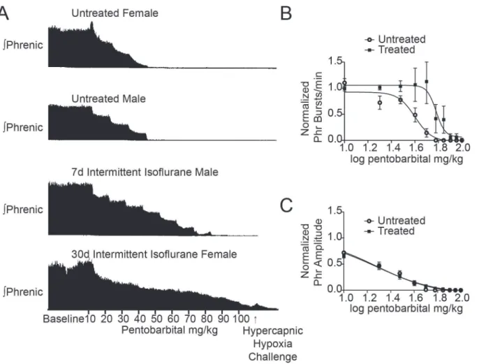
A dose response analysis to pentobarbital was performed using untreated (n = 6 male and 6 female) and treated (n = 4 male and 8 female) rats (Fig. 1). For untreated control male and fe-male rats, phrenic burst frequency and amplitude decreased during sequential pentobarbital injections (top two traces inFig. 1A) with all control rats not producing phrenic bursts by the sixth injection (60 mg/kg administered). The Hill slope of the burst frequency IC50 curve was−6.0 ± 1.7 (Fig. 1B), and the IC50 was 39.9 mg/kg (95% CI 36.0 to 44.2 mg/kg).
Following isoflurane treatment, rats continued to produce measurable phrenic motor bursts under higher doses of pentobarbital than control animals. One animal continued to produce phrenic output at the maximal dose (100 mg/kg; lowest trace inFig. 1A). The mean dose to si-lence phrenic output in isoflurane-treated rats was 53 ± 7 mg/kg versus 38 ± 2 mg/kg in un-treated control rats (p<0.05). The Hill slope increased to−11.4 ± 9.1 (Fig. 1B) and the burst
frequency IC50 significantly increased to 60.3 mg/kg (95% CI 51.9 to 70.0 mg/kg; p<0.01). To
normalize the lethal dose of pentobarbital across rats, individual approximate Hill slopes were calculated for burst frequency and averaged for treated and untreated groups. This revealed that the slope in isoflurane-treated rats (−218.4 ± 71.1) was significantly steeper than in un-treated control rats (−57.6 ± 30.4, p<0.05).
parameters defining the best-fit models for the phrenic nerve amplitude dose response curve did not differ by condition (p = 0.35;Fig. 1C), thus one IC50 (19.1 mg/kg; 95% CI 17.3 to 21.1 mg/kg) and one Hill slope (−1.68 ± 0.15) described the entire amplitude dataset. These data indicate that following isoflurane treatment, pentobarbital was still inhibitory on neurons contributing to phrenic nerve amplitude but not burst frequency.
To assess whether respiratory networks were truly silenced by 100 mg/kg pentobarbital, we sought to provide maximal stimulus for breathing to prompt the system to surmount barbitu-rate-induced depression. There was notable variability in the dose of pentobarbital required to silence the phrenic nerve. While60 mg/kg of pentobarbital was sufficient to depress phrenic nerve activity in all untreated control rats, not all isoflurane-treated rats were depressed by the maximal dose of 100 mg/kg. To assess whether the degree of inhibition differed between groups, we challenged rats with a strong stimulus (hypercapnic-hypoxia challenge) at the end of each experiment (Fig. 1A). Six of 12 isoflurane-treated rats mounted a detectable phrenic re-sponse to the challenge, while only two of 12 untreated control rats responded (p = 0.08). Fig 1. Phrenic nerve motor burst resistance to pentobarbital injections in isoflurane-treated rats.A, Representative phrenic neurograms from an untreated female rat, untreated male rat, 7d isoflurane-treated male rat, and 30d isoflurane-treated female rat. After establishing baseline conditions, 10 mg/kg of pentobarbital was administered every 5 min up to a maximum total dose of 100 mg/kg. The protocol ended with 5 min of hypercapnic-hypoxia (PET CO2= 80 mmHg, 13% inspired O2) to assess phrenic nerve response to maximal chemosensory input under 100 mg/kg of pentobarbital. The upper two
panels show pentobarbital-mediated inhibition of phrenic nerve bursts in untreated female and male rats. The bottom two tracings demonstrate significant resistance of phrenic nerve bursts to pentobarbital in a 7d isoflurane-treated male rat and a 30d isoflurane-treated female rat.C, Pentobarbital IC50 curve of phrenic nerve burst frequency demonstrates a significant rightward shift after isoflurane treatment compared untreated rats.D, Pentobarbital IC50 curve of phrenic nerve amplitude demonstrates no change following isoflurane treatment.
Pentobarbital Sensitivity of Spontaneous Neuronal Activity in Medulla
and Cortex
During sequential pentobarbital injections, respiratory phrenic burst frequency was sustained, thereby suggesting that respiratory rhythm-generating neurons in medulla express pentobarbi-tal-insensitive GABAARs. In addition, it is not clear whether isoflurane exposures increase the
expression of pentobarbital-insensitive GABAARs in other parts of the CNS. To answer these
questions, pentobarbital sensitivity was examined in the neural regions responsible for rhythm generation (VRC, preBötC) as well as in cortical neurons, which exhibit a GABAAR mediated
tonic current [38]. Four groups of rats were tested: 1) untreated male and female rats, 2) 7d iso-flurane-treated male and female rats, 3) 30d isoiso-flurane-treated male and female rats, and 4) male and female rats that were allowed 7d of recovery following 7d isoflurane treatment (data from the untreated groups are reported in [20]).
Consistent with prior data [39,40,20], in slices from untreated control male (n = 6) and fe-male rats (n = 6), 60 min exposure to bath-applied 300μM pentobarbital inhibited
spontane-ous activity in VRC neurons to 44 ± 15% of baseline (n = 103 neurons, p<0.05) and cortical
(CTX) neurons to 24 ± 8% of baseline (n = 84 neurons, p<0.05) compared to time controls
(n = 5 animals, 56 VRC neurons and 48 CTX neurons, 136 ± 19% and 123 ± 19% of baseline, respectively) (Fig. 2A, B). There were no significant differences between untreated male and female animals.
The 7d intermittent isoflurane treatment (n = 12 rats) reduced the inhibitory effects of 1h pentobarbital on VRC (n = 102 neurons, 91 ± 21% of baseline, p<0.05) and CTX neurons (n =
84 neurons, 49 ± 10% of baseline, p<0.005) compared to untreated control rats (Fig. 2C, D).
There were no significant differences between male and female animals. The effects of 7d iso-flurane treatment were reversible in the VRC, as 7d recovery significantly shifted the effect of 1h pentobarbital towards the response of untreated control rats (VRC, n = 27 neurons, 44 ± 14% of baseline, p<0.01) (Fig. 2E). There was no significant difference between spontaneous
activity after pentobarbital application in CTX neurons from 7d isoflurane-treated rats and 7d recovery rats.
The 30d of intermittent isoflurane treatment rendered VRC neurons insensitive to pento-barbital application (n = 163 neurons, 104% of baseline, p>0.05 compared to time control)
(Fig. 2F, G). The 30d intermittent isoflurane treatment differentially affected CTX neurons in males and females (p<0.05). In males, CTX neurons maintained 83 ± 18% of baseline activity
(n = 33 neurons) (Fig. 2F) while in females, CTX neurons maintained 48 ± 9% of baseline activ-ity (Fig. 2G). In both males and females, CTX neurons were significantly less inhibited by pen-tobarbital after 30d of intermittent isoflurane than untreated animals (female, p<0.05; male,
p<0.005).
The persistence of spontaneous neuronal activity following pentobarbital application to the CTX and VRC of 30d treated animals could reflect either a lack of GABAARs on neurons, or
the presence of pentobarbital-insensitive GABAARs. To address this question, 20μM muscimol
(GABAAR agonist) was bath-applied to from slices from isoflurane-treated rats after 1h of 300 μM pentobarbital (Fig. 2F, G). After 1h of exposure to 300μM pentobarbital, CTX and VRC
neurons from isoflurane-treated rats were rapidly (<5 min) inhibited by muscimol, confirming
the presence of functional, pentobarbital-insensitive GABAARs. The response of CTX and
VRC neurons to muscimol did not differ between isoflurane-treated rats and time control rats (p>0.05), indicating the presence of pentobarbital-insensitive GABAARs in CTX and VRC
after 30d of isoflurane.
Fig 2. Spontaneous neuronal activity resistance to bath-applied pentobarbital in medullary and cortical slices from 7d isoflurane-treated rats.Spontaneous activity of neurons in the region anatomically consistent with the VRC and PreBötC (all labeled as VRC neurons) are insensitive to bath-applied
non-rats; 2 male, 2 female) prior to co-application of 100μM bicuculline and 300μM pentobarbital
prevented the inhibition of spontaneous activity of both CTX and VRC neurons by pentobarbi-tal observed in virgin female and male slices (data not shown).
A summary of the steady-state pentobarbital responses in VRC and CTX neurons in slices from male and female rats shows that isoflurane exposures increased pentobarbital resistance after 7d and 30d (Fig. 2H, 2I). The response in female rats was greater compared to male rats. Together, these data suggest that intermittent isoflurane treatment is sufficient to increase the expression of a pentobarbital-insensitive GABAAR subtype in VRC and CTX neurons.
Immunohistochemical Localization of
ε
Subunit Expression in Medulla
and Cortex
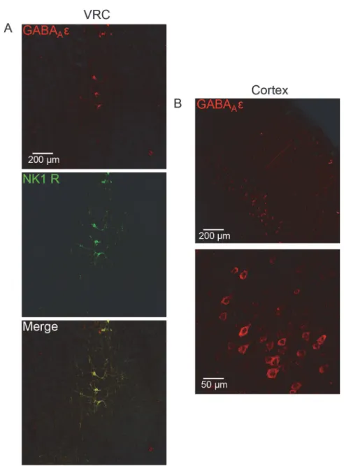
To confirm the presence or absence of GABAARεsubunit expression at thein vitrorecording
sites, medullary slices from recording experiments were treated with an antibody against the GABAARεsubunit. Medullary sections were co-labeled for GABAARεsubunit and for NK1-R
(Substance P receptor) that is used as a marker for putative respiratory neurons in the VRC and preBötC [41,42]. NK1-R and the GABAARεsubunit exhibited>90% colocalization, both
in the nucleus ambiguus (subcompact and compact; Lin et al., 2008), which was used as a land-mark for electrode placement, and in the VRC (Fig. 3A; [36]). Neurons in the VRC displayed enriched somaticεsubunit staining as well as in primary neuronal processes (Fig. 3A). The
pentobarbital-insensitive GABAARs identifiedin vitrowere functionally consistent with
immu-nohistochemically identifiedεsubunit-containing GABA
ARs in the VRC. To our surprise, the
GABAARεsubunit was detectable in CTX neurons in all groups as well. Layer 5 pyramidal
cells demonstrated the most robust expression, and staining appeared to be largely restricted to the soma (Fig. 3B).
ε
Subunit mRNA Expression in Medulla and Cortex
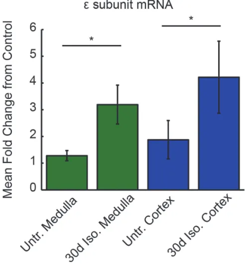
To quantify whether intermittent isoflurane treatment altered the transcriptional regulation of the GABAARεsubunit, cortical and medullary samples from 30d isoflurane-treated rats (n =
5) and untreated control rats (n = 12) were harvested for GABAARεsubunit mRNA analysis
(Fig. 4). The 30d isoflurane treatment increased GABAARεsubunit mRNA in medullary
sam-ples compared to untreated samsam-ples (3.9 ± 1.39-fold increase, p<0.05). Cortical samples from
30d isoflurane-treated rats also demonstrated increased GABAARεsubunit mRNA levels
com-pared to controls (4.2 ± 1.35-fold increase, p<0.05, Wilcoxon Rank Sum). These data
demon-strate a correlation between the emergence of barbiturate-insensitive GABAARs in VRC and
cortex and the transcriptional upregulation of GABAARεsubunit mRNA.
significantly shifted in the same direction.D, Likewise, in female rats treated with isoflurane for 7d,
pentobarbital failed to inhibit spontaneous neuronal activity in the VRC neurons (n = 40). CTX neurons (n = 51) trended non-significantly towards an increase in activity compared to untreated control female rats.E, This effect was reversible, as 7d of recovery following 7d of intermittent isoflurane shifted spontaneous activity of VRC (n = 27) and CTX neurons (n = 38) in rats toward baseline values.F., 30d of intermittent
isoflurane significantly attenuated the pentobarbital response in VRC (n = 40) and CTX neurons (n = 32) in male rats. To confirm the presence of pentobarbital-insensitive GABAARs, muscimol (20μM) was applied
at 90 min (dashed vertical line). Nearly all neurons that were resistant to pentobarbital were inhibited by muscimol.G, In female rats exposed to 30d isoflurane treatment, the response of VRC (n = 123) and
CTX (n = 83) neurons to pentobarbital was significantly attenuated. These neurons were also silenced by muscimol (dashed vertical line). (H,I)The average steady-state responses to bath-applied pentobarbital are quantified for the different isoflurane treatments and recovery for male rats (C) and female rats (D). The asterisk indicates p<0.05 for comparison to control while the pound sign indicates p<0.05 for comparison to
7d treatment.
Discussion
Multiple forms of neuronal plasticity must be carefully coordinated to produce a stable and functional nervous system. The mechanisms that are actively expressed are regulated both de-velopmentally and spatially, and depend upon external and internal environments. This study shows that repeated exposure to brief episodes of an inhaled anesthetic is sufficient to function-ally reorganize medullary and cortical neuronal circuitry. Following 7 days of 5 min/day expo-sure to isoflurane, bothin vivoandin vitromeasures of respiratory neuronal output revealed a Fig 3. GABAARεsubunit protein expression in medulla and cortex of isoflurane-treated rats.A,
Medullary slices used forin vitroelectrophysiology were immunohistochemically labeled for the GABAARε
subunit (red, top) and NK1-R (green, middle). Images were acquired in the location of electrode placement. The GABAARεand NK1-R exhibited>90% colocalization on neurons in the preBötC of the medulla (merge,
bottom). Example image was acquired in the preBötC of a virgin female rat.B, Cortical slices previously used forin vitroelectrophysiology were sectioned and immunohistochemically labeled for the GABAARεsubunit.
Example image was acquired in the cortex of a 30d isoflurane-treated male rat.
significantly increased barbiturate tolerance. After 30 days of treatment, cortical networks were similarly affected.
This study initially sought to test whether non-pregnant animals could be induced to in-creaseεsubunit expression and barbiturate tolerance in VRC neurons. Our findings make it
clear that gestational hormones are unnecessary for this effect, as both virgin female and male rats have the capacity to express this form of plasticity. We cannot rule out the effects of neuro-steroids, as there is evidence that GABAergic modulators may increase neurosteroid levels in in vitrohippocampal preparations [43]. Surprisingly, our data also reveal that this phenome-non is not restricted to medullary neurons, as cortical neurons displayed a similar pharmaco-logical profile following longer treatment periods. Whether this is a global feature of the cortex is unclear, but these data suggest that the incorporation of pharmacologically distinct GABAAR
configurations is a general mechanism of neuronal plasticity.
If this plasticity is not gated by the interaction of hormones and nuclear receptors (one of our initial hypotheses), how might this process be triggered in neurons? Long duration (min-utes to hours) application of GABA to cultured neurons leads to an increase in receptor Fig 4. Increased GABAARεsubunit mRNA levels in medulla and cortex in 30d isoflurane-treated rats.
Medullary GABAARεmRNA levels were more than 3-fold greater in 30d isoflurane-treated rats compared to
untreated control rats. Likewise, cortical GABAARεmRNA levels were more than 4-fold increased in 30d
isoflurane-treated rats. Statistics were conducted on delta CT values. Asterisk denotes p<0.05. Error bars
indicate SEM.
internalization, an uncoupling of the allosteric modulatory site from the GABA binding site, and a decrease in the expression of specific subunits [33]. The mechanism linking increased al-losteric modulatory pressure and the regulation of theεsubunit is unclear. This will be a
criti-cal question to answer in future studies.
The demonstration that the frequency of phrenic nerve outputin vivois highly resistant to sequential pentobarbital injections in isoflurane-treated rats strongly suggests that rhythm-generating neurons in the brainstem increaseεsubunit expression in GABAARs. Since it is
hy-pothesized that inspiratory activity produced mainly by preBötC neurons in the VRC [44,34,
35], pentobarbital sensitivity of spontaneous neuronal activity was tested in medullary slices from control and isoflurane-treated rats. None of the recorded neurons in the medullary slice experiments could be defined as being respiratory-related because adult medullary slices do not produce spontaneous respiratory rhythmic motor activity similar to that produced in neo-natal rodent medullary slice [44]. However, it is likely most of the recorded neurons were respi-ratory-related preBötC neurons because the preBötC region has distinctive surrounding neural landmarks that define the region and are easily visualized in medullary slicesin vitro. Further-more,εsubunit expression was increased in NK1-R-positive neurons within the preBötC
re-gion in isoflurane-treated rats. Since the NK1-R is one of several markers for preBötC neurons [45], this finding is consistent with the hypothesis thatεsubunit expression increased in
pre-BötC neurons to compensate for the isoflurane-dependent decrease in respiratory motor activi-ty. This study does not address the question as to whether intermittent isoflurane exposures alteredεsubunit expression in other respiratory-related brainstem regions controlling
expira-tion, postinspiraexpira-tion, or central chemosensation. Furthermore, modulatory neurons (e.g., sero-tonergic, noradrenergic, dopaminergic) projecting to the preBötC may also have increased expression ofεsubunits in GABA
ARs, and thereby contributed to the altered the
responsive-ness of the respiratory control network to pentobarbital.
Thein vivoand slice physiology data presented here provide measures of network activity as opposed to cell autonomous responses. Our data demonstrate the presence of pentobarbital insensitive GABAARs in the cortex and the brainstem, but it is difficult to predict specific
expression patterns from these experiments. For example, the transient increases in cortical neuronal activity of 30d isoflurane treated male rats (Fig. 2F) likely represent pentobarbital-mediated inhibition of a subclass of inhibitory interneuron, resulting in disinhibition of excit-atory cells. Subunit distribution rules will be important to understand in the future.
εsubunit-containing GABAARs are insensitive to most allosteric positive modulators [18,
19]; both the human and rat isoform of theεsubunit confers anesthetic insensitivity to neurons
[46], and GABAARs containing theεsubunit are upregulated on VRC neurons during
preg-nancy when pentobarbital sensitivity decreases [20]. Our data fall short of establishing a mech-anistic link between the induced expression ofεsubunit-containing GABAARs and barbiturate
insensitivity. We do, however, demonstrate the conditional presence of fully functional, barbi-turate-insensitive GABAARs in brain regions that are immunohistologically enriched with the
εsubunit and show increasedεsubunit mRNA transcription following treatment.
Further-more, these receptors are rapidly inhibited by muscimol, and the inhibitory actions of pento-barbital are blocked by bicuculline (indicating the involvement of GABAARs). These data are
difficult to understand without the specific pharmacological properties imbued by theε
sub-unit, thus this is the most likely explanation for our findings. However, because the connection between theεsubunit and the respiratory plasticity described here is correlative, it will take
fur-ther investigation to unequivocally confirm a molecular mechanism.
neuronal input/output ratio is necessary for survival, and gain modulation of respiratory rhythm generating neurons is provided by a tonic GABAAR current [49]. Neurosteroids, like
barbiturates, act on these currents and suppress activity in medullary slices from naïve animals [50]. Thus, these data suggest a compensatory mechanism of gain modulation that counteracts the hyperpolarizing properties of chronic exposure to allosteric positive GABAAR modulators,
such as neurosteroids, ethanol, and in some cases, anesthetics. Without a compensatory re-sponse to allosteric modulators, respiratory pathologies would arise as a result of pregnancy, regular alcohol consumption, or multiple exposures to anesthetics. To our knowledge, these are the first data to demonstrate reorganization of the neural control of respiration by repeated anesthetic exposure. Prior data demonstrate the ability of anesthetics to alter GABAAR
expres-sion patterns. Sekine et al. (2006) described changes in forebrain mRNA for the GABAARα4
subunit during anesthesia with propofol or isoflurane [51]. Similar to our findings, chronic eth-anol exposure is associated with a cross-tolerance to many GABAergic anesthetics [26,52,53]. It is likely that cross-tolerance is a feature of tonic GABAAR plasticity, as we have previously
described VRC insensitivity to both ETOH and barbiturates in hibernating ground squirrels [40].
Significance
This study presents a novel, clinically relevant, inducible model of neuroplasticity that was clearly demonstrated in an intact animal with measurable, functional spinal respiratory motor output. We provide evidence that this isoflurane-induced neuroplasticity likely involves the ex-pression of a relatively unknown, yet highly conserved, GABAAR subunit that confers
insensi-tivity to allosteric modulation by neurosteroids, anesthetics, ethanol, barbiturates, and benzodiazepines [18,19,39,40,20]. Here we demonstrate that this plasticity is restricted nei-ther to specific classes of animals (i.e. pregnant or hibernating), nor respiratory neurons. These data raise the possibility that this type of respiratory neuroplasticity, which mirrors some of the changes associated with hibernation and pregnancy, is inducible and may be harnessed to treat pathological conditions.
Many long-termin vivopreparations in basic neurobiology research rely on regular admin-istration of isoflurane and other GABAergic anesthetics in order to prepare an animal for data collection. The findings presented here suggest that these protocols may be inadvertently ma-nipulating the very networks being studied. The impact of these methods needs further study.
Author Contributions
Conceived and designed the experiments: KBH SMJ GSM MB. Performed the experiments: KBH NRN KMS SMS. Analyzed the data: KBH NRN SMS. Contributed reagents/materials/ analysis tools: JJW SMJ MB GSM. Wrote the paper: KBH.
References
1. Rudolph U, Möhler H. GABA-based therapeutic approaches: GABAAreceptor subtype functions. Curr
Opin Pharmacol. 2006; 6(1):18–23. PMID:16376150
2. Stell BM, Brickley SG, Tang CY, Farrant M, Mody I. Neuroactive steroids reduce neuronal excitability by selectively enhancing tonic inhibition mediated by delta subunit containing GABAA receptors. Proc Natl Acad Sci. 2003; 100(24):14439–14444. PMID:14623958
3. Belelli D, Lambert JJ. Neurosteroids: endogenous regulators of the GABA(A) receptor. Nat Rev Neu-rosci. 2005; 6(7):565–575. PMID:15959466
5. Jia F, Yue M, Chandra D, Homanics GE, Goldstein PA, Harrison NL. Isoflurane is a potent modulator of extrasynaptic GABA(A) receptors in the thalamus. J Pharmacol Exp Ther. 2008; 323(3):1127–1135. 6. Orser BA. Extrasynaptic GABAA receptors are critical targets for sedative-hypnotic drugs. J Clin Sleep
Med. 2006; 2(2):S12–S18. PMID:17557502
7. Feng HJ, Macdonald RL. Barbiturates require the N terminus and first transmembrane domain of the delta subunit for enhancement of alpha1beta3delta GABAA receptor currents. J Biol Chem 2010; 285 (31):23614–21. doi:10.1074/jbc.M110.122564PMID:20525684
8. Bäckström T, Andersson A, Andreé L, Birzniece V, Bixo M, Björn I, et al. Pathogenesis in menstrual cycle-lined CNS disorders. Ann NY Acad Sci. 2003; 1007:42–53. PMID:14993039
9. Herd MB, Belelli D, Lambert JJ. Neurosteroid modulation of synaptic and extrasynaptic GABAA
recep-tors. Pharmacol Ther. 2007; 116:20–34. PMID:17531325
10. Maguire J, Mody I. Steroid hormone fluctutations and GABA(A)R plasticity. Psychoneuroendocrinology 2009; 34 Suppl 1:S84–90. doi:10.1016/j.psyneuen.2009.06.019PMID:19632051
11. Maguire J, Mody I. Neurosteroid synthesis-mediated regulation of GABA(A) receptors: relevance to ovarian cycle and stress. J Neurosci. 2007; 27(9):2155–2162. PMID:17329412
12. Marutha Ravindran CR, Mehta AK, Ticku MK. Effect of chronic administration of ethanol on the regula-tion of the delta-subunit of GABA(A) receptors in the rat brain. Brain Res. 2007; 1174:47–52. PMID: 17854781
13. Mackenzie G, Maguire J. The role of ovarian hormone-derived neurosteroids on the regulation of GABAA receptors in affective disorders. Psychopharmacology (Berl), 2014; 231(17):3333–42. doi:10. 1007/s00213-013-3423-zPMID:24402140
14. Maguire J, Mody I. GABAAR plasticity during pregnancy: Relevance to postpartum depression. Neuron.
2008; 59(2):207–213. doi:10.1016/j.neuron.2008.06.019PMID:18667149
15. Maguire J, Ferando I, Simonsen C, Mody I. Excitability changes related to GABAA receptor plasticity during pregnancy. J Neurosci. 2009; 29(30):9592–9601. doi:10.1523/JNEUROSCI.2162-09.2009 PMID:19641122
16. Sanna E, Mostallino MC, Murru L, Carta M, Talani G, Zucca S, et al. Changes in expression and func-tion of extrasynaptic GABAA receptors in the rat hippocampus during pregnancy and after delivery. J Neurosci. 2009; 29(6):1755–65. doi:10.1523/JNEUROSCI.3684-08.2009PMID:19211882
17. Davies PA, Hanna MC, Hales TG, Kirkness EF. Insensitivity to anaesthetic agents conferred by a class of GABAAreceptor subunit. Nature. 1997; 385:820–823. PMID:9039914
18. Irnaten M, Walwyn WM, Wang J, Venkatesan P, Evans C, Chang KS, et al. Pentobarbital enhances GABAergic neurotransmission to cardiac parasympathetic neurons, which is prevented by expression of GABA(A) epsilon subunit. Anesthesiology 2002; 97: 717–724. PMID:12218540
19. Wagner DA, Goldschen-Ohm MP, Hales TG, Jones MV. Kinetics and spontaneous open probability conferred by the epsilon subunit of the GABAA receptor. J Neurosci. 2005; 25(45):10462–10468. PMID:16280584
20. Hengen KB, Nelson NR, Stang KM, Johnson SM, Crader SM, Watters JJ, et al. Increased GABAA
Re-ceptorεsubunit Expression on Ventral Respiratory Column Neurons Protects Breathing during Preg-nancy. PLoSONE 2012; 7(1): e30608. doi:10.1371/journal.pone.0030608PMID:22303446
21. Lesch A, Rubil S, Thiel G. Activation and inhibition of transient receptor potential TRPM3-induced gene transcription. Br J Pharmacol. 2014; 171(10):2645–58. PMID:24895737
22. Abramian AM, Comenencia-Ortiz E, Modgil A, Vien TN, Nakamura Y, Moore YE, et al. Neurosteroids promote phosphorylation and membrane insertion of extrasynaptic GABAA receptors. Proc Natl Acad Sci. USA 2014; 111(19):7132–7. doi:10.1073/pnas.1403285111PMID:24778259
23. Lambo ME, Turrigiano GG. Synaptic and intrinsic homeostatic mechanisms cooperate to increase L2/3 pyramidal neuron excitability during a late phase of critical period plasticity. J Neurosci. 2013; 33 (20):8810–9. doi:10.1523/JNEUROSCI.4502-12.2013PMID:23678123
24. Hengen KB, Lambo ME, Van Hooser SD, Katz DB, Turrigiano GG. Firing rate homeostasis in visual cortex of freely behaving rodents. Neuron 2013; 80(2):335–42. doi:10.1016/j.neuron.2013.08.038 PMID:24139038
25. Chen X, Shu S, Schwartz LC, Sun C, Kapur J, Bayliss DA. Homeostatic regulation of synaptic excitabil-ity: tonic GABA(A) receptor currents replace I(h) in cortical pyramidal neurons of HCN1 knock-out mice. J Neurosci. 2010; 30(7):2611–2622. doi:10.1523/JNEUROSCI.3771-09.2010PMID:20164346 26. Liang J, Spigelman I, Olsen RW. Tolerance to sedative/hypnotic actions of GABAergic drugs correlates
27. Wang X. Propofol and isoflurane of tonic gamma-aminobutryic acid type a current in cardiac vagal neu-rons in the nucleus ambiguus. Anesth Analg. 2009; 108(1):142–8. doi:10.1213/ane.
0b013e31818d8b79PMID:19095842
28. Grasshoff C, Antkowiak B. Effects of isoflurane and enflurane on GABAA and glycine receptors contrib-ute equally to depressant actions on spinal ventral horn neurones in rats. Br J Anaesth. 2006; 97 (5):687–94. PMID:16973644
29. Wang X, Gorini C, Sharp D, Bateman R, Mendelowitz D. Anaesthetics differentially modulate the trige-minocardiac reflex excitatory synaptic pathway in the brainstem. J Physiol. 2011; 589(Pt 22):5431–42. 30. West AE, Chen WG, Dalva MB, Dolmetsch RE, Kornhauser JM, Shaywitz AJ, et al. Calcium regulation of neuronal gene expression. Proc Natl Acad Sci USA. 2001; 98(20):11024–11031. PMID:11572963 31. Pintchovski SA, Peebles CL, Kim HJ, Verdin E, Finkbeiner S. The serum response factor and a putative
novel transcription factor regulate expression of the immediate-early gene Arc/Arg3.1 in neurons. J Neurosci. 2009; 29(5):1525–1537. doi:10.1523/JNEUROSCI.5575-08.2009PMID:19193899 32. Gao M, Sossa K, Song L, Errington L, Cummings L, Hwang H, et al. A specific requirement of Arc/
Arg3.1 for visual experience-induced homeostatic synaptic plasticity in mouse primary visual cortex. J Neurosci. 2010; 30(21):7168–7178. doi:10.1523/JNEUROSCI.1067-10.2010PMID:20505084 33. Gutiérrez ML, Ferreri MC, Gravielle MC. GABA-induced uncoupling of GABA/benzodiazepine site
inter-actions is mediated by increased GABAA receptor internalization and associated with a change in sub-unit composition. Neuroscience, 2014; 257:119–29. doi:10.1016/j.neuroscience.2013.10.077PMID: 24215979
34. Feldman JL, Del Negro CA. Looking for inspiration: new perspectives on respiratory rhythm. Nat Rev Neurosci. 2006; 7:232–242. PMID:16495944
35. Feldman JL, Del Negro CA, Gray PA. Understanding the rhythm of breathing: so near, yet so far. Annu Rev Physiol. 2013; 75:423–452. doi:10.1146/annurev-physiol-040510-130049PMID:23121137 36. Paxinos G, Watson C. The rat brain in stereotaxic coordinates. Academic Press. 2004; San Diego,
CA.
37. Livak KJ, Schmittgen TD. Analysis of relative gene expression data using real-time quantitative PCR and the 2(-Delta Delta C(T)) method. Methods, 2001; 4:402–8.
38. Sebe JY, Looke-Stewart EC, Estrada RC, Baraban S. Robust tonic GABA current can inhibit cell firing in mouse newborn neocortical pyramidal cells. Eur J Neurosci. 2010; 32(8):1310–1318. doi:10.1111/j. 1460-9568.2010.07373.xPMID:20846324
39. Hengen KB, Behan M, Carey HV, Jones MV, Johnson SM. Hibernation induced pentobarbital insensi-tivity in medulla but not cortex. Am J Physiol Regul Integr Comp Physiol. 2009; 297:1028–1036. doi: 10.1152/ajpregu.00239.2009PMID:19675281
40. Hengen KB, Gomez TM, Stang KM, Johnson SM, Behan M. Changes in ventral respiratory column GABAARεandδsubunits during hibernation mediate resistance to depression by ETOH and pentobar-bital. Am J Physiol Regul Integr Comp Physiol. 2011;doi:10.1152/ajp regu.0067.2010
41. Wang H, Stornetta RL, Rosin DL, Guyenet PG. Neurokinin-1 receptor-immunoreactive neurons of the ventral respiratory group in the rat. J Comp Neurol. 2001; 434(2):128–146. PMID:11331521
42. Bouvier J, Thoby-Brisson M, Renier N, Dubreuil V, Ericson J, Champagnat J, et al. Hindbrain interneu-rons and axon guidance signaling critical for breathing. Nat Neurosci. 2010; 13(9):1066–1074. doi:10. 1038/nn.2622PMID:20680010
43. Tokuda K, Izumi Y, Zorumski CF. Ethanol enhances neurosteroidogenesis in hippocampal pyramidal neurons by paradoxical NMDA receptor activation. J Neurosci. 2011; 31(27):9905–9. doi:10.1523/ JNEUROSCI.1660-11.2011PMID:21734282
44. Smith JC, Ellenberger HH, Ballyani K, Richter DW, Feldman JL. PBötzinger complex: a brainstem re-gion that may generate respiratory rhythm in mammals. Science. 1991; 254:726–729. PMID:1683005 45. Gray PA, Rekling JC, Bocchiaro CM, Feldman JL. Modulation of respiratory frequency by peptidergic
input to rhythmogenic neurons in the preBötzinger complex. Science. 1999; 286:1566–1568. PMID: 10567264
46. Sergeeva OA, Andreeva N, Garret M, Scherer A, Haas HL. Pharmacological properties of GABAA re-ceptors in rat hypothalamic neurons expressing the epsilon-subunit. J Neurosci. 2005; 25(1):88–95. PMID:15634770
47. Maffei A, Turrigiano G. Multiple modes of network homeostasis in visual cortical layer 2/3. J Neurosci. 2008; 28(17):4377–4384. doi:10.1523/JNEUROSCI.5298-07.2008PMID:18434516
49. Zuperku EJ, McCrimmon DR. Gain modulation of respiratory neurons. Respir Physiol and Neurobiol. 2002; 131:121–133. PMID:12107000
50. Ren J, Greer JJ. Neurosteroid modulation of respiratory rhythm in rats during the perinatal period. J Physiol. 2006; 574(Pt 2):535–546.
51. Sekine S, Matsumoto S, Issiki A, Kitamura T, Yamada J, Watanabe Y. Changes in expression of GABAA alpha4 subunit mRNA in the brain under anesthesia induced by volatile and intravenous anes-thetics. Neurochem Res. 2006; 31(3):439–448. PMID:16733821
52. Cagetti E, Liang J, Spigelman I, Olsen RW. Withdrawal from chronic intermittent ethanol treatment changes subunit composition, reduces synaptic function, and decreases behavioral responses to posi-tive allosteric modulators of GABAA receptors. Mol Pharmacol 2003; 63: 53–64. PMID:12488536 53. Kang MH, Spigelman I, Olsen RW. Alteration in the sensitivity of GABAA receptors to allosteric