Male-Specific Alleviation of Iron-Induced
Striatal Injury by Inhibition of Autophagy
Li-Fang Wang1, Kazunari K. Yokoyama2, Tzu-Yin Chen3, Hsiu-Wen Hsiao3, Pei-Chi Pei-Chiang3, Ya-Ching Hsieh4, Steven Lo4,5, Chin Hsu3*
1Department of Medicinal and Applied Chemistry, College of Life Science, Kaohsiung Medical University, Kaohsiung, Taiwan,2Graduate Institute of Medicine, Kaohsiung Medical University, Kaohsiung, Taiwan,
3Department of Physiology, Faculty of Medicine, College of Medicine; Kaohsiung Medical University, Kaohsiung, Taiwan,4Department of Medical Research, E-Da Hospital, I-Shou University, Kaohsiung, Taiwan,5Canniesburn Plastic Surgery Unit, Royal Infirmary, Glasgow, United States of America
*chinhsu@kmu.edu.tw
Abstract
Men exhibit a worse survival rate than premenopausal women after intracerebral hemor-rhage (ICH), however, no sex-specific management has been concerned. In a rat model involving infusion of ferrous citrate (FC) that simulates iron accumulation after hemorrhage, a higher degree of autophagy associated with higher injury severity was observed in stria-tum of males than in females. Since the imbalance between the levels of autophagy and energy demand may lead to cell death, we proposed that FC-induced autophagy is detri-mental in a male specific manner and autophagy modulation affects injury severity in a sex-dependent manner. Rapamycin, an autophagy inducer, and conditional knockout gene of autophagy-related protein 7 (Atg7) in dopamine receptor D2 (DRD2) neurons were used to test our hypothesis using a mouse model with striatal FC infusion. The result showed that the levels of autophagic cell death and injury severity were higher in male than in female mice. Pre-treatment of FC-infused females with rapamycin increased the FC-induced behavioral deficit and DRD2 neuron death. However, DRD2 neuron-specific knockout of Atg7decreased FC-induced injury severity and the number of TUNEL(+) DRD2 neurons in males. These results suggest that autophagy in FC-infusion males is overactive with mal-adaptive consequences and inhibition of autophagy decreases the severity of FC-induced striatal injury in males. These findings present prospects for male-specific therapeutic strat-egy that targets autophagy in patients suffering from iron overload.
Introduction
Primary intracerebral hemorrhage (ICH) results in the highest mortality of all stroke subtypes and less than 40% of ICH survivors regain their independence [1]. The incidence of ICH is higher among man, and their survival rate is lower compared with that of premenopausal women [2,3]. While current treatments for stroke are primarily limited to supportive manage-ment [4,5]. Previous reports indicated that men exhibit worse free radical homeostasis and OPEN ACCESS
Citation:Wang L-F, Yokoyama KK, Chen T-Y, Hsiao H-W, Chiang P-C, Hsieh Y-C, et al. (2015) Male-Specific Alleviation of Iron-Induced Striatal Injury by Inhibition of Autophagy. PLoS ONE 10(7): e0131224. doi:10.1371/journal.pone.0131224
Editor:Richard Keep, University of Michigan, UNITED STATES
Received:January 6, 2015
Accepted:May 30, 2015
Published:July 6, 2015
Copyright:© 2015 Wang et al. This is an open access article distributed under the terms of the
Creative Commons Attribution License, which permits unrestricted use, distribution, and reproduction in any medium, provided the original author and source are credited.
Data Availability Statement:All relevant data are within the paper.
Funding:CH received research grants from the National Science Council of Taiwan (101-2629-B-037-001, 102-2320-B-037-015, and 103-2320-B-037-028). LW received research grants from“Aim for the Top Journals Grant,”grant No. KMU-DT103007 from Kaohsiung Medical University. The funders had no role in study design, data collection and analysis, decision to publish, or preparation of the manuscript.
weaker defense capacities against oxidative brain damage than women [6]. Our previous study also revealed that the levels of neuroprotective thioredoxin in the caudate nucleus of male rats were less than those in females after FC infusion and thioredoxin mediates the neuroprotective effect of estradiol in females but not in males [7]. Recent reports indicates that sex specific dif-ferences exist in both constitutive autophagy [8] and autophagy in the brain following hyp-oxia-ischemia [9]. These results imply a sexual dimorphism in the mechanism by which neurons respond to stress or to neuroprotectant. However, no sex-specific therapies have been considered for patients suffering from neurodegeneration following hemorrhage or iron intoxication.
Following ICH, accumulation of ferrous iron causes oxidative damage and contributes to the long-term neurological deficits [10,11] and striatum is the area where ICH common occurred [12]. Oxidative stress and organelle damage induce autophagy that acts as an essential recycling mechanism for survival [13]. However, over-activation of autophagy has been recog-nized in conditions of severe oxidative stress following hypoxic/ischemic brain injury [14]. Pre-vious results showed that striatal infusion of FC, which simulates iron accumulation after ICH, causes more autophagy and greater injury severity in male rats than in females [15]. Whether autophagy plays a sex-specific detrimental role in striatal injury caused by iron overload is unknown. In the present study, we infused FC into striatum of mice to mimic ferrous iron accumulation after ICH and used conditional knockout ofAtg7to address the issue whether autophagy inhibition is beneficial for patients suffering from neurodegeneration caused by iron overload in a male-specific manner.
Materials and Methods
Intact and castrated mice
A total of 132 age-matched (12-week-old) [16] male and female C57BL/6 mice (National Labo-ratory Animal Center, Taipei, Taiwan) were used to examine the sex difference in FC-induced autophagy and the effect of rapamycin on FC-induced striatal injury. No mice were excluded. Mice implanted with estradiol (E2) after castration were used to study the contribution of E2
to the sex difference in FC-induced autophagy and injury in striatum. For the female mice, ovariectomy involved a small incision in both flanks after which the ovaries were removed. In the male mice, the skin and body wall caudal to the penis were incised with a scissor, and the testicles were retracted through the incision. The exposed testicular artery and vein were dou-ble-ligated and cut.[17] A Silastic tube (2 mm inner diameter, 20 mm in length) containing 0.38 mM E2(Sigma-Aldrich, E8515) was implanted subcutaneously 24 h before FC infusion.
Mice deficient for
Atg7
in DRD2 neurons
TheAtg7-conditional knockout mice (Atg7F/F) (a gift from Dr. Masaaki Komatsu, Laboratory of Frontier Science, Tokyo Metropolitan Institute of Medical Science, Bunkyo-ku, Tokyo, Japan) were cross-bred with transgenic mice expressingCrerecombinase under the control of theDrd2 promoter (Drd2-Cre) (BAC-CRE drd2-44, Mutant Mouse Regional Resource Centers) to pro-duce mice deficient forAtg7in the DRD2 neurons, which are abundant in the striatum [18]. The F1 heterozygote male (n = 20) and female (n = 20) littermates (Atg7+/–Drd2-Cre) were used.
Rapamycin pre-treatment and FC infusion
In mouse striatum, the caudate nucleus and the putamen are not distinguishable. Therefore, three microliters of rapamycin (50 nmol/L; LC Laboratories, Woburn, USA)[19], was infused into the right striatum (coordinates: 0.2 mm anterior, 2.5 mm lateral, and 3.5 mm ventral to bregma) using a microinfusion pump (CMA Microdialysis, Kista, Sweden) at a rate of 1μL/min [15]. Five hours after rapamycin-pre-treatment [15], 3μL of FC (1 mmol/L) (Sigma-Aldrich, St. Louis, USA) was infused into the right striatum at the same stereotaxic coordinates as the rapamycin injection. Two days later, the forelimb use asymmetry test was performed to evaluate FC-induced functional deficits. Then, mice were sacrificed and the brain tissue containing stria-tum was sampled for histological examination and immunihistochemical analysis. The samples for Western blot were dissected at 2 mm anterior and 2 mm posterior of the injection site, then separated the outer cortex and isolated the striatum. The iron overload was confirmed by Prus-sian blue assay on brain sections from mice infused with FC as shown inS1 Fig.
Forelimb use asymmetry test
Individual mice were placed in a transparent cylinder in the dark, and the use of the ipsilateral limbs (I), contralateral limbs (C), and simultaneous use of both forelimbs (B) were observed for a 5-min period. The test was randomized, blinded, and repeated twice for each mouse. The forelimb use asymmetry score = I/(I+C+B)-C/(I+C+B) [20].
All of the above animal manipulations were approved by the Kaohsiung Medical University Committee for the Use of Experimental Animals. The IACUC approval numbers are 101001; 101106; and 102117.
Western blot analysis
An equal amount of protein from each sample was separated by SDS-PAGE, transferred onto a PVDF membrane (NEN Life Science Products, Massachusetts, USA), recognized by rabbit LC3 (microtubule-associated protein light chain 3) antibody (1:2000) (Sigma), rabbit
anti-α-II spectrin antibody (1:1000, Santa Cruz), or mouse anti-β-actin antibody (1:5000) (Chemi-con, CA, USA), followed by secondary antibody (goat anti-rabbit 1:5000 or goat anti-mouse 1:10,000); then visualized by ECL chemiluminescence (Perkin Elmer, Massachusetts, USA). The relative level of LC3-II to LC3-I depicted the level of autophagy [21]. The ratio of spectrin breakdown products with molecular weights of 145 or 150 kDa (SBDP 145/150) to spectrin served as an index of injury severity [22].
Histological lesion
The paraffin-embedded tissues were serially sectioned into 10-μm thick slices. After hematoxy-lin and eosin (HE) staining, the extents of the histological lesions in every fifteenth section of the striatum were analyzed using Image-proPlus software (Universal Imaging Corp., Pennsyl-vania, USA), according to the staining intensity. The ratio of ipsilateral hemispheric volume of the striatum to the contralateral hemispheric volume served as an index of histological lesion [15].
Immunostainning and TUNEL staining
Sigma) and mouse anti- beclin-1(BECN1) antibody (1:50; BD), respectively. DAPI (Sigma) was used for nuclear labeling. Terminal deoxynucleotidyl transferase-mediated nick end labeling (TUNEL) solution containing FITC-dUTP (Roche, Basel Schweiz, Switzerland) was used for the detection of DNA fragmentation. Autophagic cell death was identified by the presence of TUNEL (+) BECN1 immunoreactive cells with intact nuclei [23]. The fields selected for quanti-fication are around the lesion site resided in striatum as shown in panel (C) ofS2 Fig, because the cells in the injection site are scarce as shown in the panel (B) ofS2 Fig. Five fields per sec-tion and night secsec-tions per animal were selected for quantificasec-tion.
Automated quantification of TUNEL (+) DRD2 immunoreactive cells
After TUNEL staining and DRD2 immunostaining, the images of the entire striatum were scanned using a TissueFAXS, followed byin situquantification using microscopy-based multi-color tissue cytometry (MMTC). Then, TissueQuest analysis software (TissueGnostics) was used to analyze TUNEL (+) DRD2 immunoreactive cells. The fluorescence intensities of the negative control samples defined the levels of unspecific staining and allowed us to determine the individual cut-off value for each sample.Statistical analyses
FC-induced injury severity was compared between brains of males and females using two-way ANOVA, followed by Scheffépost hoctest. Data related to the effects of rapamycin andAtg7 knockout on FC-induced injury severity, autophagy, and DNA fragmentation were analyzed using multi-way ANOVA. Significance was accepted at the level of p<0.05.
Results
Levels of FC-induced autophagy, autophagic cell death and injury
severity in the striatum of male and female mice
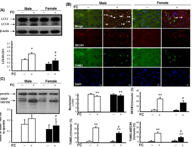
To examine the sex differences in FC-induced autophagy and injury severity, the levels of LC3-II, autophagic cell death and SBDP 145/150 were detected in striatum of C57BL/6 mice after FC infusion. The results revealed that FC infusion caused increases of LC3-II (Fig 1A) and autophagic cell death, which is simultaneously TUNEL positive and BECN1 immunoreac-tive (Fig 1B), in male and female mice. After quantification, the numbers of nuclei in the stria-tums of both males and females were decreased following FC infusion (Fig 1). Additionally, FC infusion significantly increased the percentages of TUNEL(+) nuclei, BECN1(+) cells, and TUNEL(+) BECN1 immunoreactive cells by 32%, 27%, and 19% in males, respectively, and 22%, 19%, and 10% in females, respectively (Fig 1B). Moreover, FC infusion caused increases of 33% and 24% in the ratios of SBDP 145/150 to spectrin (Fig 1C) in male and female mice, respectively.
Role of E
2in sex differences in FC-induced autophagy and injury
severity
To determine whether E2contributed to the sex differences in FC-induced autophagy and
injury severity in the striatum, both male and female C57BL/6 mice were sterilized prior to the implantation of E2to exclude the influence of endogenous sex hormones. The results revealed
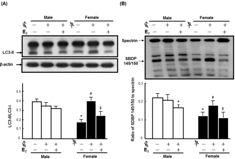
that no significant effect of orchidectomy on FC-induced LC3 lipidation was observed, while ovariectomy increased the levels of FC-induced LC3 lipidation by 1.57-fold. Although E2
lower than that of intact males by 25%. Moreover, E2decreased FC-induced LC3 lipidation in
the ovariectomized females by 39%, whereas no significant difference of the level of LC3-II between E2implanted ovariectomized females and intact females was observed (Fig 2A). No
significant effect of orchidectomy on FC-induced injury severity was observed in the male group, while ovariectomy markedly increased the FC-induced injury severity. E2implantation
decreased the ratios of SBDP 145/150 to spectrin in both sterilized male and female mice by 13% and 42%, respectively (Fig 2B).
Fig 1. FC induced higher levels of autophagy, autophagic cell death, and injury severity in males. (A) FC-induced LC3 lipidation.White column: male group; black column: female group.(B) Upper panel: Representative images of TUNEL(+) BECN1-immunoreactive cells.The yellow spots indicated by arrows depicted TUNEL(+) BECN1-immunoreactive cells with round (intact) nuclei, which were regarded as indicators of autophagic cell death.
Lower panel: Numbers of DAPI-positive nuclei, TUNEL(+) nuclei, BECN1(+) cells, and TUNEL(+) BECN1-immunoreactive cells.The number of immunoreactive cells was counted in every fifteenth section of the striatum under fluorescent microscope. Five fields in each tissue section were randomly selected for counting. Male: white column; female: black column. Data are presented as the means±SDs (6 mice in each group).(C) FC-induced injury severity.The ratio of SBDP 145/150 to spectrin depicts injury severity. Data are presented as the means±SDs (n = 6),*p<0.05,**p<0.01 compared with sex-matched saline-infusion controls; #p<0.05 compared with the FC-infusion males.
Female-specific effects of rapamycin on FC-induced injury severity and
DNA fragmentation in DRD2 neurons
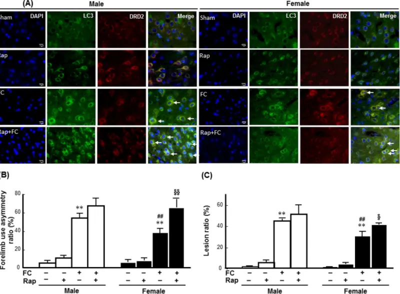
To examine whether autophagy induction affected FC-induced striatal injury in a sex-depen-dent manner, the effects of rapamycin on FC-induced injury, behavioral deficits, histological lesion, and the numbers of TUNEL(+) DRD2 neurons were compared between the intact male and female C57BL/6 mice. As shown inFig 3A, immunoreactivity of LC3 was co-localized with the DRD2 neurons in both male and female mice with or without FC infusion. The histological changes after FC infusion and the images of brain sections after HE stain were showed in the
S4 Fig. The quantitative result showed that rapamycin increased both the FC-induced forelimb use asymmetry scores and the lesion ratios by 63% (Fig 3B) and 33% (Fig 3C), respectively, in female mice but did not affect these measurements in the males.
To examine whether the increase in autophagic activity correlates with cell death in neu-rons, we examined LC3 staining along with neutron marker (NeuN) staining, while the result showed that LC3 immunoreactivity exhibited in both neuron and glial cells and the LC3 anti-body reacted with both LC3-I and LC3-II(S2 Fig). Moreover, TUNEL staining along with Fig 2. E2contributed to the sex differences in FC-induced autophagy and injury severity in the striatum.All of the mice in every groups were infused with 3μL of FC (1 mmol/L) into the right striatum.(A) E2reversed the increase of FC-induced LC3 lipidation by ovariectomy. (B) E2decreased the
ratios of SBDP 145/150 to spectrin in both sterilized female (♀with a slash) and male (♂with a slash) mice after FC infusion. The data are expressed as the means±SDs (n = 6),*p<0.05 compared with intact males; #p<0.05 compared with intact females; §p<0.05 compared with ovariectomized females.
neutron marker (NeuN) or astrocyte marker (GFAP) staining were performed. The result showed that DNA fragmentation also exhibited in both neuron and astrocyte(S3 Fig). There-fore, we quantified the number of TUNEL positive DRD2 neuron that is more correlative to behavioral deficit due to striatal injury using automated TissueFAXS followed by TissueQuest analysis. The results revealed that FC infusion increased the numbers of TUNEL(+) DRD2 neurons in the striatums of both the male and female mice (Fig 4A). Rapamycin did not change the percentages of DRD2 neurons in the total nuclei of either male or female mice (Fig 4B). Whereas, rapamycin increased the percentages of TUNEL(+) cells and TUNEL(+) DRD2 neu-rons by 63% and 73%, respectively, in the striatum of the FC infusion female mice but not in males following FC infusion (Fig 4C & 4D).
Fig 3. Rapamycin exaggerated FC-induced striatal injury in female mice. (A) Rapamycin (Rap) enhanced the LC3 immunoreactivity in DRD2 neurons of both male and female striatum. The pictures were captured under fluorescent microscope after immunostaining. Arrows indicated the merged nuclei surrounded by LC3 and DRD2 immunoreactivity depicted DRD2 neurons with autophagic features.(B) Rapamycin exaggerated FC-induced behavioral deficits in the female group. (C) Rapamycin increased the histological lesion ratio caused by FC infusion in the females. The data are expressed as the means±SDs (n = 6).**p<0.01 compared with sex-matched saline-infusion control. ##p<0.01 compared with the FC-infusion males.
§p<0.05, §§p<0.01 compared with the FC-infusion females.
Male-specific effects of
Atg7
knockout (KO) in DRD2 neurons on
FC-induced injury severity and DNA fragmentation in DRD2 neurons
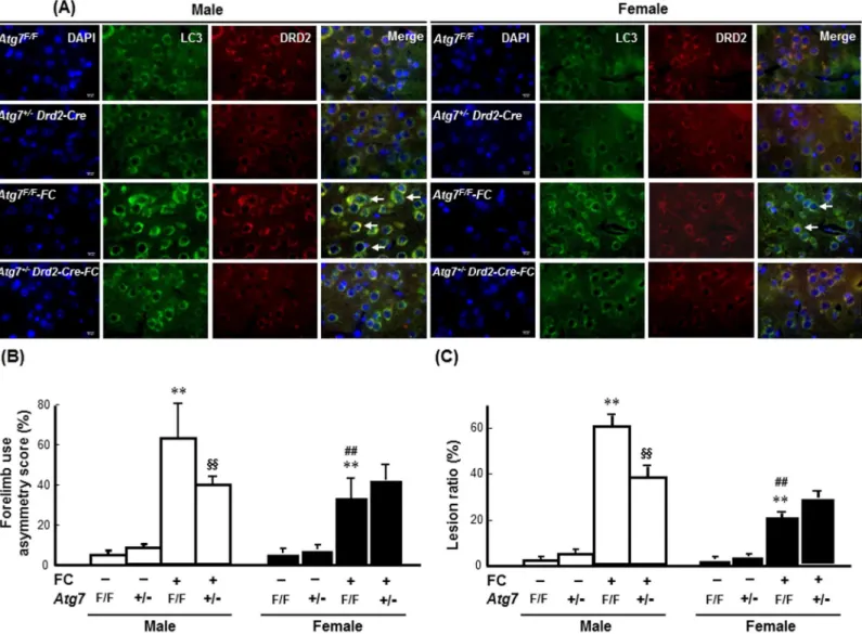
To address the effects of neuron-specific autophagy inhibition on iron toxicity, heterozygote-conditional knockout mice in whichAtg7was deleted in the DRD2 neurons (Atg7+/-Drd2-Cre) were used. The knockdown efficiency ofAtg7in the DRD2 neurons was confirmed by counting the numbers of ATG7-immunoreactive DRD2 neurons. As shown inFig 5A, in both the male and female groups, the numbers of ATG7-immunoreactive DRD2 neurons were decreased in the striatums of theAtg7+/-Drd2-Cremice compared with those of theAtg7F/Fmice. The repre-sentative images of striatum containing brain sections after HE stain were showed in theS5 Fig. The quantitative result showed that there were no differences in the total numbers of nuclei or the percentages of DRD2 neurons in the total nuclei between theAtg7+/-Drd2-Cre and wild-typeAtg7F/Fmice (Fig 5B and 5C). The percentages of ATG7-immunoreactive DRD2 neurons in the striatums of the male and femaleAtg7+/-Drd2-Cremice were significantly lower than those in the male and femaleAtg7F/Fmice by 44% and 33%, respectively (Fig 5D).Both the male and femaleAtg7+/-Drd2-Cremice exhibited lower levels of FC-induced LC3 aggregation in the striatum compared with theAtg7F/Fmice (Fig 6A). In males,Atg7knockout decreased both FC-induced behavioral deficits (Fig 6B) and histological lesions (Fig 6C) by 36% (p<0.01) and 36% (p<0.01), respectively. By contrast, in females, no significant effect of
Atg7KO on the FC-induced behavioral deficits and histological lesions was observed (Fig 6B and 6C).
Moreover, the results from TissueQuest analysis showed that no significant sex differences in the numbers of DRD2 neurons were found between theAtg7F/FandAtg7+/-Drd2-Cremice (Fig 7A & 7B). FC infusion caused a significantly higher level of DNA fragmentation in male than in the femaleAtg7F/Fmice. Interestingly, in males,Atg7knockout decreased the percent-ages of TUNEL(+) cells and TUNEL(+) DRD2 neurons by 70% and 69%, respectively, follow-ing FC infusion. By contrast, no significant differences in the percentages of TUNEL(+) cells or TUNEL(+) DRD2 neurons between theAtg7+/-Drd2-CreandAtg7F/Ffemales following FC infusion were observed (Fig 7C and 7D).
Discussion
The present study demonstrated that the striatum of male mice were more susceptible to FC-infusion that simulates iron accumulation after ICH. Enhancement of autophagy exaggerated the FC-induced striatal injuries and behavioral deficits in females but not males, and inhibition of neuronal autophagy decreased FC-induced striatal injuries and behavioral deficits in males but not in females. These results suggest that the over-activation of autophagy plays a harmful response to striatal iron overload in males but not in females. For the first time, this study explored the sex-dimorphic role of autophagy induction in striatal injury caused by iron overload.
Men are more vulnerable to ICH [24] and exhibit worse survival rates than premenopausal women following ICH [3]. Previously, a rat models of ICH with autologous whole blood infu-sion into basal ganglia, both brain edema and behavioral deficits were more severe in males than in females [25]. Recently, a rat model involving infusion with ferrous iron, which contrib-utes to the neurological deficit following ICH, into the caudate nucleus showed that males are more vulnerable to iron overload [7,15]. Presently, a mouse model of striatal FC infusion
sections were scanned using a TissueFax cytometer, and the number of TUNEL(+) and/or DRD2 immunoreactive neurons were analyzed by TissueQuest. The data are expressed as the means±SDs (n = 6).*p<0.05 compared with FC-infusion male; #p<0.05 compared with female mice without rapamycin
pre-treatment.
Fig 5. HeterozygousAtg7knockout in striatal DRD2 neurons. (A) Representative images of ATG7 immunoreactive DRD2 neurons. The pictures were captured under fluorescent microscope after immunostaining. Arrows indicate blue nuclei surrounded by immunoreactivities of ATG7 and DRD2.(B) total number of DAPI(+) nuclei. (C) percentage of DRD2 neurons in total nuclei. (D) percentage of ATG7 immunoreactive DRD2 neurons in total nuclei. The number of immunoreactive cells was counted in every fifteenth section of the striatum. Five fields in each tissue section were randomly selected for counting. Data are presented as the means±SDs (number of mice was 4 in each group).Atg7 KOsignificantly decreased the percentage of ATG7(+) DRD2 neurons in the striatum of both male (white column) and female (black column) mice.**p<0.01 compared with sex-matchedAtg7F/Fgroups.
shows a higher level of autophagy associated with a higher injury severity in males than in females, implicating a possible harmful role of FC-induced autophagy in males. Additionally, ovariectomy, which simulates menopause in women, increased FC-induced striatal autophagy and injury severity, while E2implantation decreased FC-induced autophagy and injury severity
in the ovariectomized mice. These results supported the supposition that E2contributes to the
lower level of FC-induced autophagy and injury severity in females. Lacking of the protection conferred by endogenous estrogen may explain their vulnerability to FC-infusion in males and ovariectomized-females.
After ICH, degradation of hemoglobin releases ferrous iron, which undergoes a redox cycle such as Haber-Weiss reaction and/or a Fenton reaction in the presence of citric acid, causes persistent conversion of oxygen into reactive oxygen species that trigger cell death. In addition to apoptosis and necrosis, which contribute to the ICH/FC-induced neuronal death [26] [27], Fig 6. Knockout ofAtg7in DRD2 neurons decreased FC-induced striatal injury in male and female mice. (A)Atg7KO decreased LC3
immunoreactivity in the DRD2 neurons. The images were captured using TissueFAXS. Arrows indicate LC3 immunoreactive DRD2 neurons.(B)Atg7KO decreased FC-induced behavioral deficits in males but not in females. (C)Atg7KO decreased the lesion ratios caused by FC infusion in males only. The data are expressed as the means±SDs (n = 6).**p<0.01 compared with the sex-matchedAtg7F/Fgroups; ##p<0.01 compared with the
FC-infusedAtg7F/Fmale mice. §§p<0.01 compared with the FC-infusedAtg7F/Fmale mice.
Fig 7.Atg7KO mice exhibited a male-specific benefit in FC-induced DNA fragmentation in the DRD2 neurons in the striatum. (A) Representative images of the TUNEL(+) DRD2 neurons. The bright green-yellow dots depict DRD2 neurons with DNA fragmentation, as indicated by the arrows. Quantitative results of the percentages of DRD2 neurons(B), TUNEL(+) cells (C), and TUNEL(+) DRD2 neurons (D). The data are expressed as the means±SDs (n = 6).**p<0.01,Atg7F/Ffemale vs.Atg7F/Fmale; ##p<0.01Atg7F/Fvs.Atg7+/-Drd2-Cre.
ROS generated by ferrous ions also triggers autophagy and/or autophagic cell death [28,29]. The induction of autophagy, a process for organelle turnover and bulk protein degradation [13], has been reported in patients with critical illnesses [30] and in the rat model of ICH [31]. The beneficial or harmful role of autophagy under stress might be determined by the balance/ imbalance between the levels of autophagy and energy demand. In the present result, rapamy-cin pre-treatment slightly but not significantly increased the FC-induced autophagic cell death and injury severity in males. It is possible that the combination of rapamycin pre-treatment and FC-infusion reached the plateau of autophagy induction. Because, autophagy is an ATP-dependent process and the energetic dysfunction existed under oxidative stress due to iron overload. Interestingly, a prominent increase of autophagic cell death and injury severity was observed in FC infusion females pre-treated with rapamycin compared with FC infusion females without rapamycin pre-treatment. It suggests that pre-treatment with rapamycin diminishes the limiting effect of female endogenous E2on FC-induced autophagy, which in
turn becomes harmful au1tophagic cell death. E2has been reported to interact with ERαin
plasma membrane that is in a complex with ER-interacting protein, which facilitates estrogen activation of PI3K/Akt. Since mTOR (autophagy inhibitor) is a downstream target of PI3K/ Akt [32], rapamycin (a mTOR inhibitor) may interfere E2signaling via affecting mTOR.
How-ever, the other possible mechanism underlying the effect of rapamycin on interfering E2
signal-ing may not be excluded due to the non-specific effects of rapamycin/mTOR on downstream signaling. It actually remains unknown what factors determine whether autophagy is beneficial or pathogenic. An improved understanding of neuronal autophagy will provide novel insights into the pathogenic mechanisms of dysfunctional autophagy that underlie neurodegeneration due to iron overload and, ultimately, help develop therapeutic interventions.
On the other hand, conditional knockout ofAtg7in DRD2 neurons decreased the percent-age of TUNEL(+) DRD2 neurons and diminished FC-induced injury severity in males, while slightly increased the FC-induced injury severity in females. These results strongly suggested that, in males, the inhibition of FC-induced autophagy could switch a maladaptive or harmful autophagy to a response in favor of neuroprotection. Whereas, in females, the moderate level of FC-induced autophagy might be sufficient for supplying energy demand, thus inhibition of autophagy deprived the protective machinery under oxidative stress. Accordingly, male-spe-cific inhibition of autophagy might be a useful therapeutic strategy for patients with iron overload.
In conclusion, over-activation of the autophagic machinery plays a maladaptive and harm-ful role in FC-induced striatal injury in males but not in females. Inhibition of autophagy could represent a useful therapeutic target for iron-induced brain injury in men or postmenopausal women who have lost the limitation of autophagy by estrogen. Therefore, this study presents new paths for exploring sex-specific therapeutic strategies that address the maladaptive over-activation of autophagy, such as gene therapy targeting autophagy-related genes.
Supporting Information
S1 Fig. The Prussian blue assay on brain section from mice infused with or without ferrous citrate (FC).Two days after FC infusion, the brain tissue containing striatum was sectioned and stained by working solution which is a mixture of equal parts of hydrochloric acid and potassium ferrocyanide prepared immediately before use. Arrows in the lower left panel indi-cate dark blue deposits that confirmed the iron overload after FC infusion.
(TIF)
(B) Lesion site; (C) Around lesion site. Bal/c mice were stereotaxically infused with 3μL FC into the right striatum. Two days after FC infusion, the brain tissue containing striatum was sectioned and stained by LC3 antibody for LC3-II aggregation (green dots) as an autophagic marker; NeuN for neuron marker; and DAPI for nuclei. Bright green spots (arrow) showed in the lower raw of right panel indicate LC3 aggregation, which are exhibited in both neuron and non-neuronal cells.
(TIF)
S3 Fig. Ferrous citrate (FC) induced DNA fragmentation in both neuron and astrocyte.
Bal/c mice were stereotaxically infused with 3μL FC into the right striatum. Two days after FC infusion, the brain tissue containing striatum was sectioned and stained by LC3 Ab (Green), in the left panel; TUNEL (Green) and GFAP Ab (Red) in the left panel; and TUNEL (Green) and NeuN Ab (Red) in the right panel. DAPI (Blue) was used to identify nucleus simultaneously. Bright green spots showed in the left panel indicate LC3 aggregation, which is a detection marker of autophagy. Arrow showed in the lower raw of right panel indicates the TUNEL posi-tive astrocyte; arrowhead showed in the lower raw of right panel indicates the TUNEL posiposi-tive neuron.
(TIF)
S4 Fig. Rapamycin pre-treatment exaggerated the histological lesion in striatum of females after FC infusion.The paraffin-embedded tissues were serially sectioned into 10-μm thick slices. After hematoxylin and eosin (HE) staining, the extents of the histological lesions were analyzed using Image-proPlus according to the staining intensity in the enclosed area. The ratio of ipsilateral hemispheric volume of the striatum to the contralateral hemispheric volume served as an index of histological lesion. The images at the upper panel indicate the higher magnifications the histological injury is more obvious. The scale bars represent the different magnifications under microscope.
(TIF)
S5 Fig.Atg7knockout diminished the histological lesion in striatum of males after FC infu-sion.The paraffin-embedded tissues were serially sectioned into 10-μm thick slices. After hematoxylin and eosin (HE) staining, the extents of the histological lesions in every fifteenth section of the striatum were analyzed using Image-proPlus software, according to the staining intensity in striatum. The ratio of ipsilateral hemispheric volume of the striatum to the contra-lateral hemispheric volume served as an index of histological lesion.
(TIF)
Acknowledgments
The authors thank the help from the Statistical Analysis Laboratory, Department of Medical Research, Kaohsiung Medical University Hospital, Kaohsiung Medical University.
Author Contributions
Conceived and designed the experiments: CH LW. Performed the experiments: TC HH PC. Analyzed the data: YH. Contributed reagents/materials/analysis tools: CH LW KKY. Wrote the paper: CH SL YH LW.
References
2. Lewsey JD, Gillies M, Jhund PS, Chalmers JW, Redpath A, Briggs A, et al. Sex differences in inci-dence, mortality, and survival in individuals with stroke in Scotland, 1986 to 2005. Stroke. 2009; 40 (4):1038–43. Epub 2009/02/13. STROKEAHA.108.542787 [pii] doi:10.1161/STROKEAHA.108.
542787PMID:19211485.
3. Zia E, Engstrom G, Svensson PJ, Norrving B, Pessah-Rasmussen H. Three-year survival and stroke recurrence rates in patients with primary intracerebral hemorrhage. Stroke. 2009; 40(11):3567–73.
Epub 2009/09/05. STROKEAHA.109.556324 [pii] doi:10.1161/STROKEAHA.109.556324PMID: 19729603.
4. Mendelow AD, Gregson BA, Fernandes HM, Murray GD, Teasdale GM, Hope DT, et al. Early surgery versus initial conservative treatment in patients with spontaneous supratentorial intracerebral haemato-mas in the International Surgical Trial in Intracerebral Haemorrhage (STICH): a randomised trial. Lan-cet. 2005; 365(9457):387–97. Epub 2005/02/01. doi: S0140-6736(05)17826-X [pii] doi:10.1016/
S0140-6736(05)17826-XPMID:15680453.
5. Mendelow AD, Gregson BA, Rowan EN, Murray GD, Gholkar A, Mitchell PM. Early surgery versus ini-tial conservative treatment in patients with spontaneous supratentorial lobar intracerebral haematomas (STICH II): a randomised trial. Lancet. 2013. Epub 2013/06/04. doi: S0140-6736(13)60986-1 [pii] doi: 10.1016/S0140-6736(13)60986-1PMID:23726393.
6. Ali SS, Xiong C, Lucero J, Behrens MM, Dugan LL, Quick KL. Gender differences in free radical homeo-stasis during aging: shorter-lived female C57BL6 mice have increased oxidative stress. Aging Cell. 2006; 5(6):565–74. Epub 2006/11/30. doi: ACE252 [pii] doi:10.1111/j.1474-9726.2006.00252.xPMID:
17129217.
7. Chen TY, Tsai KL, Lee TY, Chiueh CC, Lee WS, Hsu C. Sex-specific role of thioredoxin in neuroprotec-tion against iron-induced brain injury conferred by estradiol. Stroke. 2010; 41(1):160–5. Epub 2009/11/
27. doi: STROKEAHA.109.562850 [pii] doi:10.1161/STROKEAHA.109.562850PMID:19940280.
8. Olivan S, Calvo AC, Manzano R, Zaragoza P, Osta R. Sex differences in constitutive autophagy. BioMed research international. 2014; 2014:652817. doi:10.1155/2014/652817PMID:24719882; PubMed Central PMCID: PMC3955681.
9. Weis SN, Toniazzo AP, Ander BP, Zhan X, Careaga M, Ashwood P, et al. Autophagy in the brain of neonates following hypoxia-ischemia shows sex- and region-specific effects. Neuroscience. 2014; 256:201–9. doi:10.1016/j.neuroscience.2013.10.046PMID:24184979.
10. Hua Y, Nakamura T, Keep RF, Wu J, Schallert T, Hoff JT, et al. Long-term effects of experimental intra-cerebral hemorrhage: the role of iron. J Neurosurg. 2006; 104(2):305–12. doi:10.3171/jns.2006.104.2.
305PMID:16509506.
11. Keep RF, Hua Y, Xi G. Intracerebral haemorrhage: mechanisms of injury and therapeutic targets. Lan-cet neurology. 2012; 11(8):720–31. doi:10.1016/S1474-4422(12)70104-7PMID:22698888; PubMed
Central PMCID: PMC3884550.
12. Inagawa T, Ohbayashi N, Takechi A, Shibukawa M, Yahara K. Primary intracerebral hemorrhage in Izumo City, Japan: incidence rates and outcome in relation to the site of hemorrhage. Neurosurgery. 2003; 53(6):1283–97; discussion 97–8. PMID:14633295.
13. Yoshimori T. Autophagy: a regulated bulk degradation process inside cells. Biochem Biophys Res Commun. 2004; 313(2):453–8. Epub 2003/12/20. S0006291X03023489 [pii]. PMID:14684184. 14. Koike M, Shibata M, Tadakoshi M, Gotoh K, Komatsu M, Waguri S, et al. Inhibition of autophagy
pre-vents hippocampal pyramidal neuron death after hypoxic-ischemic injury. Am J Pathol. 2008; 172 (2):454–69. Epub 2008/01/12. doi: S0002-9440(10)61811-1 [pii] doi:10.2353/ajpath.2008.070876
PMID:18187572; PubMed Central PMCID: PMC2312361.
15. Chen CW, Chen TY, Tsai KL, Lin CL, Yokoyama KK, Lee WS, et al. Inhibition of autophagy as a thera-peutic strategy of iron-induced brain injury after hemorrhage. Autophagy. 2012; 8(10):1510–20. Epub
2012/08/23. doi: 21289 [pii] doi:10.4161/auto.21289PMID:22909970.
16. Gupta M, Mazumder UK, Pal DK, Bhattacharya S. Onset of puberty and ovarian steroidogenesis follow-ing adminstration of methanolic extract of Cuscuta reflexa Roxb. stem and Corchorus olitorius Linn. seed in mice. Journal of ethnopharmacology. 2003; 89(1):55–9. PMID:14522432.
17. Lofgren JL, Erdman SE, Hewes C, Wong C, King R, Chavarria TE, et al. Castration eliminates conspe-cific aggression in group-housed CD1 male surveillance mice (Mus musculus). Journal of the American Association for Laboratory Animal Science: JAALAS. 2012; 51(5):594–9. PMID:23312088; PubMed
Central PMCID: PMC3447448.
18. Missale C, Nash SR, Robinson SW, Jaber M, Caron MG. Dopamine receptors: from structure to func-tion. Physiological reviews. 1998; 78(1):189–225. PMID:9457173.
(34):11388–97. Epub 2010/08/27. doi: 30/34/11388 [pii] doi:10.1523/JNEUROSCI.1356-10.2010
PMID:20739560.
20. Hua Y, Schallert T, Keep RF, Wu J, Hoff JT, Xi G. Behavioral tests after intracerebral hemorrhage in the rat. Stroke. 2002; 33(10):2478–84. Epub 2002/10/05. PMID:12364741.
21. Mizushima N, Yoshimori T. How to interpret LC3 immunoblotting. Autophagy. 2007; 3(6):542–5. Epub
2007/07/06. 4600 [pii]. PMID:17611390.
22. Weiss ES, Wang KK, Allen JG, Blue ME, Nwakanma LU, Liu MC, et al. Alpha II-spectrin breakdown products serve as novel markers of brain injury severity in a canine model of hypothermic circulatory arrest. Ann Thorac Surg. 2009; 88(2):543–50. Epub 2009/07/28. doi: S0003-4975(09)00645-6 [pii] doi:
10.1016/j.athoracsur.2009.04.016PMID:19632410; PubMed Central PMCID: PMC3412404.
23. Yu L, Alva A, Su H, Dutt P, Freundt E, Welsh S, et al. Regulation of an ATG7-beclin 1 program of autop-hagic cell death by caspase-8. Science. 2004; 304(5676):1500–2. Epub 2004/05/08. doi:10.1126/
science.10966451096645[pii]. PMID:15131264.
24. Hu YZ, Wang JW, Luo BY. Epidemiological and clinical characteristics of 266 cases of intracerebral hemorrhage in Hangzhou, China. J Zhejiang Univ Sci B. 2013; 14(6):496–504. Epub 2013/06/05. doi:
10.1631/jzus.B1200332PMID:23733426; PubMed Central PMCID: PMC3682165.
25. Nakamura T, Hua Y, Keep RF, Park JW, Xi G, Hoff JT. Estrogen therapy for experimental intracerebral hemorrhage in rats. J Neurosurg. 2005; 103(1):97–103. Epub 2005/08/27. doi:10.3171/jns.2005.103.
1.0097PMID:16121980.
26. Han N, Ding SJ, Wu T, Zhu YL. Correlation of free radical level and apoptosis after intracerebral hemor-rhage in rats. Neuroscience bulletin. 2008; 24(6):351–8. doi:10.1007/s12264-008-0711-4PMID:
19037320.
27. Zheng M, Du H, Ni W, Koch LG, Britton SL, Keep RF, et al. Iron-Induced Necrotic Brain Cell Death in Rats with Different Aerobic Capacity. Translational stroke research. 2015. doi: 10.1007/s12975-015-0388-8PMID:25649272.
28. Scherz-Shouval R, Elazar Z. ROS, mitochondria and the regulation of autophagy. Trends Cell Biol. 2007; 17(9):422–7. Epub 2007/09/07. doi: S0962-8924(07)00168-7 [pii] doi:10.1016/j.tcb.2007.07.009
PMID:17804237.
29. Scherz-Shouval R, Shvets E, Fass E, Shorer H, Gil L, Elazar Z. Reactive oxygen species are essential for autophagy and specifically regulate the activity of Atg4. EMBO J. 2007; 26(7):1749–60. Epub 2007/
03/10. doi: 7601623 [pii] doi:10.1038/sj.emboj.7601623PMID:17347651; PubMed Central PMCID: PMC1847657.
30. Clark RS, Bayir H, Chu CT, Alber SM, Kochanek PM, Watkins SC. Autophagy is increased in mice after traumatic brain injury and is detectable in human brain after trauma and critical illness. Autophagy. 2008; 4(1):88–90. Epub 2007/10/25. doi: 5173 [pii]. PMID:17957135.
31. He Y, Wan S, Hua Y, Keep RF, Xi G. Autophagy after experimental intracerebral hemorrhage. J Cereb Blood Flow Metab. 2008; 28(5):897–905. Epub 2007/11/08. doi: 9600578 [pii] doi:10.1038/sj.jcbfm.
9600578PMID:17987045.
32. Raz L, Khan MM, Mahesh VB, Vadlamudi RK, Brann DW. Rapid estrogen signaling in the brain. Neuro-signals. 2008; 16(2–3):140–53. Epub 2008/02/07. doi: 000111559 [pii] doi:10.1159/000111559PMID: