Pak. J. Anal. Environ. Chem. Vol. 17, No. 1 (2016) 72 – 76
Determination of Lead in Human Calculi and Its Effects on
Renal Function of Lead Occupational Workers
F. Memon
1, N.T. Narejo
3⃰, A. G. M. Vasandani
2, K. P. Kunbhar,
S. Jalbani
3and P. Khan
31Department of Physiology, University of Sindh Jamshoro, Pakistan 2
M.A.Kazi Institute of Chemistry, University of Sindh Jamshoro, Pakistan 3
Department of Fresh Water Biology and Fisheries, University of Sindh Jamshoro, Pakistan *Corresponding Author Email: [email protected]
Received 26 January 2016, Revised 27 June 2016, Accepted 28 June 2016
---Abstract
Seventy five samples of renal and eighteen samples of supra gingival calculi of lead recycling workers were collected over the period of seven years (2008-2014) and studied for the accumulation of lead. The results were compared with those of non exposed subjects. The lead content of calculi was investigated for its dependence on type and composition of calculi, blood lead, job status and duration of exposure. The effect of blood lead and renal calculi was also investigated in relation to kidney function of respective subjects. The mean lead levels of various types of calculi were found to follow the order as phosphate > oxalate > urate .> cystine while single principal group of supra gingival calculi resulted in lower levels of metal. The lead content of calculi positively correlated with phosphate content of both of the renal (r = 0.655) and supra gingival calculi (r= 0.866). Impaired renal function was more pronounced in active workers and depended on blood lead levels in addition to presence of metal in renal calculi.
Keywords:Lithiasis, Lead poisoning, Creatinine, Battery recycling, Lead nephropathy
---Introduction
Lead is known for its toxic effects in humans and animals. There are different sources of exposure among which the uncontrolled battery recycling has been cited as potentially dangerous for the health of workers and general population. Due to lack of strict laws and technically simple process, the activity is reported to be common in most of the developing countries including Pakistan [1].
Few years back Pakistan introduced the relatively inexpensive compressed natural gas (CNG) as alternate fuel for petrol driven vehicles resulting in the proportional increase in the number of vehicles. Moreover the power shortage in the country has also led to installation of battery supported uninterrupted power supply (UPS) devices everywhere in offices homes and commercial buildings. The large quantity of discarded batteries thus generated is also added to scrap leading to growth of recycling facilities and consequent
number of lead recycling workers. Quite a large number of recycling cottage industries have therefore become functional in the major urban centers of country where work force is usually prone to health hazards such as anemia, disease of bones, neuropathy, renal insufficiency and variety of cancers.
During the routine medical checkup of these workers a number of renal lithiasis cases were detected which prompted the question whether lead accumulates in renal calculi like other body pools such as physiological fluids, soft tissues and bones [2]. Number of reports exist in the literature wherein lead in human calculi has been reported as part of their overall metallic composition [3-5, 15, 18- 21] but to our knowledge, no work has been reported on the accumulation of lead in calculi of occupationally
exposed workers or its etiological role in the disease. In this study we therefore report the lead content of renal (R-cal) as well as supra gingival calculi (S-cal) of battery recycling workers in order to see if metal has got specific tendency to deposit in crystalline phases of stones forming in biological systems. The dependence of lead accumulation on factors such as blood lead levels, duration of exposure, and viability of lead in human calculi as marker of exposure is explored and some preliminary work on renal health of lead exposed subjects is also reported.
Materials and Methods
Subjects
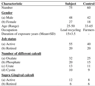
All studied subjects were occupationally involved in battery recycling activities whereas reference subjects had no history of exposure to lead. All the subjects agreed to cooperate once they were informed about the objectives of study. Some demographic characteristics and job status of subjects are given in (Table 1).
Methods
Classification of calculi and determination of lead Samples of renal calculi were collected from urology wards of different hospitals of major cities of Sindh excluding Karachi. The medical records of lithiasis patients were examined in relation to occupation and stone samples of battery recycling workers collected for study purposes. All samples were extracted from various parts of renal system by qualified surgeons of the respective hospitals using standard procedures. The reference samples of renal stones were collected after ascertaining that patients had no history of lead exposure or had never worked in any industry during their lives. The study and reference subjects were also approached in person who provided the samples of supragingival calculi through extraction by qualified dental technicians. Optical microscopy, FTIR and wet chemical analysis using Merckognist ® kits in addition to methods reported by Hodgkinson [9] were used, to identify major crystalline phases and classify stones in subgroups (Table 1). The information regarding age, job status, duration of exposure and other aspects was obtained by administering a carefully designed
questionnaire to subjects .Each of the R-cal, and S-cal samples was washed with lead free de ionized water washings preserved and calculi dried in pre heated oven at 80oC for 24 hours. The materials were then crushed with agate pestle and mortar and powder stored in polyethylene vials. 500 mg sample of the each of the calculi was processed for separation and pre concentration in an ion exchange column [6,7] and analyzed for lead by graphite furnace atomic absorption spectrophotometer (SpectrAA 220/880 Varian Australia). Twenty samples of R-cal of suitable size were also subjected to transverse section by jewelers saw. The clear ring shaped striations were carefully removed from periphery to nucleus and analyzed for lead as stated above. The lead content of washings was also adjusted in the results of respective powdered samples.
Table 1. Some demographic characteristics, job status, number and types of calculi of lithiasis patients.
Determination blood lead and Creatinine clearance (Cr-cl) 10ml venous blood was drawn from each of the subjects at the time of or before the surgical removal of stones and extraction of supra gingival calculus. The lead in blood was also determined by atomic absorption method [7]. Serum creatinine was determined by picric acid – sodium hydroxide method and the data along with height, weight and age of respective subjects, was processed to estimate Cr-cl using computer program based on Cockroft-Gault formula [8].
Characteristic Subject Control
Number 75 60
Gender
(a) Male 48 42
(b) Female 27 18
Age (Range) 25-50 33-65
Occupation Lead recycling Farmers
Duration of exposure years (Mean±SD) 15±3.5
-Job status
(a) Active 55 40
(b) Retired 20 20
Number of different calculi
(a) Oxalate 32 25
(b) Phosphate 20 15
(c) Urate 13 11
(d) Cystin 10 9
Supra Gingival calculi
(a) Active 12 8
Statistical analysis
The statistical analysis of data was performed by Minitab 13 Computer program. t- test, ANOVA, Pearson’s correlation coefficients, regression and descriptive statistics were used to characterize the data at p levels of 0.05.
Results and Discussion
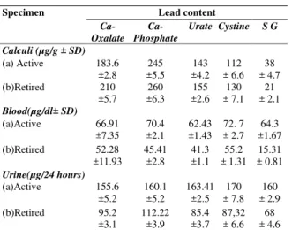
Table-1 shows the mean age, duration of exposure and job status of lead workers, whose calculi, were studied in the present work. Of the total number of renal and supra gingival calculi (N= 93), 72% were delivered by active workers where as the 28% by retired workers. The proportion of R-cal, and S- cal constituted 80.7%, and 19.3% respectively. The mean durations of exposure of active and retired workers did not significantly differ (p > 0.05) however 60% of the calculi were delivered by the subjects who had remained on job for 7 to 9 years. The mean ages of lead workers and reference group were not comparable and the age of lead workers having lithiasis ranged between 30 to 47 years which was found to be lower than that of reference subjects (40 to 65 years). The age at which the lithiasis occurred did not affect nature of calculi as both the groups yielded stones identical in terms elemental composition and dominant crystalline phases (p>0.05). On the basis of results of wet chemical analysis, FTIR and microscopic examination only four subgroups of renal and the single group of supra gingingival calculi samples could be formed. The lead content of the calculi of recycling workers was found to be very high (p<0.01) in comparison to the reference group (Table 2). Rohana et al [3] studied the bio-mineralization of kidney stone and reported lead content of renal calculi as high as 147µg/g, Yamamoto et al [4] studied renal calculi subjects having history of exposure to polluted environments and reported the lead levels as high as 204.8.µg/g, similarly Durak et al [15] reported lead in the range 3-59 µg/g in various types of renal calculi. In the present investigation the highest lead levels found in the renal calculi was 260 µg/g associated with phosphate calculi.It supports the recent report [19] that human calculi have potential to accumulate lead and its presence can be used as indicator of
human exposure to metal and increased body lead burden. The mean values of lead in calculi of active and retired workers except S-cal were found comparable but they were significantly higher than those of reference group (p< 0.05). The role of current level of lead in blood and probable internal mobilization of stored metal specially in skeleton where it is known to have long residence times.[2] cannot be ignored. In the two principal groups the mean lead levels followed the order as R-cal > S-cal while among the subgroups of R- S-cal it followed the order as phosphate > oxalate >urate .> cystine calculi. In the case of S-cal however the mean lead levels of retired workers were significantly lower (p <0.05) than those who were on job It is known that supra gingival calculus is largely composed of mixed phases of apatite and ß-tri calcium compounds formed from the deposition of calcium and phosphate in saliva [10] In view of the presence of metallic taste due to lead in saliva of lead workers [16] it is reasonable to expect that composition of S-cal is likely to be influenced by oral environment. The accumulation of lead in S-cal may therefore results from lead present in oral cavity environment and its concentration may decrease upon removal from the source of exposure.
The mean blood lead levels of active lead workers of all the principal groups were not significantly different from each other but resulted to be higher than those of retired workers (p<0.05). This was found to be true in the case of blood lead levels of active and retired S-cal groups (p<0.05) also. Statistically significant Pearson’s correla-tion coefficients (r) for lead and phosphate in S-cal of active workers and R-cal of all workers were respectively found as r= 0.886 (p < 0.01) and r=0.655 (p<0.01) which is in accordance with the earlier works [12,17] in which association of lead with phosphate has been reported.
clearance and other markers of renal function. [13,14]. In present study therefore the mean creatinine clearance (Cr-cl) of lead workers was found to be lower than those having lithiasis but no exposure to lead. (p<0.01) and appeared to be affected by both blood lead and presence of lead in renal calculi. Other markers of renal function (Table 3) including specific gravity of urine, serum creatinine and urine proteins were also found to be abnormal indicating marked renal insufficiency in lead exposed workers.
Table 2. Lead level of various calculi, blood and 24 hour urine samples of lithiasis patients.
Specimen Lead content
Ca-Oxalate
Ca-Phosphate
Urate Cystine S G
Calculi (µg/g ± SD)
(a) Active 183.6
±2.8 245 ±5.5 143 ±4.2 112 ± 6.6 38 ± 4.7 (b)Retired 210 ±5.7 260 ±6.3 155 ±2.6 130 ± 7.1 21 ± 2.1 Blood(µg/dl± SD) (a)Active 66.91 ±7.35 70.4 ±2.1 62.43 ±1.43 72. 7 ± 2.7 64.3 ±1.67 (b)Retired 52.28 ±11.93 45.41 ±2.8 41.3 ±1.1 55.2 ± 1.31 15.31 ± 0.81 Urine(µg/24 hours) (a)Active 155.6 ±5.2 160.1 ±5.2 163.41 ±2.5 170 ± 7.8 160 ± 2.9 (b)Retired 95.2 ±3.1 112.22 ±3.9 85.4 ±3.7 87,32 ± 6.6 68 ± 4.6
Table 3.Biochemical evaluation of renal function of lead exposed lithiasis subjects.
Biochemical Test Outcome for calculi type
Oxalate Phosphate Urate Cystine Cr- cl (ml/min}
(a) Active 84.3
± 2.1 81.3 ± 3.15 87.3 ± 2.2 74.3 ± 4.5
(b) Retired 80.7
± 1.13 85.7 ± 1.25 98.7 ±1.17 70.7 ± 2.53 S- Cr (mg/dl)
(a) Active 2.3
± 0.97 2.7 ± 1.77 2.8 ±0.94 3.8 ± 1.12
(b) Retired 2.8
± 1.1 2.5 ± 0.78 3.1 ± 0.64 4.8 ± 1.66 Urine Sp-G
(a) Active 1.002
± 0.001 1.003 ± 0.001 1.003 ±0.001 1.003 ± 0.001
{b} Retired 1.004
± 0.001 1.002 ± 0.002 1.001 ±0.001 1.004 ± 0.002 U-protein/24 hour (a)active 121 ± 3.6 118 ± 3.4 128 ± 2.8 135 ± 4.1
(b) Retired 131
± 2.2 122 ± 5.2 125 ± 3.2 140 ± 5.5
Cr- cl= Creatinine Clearance
S- Cr= Serum Creatinine
Uri ne Sp-G= Urine specific gravity
U-protein= Urine Protein 24 hour sample
Conclusion
The present study shows that poor control measures in recycling settings may result in the high levels of lead in the working environment and increase in the body lead burden of work force. High lead levels may further result in severe renal insufficiency in the presence of renal lithiasis and lead levels in calculi can be used as marker of exposures with reasonable certainty. The observation that despite of removal of subjects from exposure source, their high calculi lead levels suggest that lead from internal body stores such as skeleton is also mobilized and circulated in physiological fluids during development of lithogenesis and deposit in growing calculi. The aspects such as etiological role of lead in lithiasis and effects of its elevated levels in stones on the health of host organs need to be further investigated.
References
1. S. Tong, E. Yasmin, V. Schrinding and T. Prapamontol Bull. World Health Org. 78, (2000) 1068.
2. S. J. DeMichele, Comp. Biochem. Physiol. Part A: Physiology 78.3 (1984) 401.
3. C. Rohana, G. Wijewardana, C. B.
Dissanayake and A. Abeygunasekara.
Environ. Geochem. health28, (2006) 393. 4. I. M. Yamamoto, S. Itoh, N. Narimatsu, R.
Suzuki,. Demura, N. Kotani and S. Tsukada.
Bull. Environ. Contam. Toxicol. 42 (1989) 1. 5. H. T. Perk, S. Ahmet, A. Koşar, D. Nuri and S. Adnan.Urologia Internationalis68 (2002) 286.
6. P. G. Jeffery and D. Hutchison Chemical Methods of Rock Analysis. (Pergamon press, New York) 3/e (1986) 213.
7. P. J. Parsons and W. Slavin Spectrochim Acta B:48 (1993) 925.
8. Cockcroft-gault calculator.
http://nephron.com/cgi-bin/cgsi.cgi
9. A. Hodgkinson, J. Clin. Pathol. 24 (1971) 147.
10. Y. Jin and H. K. Yip. Crit. Rev. Oral Biol. Med.13 (2002) 426.
C. David Occup. Environ. Med. 51 (1994) 505.
12. J. Kuta, M. Jiří, B. Daniela, C. Rostislav, Z. Josef and P. Martinec. Environ. Geochem. Health,35 (2013) 511.
13. A. Cardenas, R. Harry, A. M. Bernard, R. Barbon, J. P. Buchet, R. R. Lauwerys, J. Rosello, I. Ramis, A. Mutti and I. Franchini.
Brit. J. Ind. Med. 50 (1993) 28.
14. R. Ehrlich, R. Thomas, J. Esme, M. Shirley, M. Simphiwe, S. Paul, W. Sinclair. Occup. Environ. Med.55 (1998) 453.
15. I. Durak, A. Yasar, Z. Yurtarslani, M. Akpoyraz and S. Tasman. Brit. J. Urol. 62, (1988) 203.
16. J. Morton, L. Elizabeth, A. H. Harding, J. Kate and S. Ovnair.Biomonitoring,1 (2014) 75.
17. D. Bazin, P. Chevallier, G. Matzen, P. Jungers and M. Daudon. Urol. Res., 35 (2007) 179.
18. A. Zarasvandi, M. Heidari, M. Sadeghi and E. Mousapoor. J. Geochem. Explor., 131 (2013) 52.
19. J. Kuta, S. Smetanová, D. Benová, T. Kořistková and J. Machát. Environ. Geochem. Health,38 (2016) 133.
20. R. Moser, F. Zaccarini, W. Moser, R. Schrittwieser and R. Kerbl, Microsc and Microanal.,21 (2015) 1167.