Lentiviral-mediated RNAi targeting
caspase-3 inhibits apoptosis induced by
serum deprivation in rat endplate
chondrocytes
in vitro
L. Ding
1, J.P. Wu
1, G. Xu
2, B. Zhu
1, Q.M. Zeng
1, D.F. Li
1and W. Lu
1 1Department of Orthopaedics, Jinshan Hospital, Fudan University, Shanghai, China 2Center Laboratory, Jinshan Hospital, Fudan University, Shanghai, ChinaAbstract
Current studies find that degenerated cartilage endplates (CEP) of vertebrae, with fewer diffusion areas, decrease nutrient supply and accelerate intervertebral disc degeneration. Many more apoptotic cells have been identified in degenerated than in normal endplates, and may be responsible for the degenerated grade. Previous findings suggest that inhibition of apoptosis is one possible approach to improve disc regeneration. It is postulated that inhibition of CEP cell apoptosis may be responsible for the regeneration of endplates. Caspase-3, involved in the execution phase of apoptosis, is a candidate for regulating the apoptotic process. In the present study, CEP cells were incubated in 1% fetal bovine serum. Activated caspases were detected to identify the apoptotic pathway, and apoptosis was quantified by flow cytometry. Lentiviral caspase-3 short hairpin RNA (shRNA) was employed to study its protective effects against serum deprivation. Silencing of caspase-3 expression was quantified by reverse transcription-polymerase chain reaction and Western blots, and inhibition of apoptosis was quantified by flow cytometry. Serum deprivation increased apoptosis of rat CEP cells through activation of a caspase cascade. Lentiviral caspase-3 shRNA was successfully transduced into CEP cells, and specifically silenced endogenous caspase-3 expression. Surviving cells were protected by the downregulation of caspase-3 expression and activation. Thus, lentiviral caspase-3 shRNA-mediated RNAi successfully silenced endogenous caspase-3 expression, preventing inappropriate or premature apoptosis.
Key words: Cartilaginous endplate; Chondrocytes; RNA interference; Apoptosis; Caspase-3; Serum deprivation
Introduction
Degeneration of the intervertebral disc (IVD) plays a critical role in the pathogenesis of spinal disorders, and is also the main cause of back or cervical pain and morbidity (1,2). Treatment or prevention of degenerative disc disease is not easily achieved because its molecular processes are poorly understood. The cartilage endplate (CEP) is the main source of nutrients for the IVD (3-6). Degeneration of the CEP dramatically decreases disc biomechanical integ-rity and nutrition, resulting in breakdown of the metabolic equilibrium of the extracellular matrix, and ultimately accelerating disc degeneration (1,3,4,7-11). Apoptotic cells have been identified in degenerated endplates, and their quantity has a positive relationship with the degeneration grade of disk diseases (7). Ariga et al. (7) reported that apoptosis was particularly noticeable in CEP of advanced age, which was more evident in a surgically treated group
than in a naturally aged group. Prevention of premature apoptosis of endplate chondrocytes is a potential thera-peutic strategy in maintaining IVD health and preventing spondylopathy.
Two major pathways of cellular apoptosis have been identified, the death-inducing signaling complex pathway and the mitochondrial pathway (12). Which of these apoptosis pathways is induced depends upon the activation of the caspase cascade, including initiators (caspases 8 and 9) and executioners (caspases 3 and 6) (13). Caspase-3, characterized as both a marker and an ultimate executioner, is a candidate for inhibiting the apoptosis process (14,15). Sudo and Minami (15) demonstrated that downregulation of endogenous caspase-3 expression can successfully pre-vent apoptosis of the nucleus pulposus bothin vitroandin vivo; the effects on CEP cells have yet to be investigated.
Correspondence: J.P. Wu, Department of Orthopaedics, Jinshan Hospital, Fudan University, 201508 Shanghai, China. Fax: + +86-021-6722-6910. E-mail: drwujp@yahoo.com
RNA interference (RNAi) is a posttranscriptional gene silencing mechanism (16) that has emerged as a powerful method for silencing gene expression, and is widely used for gene therapy (17,18). However, two major disadvan-tages of small interfering RNA (siRNA) are low delivery efficiency and transient gene silencing. Virus vectors encoding short hairpin RNAs (shRNAs) have been exploited to overcome these obstacles (19). Lentivirus vectors, which have favorable longevity, high delivery rates, and minimal immunogenicity, have been used in large-scale RNAi assays to study gene functions (20,21). RNAi has been used to explore the effects of inhibiting gene expression in nucleus pulposus cells (15,22,23).
In the present study, a lentivirus vector encoding shRNA was used to target caspase-3 and to investigate its anti-apoptosis effects in CEP cells.
Material and Methods
Cell isolation and culture
Sprague-Dawley rats, approximately 12 weeks of age and 400 g in weight, were used in the current study. The rats were killed by cervical dislocation and the lumbar spines were obtained within 1 h of death. The discs were carefully dissected under a microscope to obtain only the CEPs, which were minced into small pieces (,0.3 mm3)
under aseptic conditions. To isolate the chondrocytes, the tissues were sequentially treated with 0.25% trypsin (Sigma, USA) at 376C for 120 min followed by 0.02% collagenase (Sigma) at 376C for 24 h. After enzymatic digestion, the tissues were filtered through a 100-mm cell strainer (BD Biosciences, USA), and then washed with phosphate-buffered saline (PBS). Afterward, the cells were released from the matrix by centrifugation at 1000 g for 5 min, placed on 6-well plates at 26104cells/well, and maintained in Dulbecco’s modified Eagle’s medium (DMEM; Gibco, USA) supplemented with 10% fetal bovine serum (FBS; Gibco), 100 U/mL penicillin, and 100mg/mL streptomycin under 5% CO2in a humidified incubator at
376C. Primary chondrocytes were maintained in high-density monolayer culture for 1 week. Then, the cells were trypsinized and subcultured on 6-well plates, which were used in the following experiments as secondary cells.
Construction of lentivirus vectors
A DNA template and oligonucleotides corresponding to the caspase-3 gene (Gene ID NM_012922), which had proved to be the most efficient for use in RNAi in previous experiments, were targeted. The oligonucleotide sequences were designed and synthesized as follows: caspase-3-siRNA-F: 59-TGCTGATATCATCGTCAGTTC CACTGGTTTTGGCCACTGACTGACCAGTGGAAGACG ATGATAT-39, caspase-3-siRNA-R: 59-CCTGATATCATC GTCTTCCACTGGTCAGTCAGTGGCCAAAACCAGTGG AACTGACGATGATATC-39.
The combined sequences of the enhanced green
fluorescent protein (EGFP) gene and the caspase-3 siRNA were cloned into theAscI andPmeI sites of the pLenti6.3-MCS vector (R&S Biotechnology, China) containing a CMV-driven GFP reporter. An siRNA unrelated to human gene sequences was used as a negative control. All constructed plasmids were confirmed by sequence analy-sis. All plasmids were transfected into 293T cells using a packaging vector mix (Invitrogen, USA). Supernatants containing lentiviruses were harvested at 96 h after transduction. We performed subsequent purification by ultracentrifugation at 4000 g, 46C for 10 min, and stored the isolated lentiviruses at ––806C until use. The titer of the lentiviruses was 1.56106.
Transfection of lentivirus
The secondary cells were transferred to 6-well plates at a density of 56105cells/well in DMEM with 10% FBS without antibiotics the day before transduction proce-dures. When they had reached 80% confluence, the cells were transfected with the recombinant experimental virus or the control virus at a multiplicity of infection of 50 with polybrene (5mg/mL) for 24 h. All cells were placed in fresh DMEM containing 10% FBS without antibiotics and cultured in this complete medium for 48 h. The transduc-tion efficiency was determined by fluorescence micro-scopy.
Cell treatments
We used 1% FBS to investigate the apoptotic effects of serum deprivation on CEP cells. Apoptosis was assessed at 48 h. The cells were infected with either the caspase-3 shRNA vector (CEP-caspase-3) or the nega-tive control vector (CEP-NC) in complete medium. An additional, untransfected group (CEP-CTR) was included in the analysis. The three groups were incubated in medium containing 1% FBS for the 48 h before the apoptosis assay.
Measurement of caspase activation
Caspase-3, -8, and -9 enzymatic activities were assessed using a caspase activity kit (Beyotime, China) according to the manufacturer’s protocol. In brief, cells were replated at 16105cells/well on a 12-well plate. Cell lysates were prepared, and 2 mM caspase-3, -8, and -9 substrates (Ac-DEVD-pNA, Ac-IETD-pNA, Ac-LEHD-pNA, respectively) were added to the lysates. The mixtures were incubated on ice for 1 h, and the absorbance at 405 nm was measured with an ELISA reader (Beckman Coulter, Inc., USA). Caspase activities were calculated as the change in absorbance at 405 nm.
Detection of apoptosis
cells were stained simultaneously with FITC-labeled annexin V and PI and scored as follows: 1) annexin V––/ PI–– (viable cells); 2) annexin V++/PI–– (cells in the initial stages of apoptosis); 3) annexin V++/PI++ (cells in the advanced stages of apoptosis), and 4) annexin V––/PI++ (necrotic cells). To quantify apoptosis, the cells were washed with cold PBS and then suspended in binding buffer. The cells were stained with 5mL annexin V-FITC and 10mL PI and then analyzed using FACScan flow cytometry (FCM; Becton Dickinson, USA) at 48 h. 49 ,6-Diamidino-2-phenylindole (DAPI, Beyotime) was added to the culture medium to determine morphological changes during apoptosis; and the fragmentation of the nucleus and chromatin condensation were examined by fluores-cence microscopy.
The three groups (CEP-caspase-3, CEP-NC, and CEP-CTR) at a density of 16105 cells/well were incubated in 1% FBS for 24 and 48 h, and harvested after trypsinization. Apoptosis of cells transfected with EGFP and control group cells was determined by staining with annexin V phycoerythrin (PE) (Beyotime), and analyzed using FCM.
Reverse transcription-polymerase chain reaction (RT-PCR)
RT-PCR was performed to detect caspase-3 mRNA, and b-actin was used as the internal standard control. Briefly, total RNA was extracted with Trizol reagent (Invitrogen) following the manufacturer’s instructions. Single-strand cDNA templates were prepared from 1mg total RNA using the RT-for-PCR kit (Invitrogen). Specific cDNAs were then amplified by PCR using the following primers: caspase-3 forward: 59-GAAATTCAAGGGACG GGTCA-39, caspase-3 reverse: 59-TTCTTTGCATGGAAA GTGGC-39, b-actin forward: 59-GCTATGTTGCCCTAGA CTTCGA-39, and b-actin reverse: 59-GATGCCACAGGA TTCCATACC-39.
PCR amplification from cDNA was performed in a final volume of 20mL, cycling conditions were denaturation at 956C for 15 s, annealing at 596C for 20 s, and elongation at 726C for 20 s, and the optimal cycle number was 40 cycles. PCR products were subjected to amplification curve analysis, and quantified using SYBR Green (Invitrogen). Caspase-3 expression data were normalized to b-actin, and were shown as DDCt. Caspase-3 mRNA was quantified after incubation in 1% FBS for 48 h.
Western blot analysis
The protein expression of procaspase-3 and active caspase-3 was detected by Western blot analysis according to the kit manufacturer’s instructions. Total protein was extracted with protein-loading buffer. Total protein concentration was determined by the bicinchoninic acid (BCA) assay (Sigma). Protein extracts were sepa-rated by 8-12% sodium dodecyl sulfate-polyacrylamide gel electrophoresis and transferred to nitrocellulose
membranes. The membranes were blocked with 5% nonfat dry milk in Tris-buffered saline with Tween 20 (TBST) for 1 h at 376C, and incubated overnight at 46C in TBST with the anti-procaspase-3 or anti-active-caspase-3 antibody (dilution 1:200) and anti-b-actin antibody (dilution 1:2000). Following incubation with horseradish perox-idase-conjugated anti-rabbit secondary antibody (dilution 1:5000) for 1 h, the membranes were treated with ECL Plus (Beyotime Institute of Biotechnology, China) accord-ing to the manufacturer’s instructions.b-actin was used as a control to verify equal protein loading. All antibodies were supplied by the Beyotime Institute of Biotechnology.
Statistical analysis
All measurements were carried out using the same instrument under the same experimental conditions and independently performed at least three times to ensure consistency. Data are reported as means±SD, and significant differences were analyzed by one-way ANOVA among groups and by the Student t-test. P,0.05 was considered to be significant.
Results
Serum deprivation induces apoptosis of rat CEP cells mediated by caspase-3
Figure 1A shows that CEP cells underwent apoptosis after incubation in 1% FBS for 48 h, as determined by DAPI staining. Figure 1B shows that CEP cells displayed apoptotic cell death, as determined by double staining with annexin V-FITC and PI. The mean percentage of apoptotic cells, including early (annexin V++/PI––) and late (annexin V++/PI++) apoptosis, was notably increased in 1% FBS compared with 10% FBS (22.3±0.58 vs 10.06±0.35%, P,0.05; Figure 1B). We found that there were more early stage apoptotic cells in the 1% FBS group than in the 10% FBS group (12.2±0.37 vs 7.88±0.33%, P,0.05), and more late stage apoptotic cells were also observed in the 1% FBS group than in the 10% FBS group (10.1±0.20vs2.18±0.22%, P,0.05).
To quantify the enzymatic activation of the caspase cascade during apoptosis, we determined the changes in caspase-3, -8, and -9 activities. As shown in Figure 1C, caspase-3, -8, and -9 activities were increased by 4-, 3-, and 4-fold, respectively, after incubation in 1% FBS for 48 h. These results strongly suggest that serum depriva-tion induced CEP cellular apoptosis by caspase activadepriva-tion.
Downregulation of endogenous caspase-3 mRNA and protein expression in CEP cells by lentiviral-mediated RNAi
in 1% FBS for 48 h. As shown in Figure 3B, the levels of procaspase-3 and active caspase-3 in the CEP-NC group increased almost 4- and 2-fold, respectively, over the experimental group, according to gray scale analysis. As expected, the caspase-3 mRNA level decreased in parallel with the protein expression in the CEP-caspase-3 group, and was only 44% of that in the CEP-NC group (Figure 3A). These results show that caspase-3 gene expression was successfully knocked down by the lentivirus-mediated RNAi.
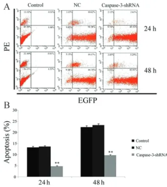
RNAi reduces apoptosis of vertebral CEP cells To determine whether silencing caspase-3 had an inhibitory effect on apoptosis of CEP cells, we qualitatively and quantitatively assessed the apoptosis rate of cells stained with PE-labeled annexin V after incubation in 1% FBS for 24 and 48 h. FCM analysis showed that the apoptosis rate of the CEP cells (annexin V––/PE++) was significantly lower after lentivirus transduction (Figure 4A and B). The apoptosis rate of the CEP-caspase-3 group
(4.73±0.35%) was significantly decreased at 24 h com-pared with the CEP-NC and CEP-CTR groups (13.67±0.42 and 13.33±0.45%, respectively, P,0.05). Similar to the results at 24 h, the apoptosis rate of the CEP-caspase-3 group was markedly lower than that of the CEP-NC (9.74±0.21vs23.32±0.62%, P,0.05) and CEP-CTR groups (9.74±0.21vs22.41±0.69%, P,0.05) at 48 h.
Discussion
Recently, many biological therapies for disc degen-erative diseases, including molecular, genetic, and cell-based strategies, have received increased attention and have been assessed for their abilities to halt and reverse disc degeneration (24,25). Unlike surgical procedures, these approaches focus on the basic pathophysiological processes of disc degeneration. Molecular therapies, including the use of growth factors (26), inflammation inhibitors (27), and proteinase inhibitors (28), have exhibited limited therapeutic durations and are not suitable for treating chronic degeneration processes. Gene therapies, using virus vectors or plasmids encoding exogenous proteins to stimulate matrix synthesis or inhibit its degradation, have overcome the limitations of mole-cular treatment (24,29-31). Cell-based therapies, includ-ing reimplantation of nucleus pulposus cells or stem cells, also have shown exciting results in animal experiments
Figure 1.Effects of serum deprivation on cartilage endplate cells after treatment for 48 h.A, DAPI staining of apoptotic bodies assessed by fluorescence microscopy (4006).B, Representative
graphs obtained by flow cytometry.C, Assay of caspase-3, -8, and -9 activities. *P,0.05vs1% fetal bovine serum (FBS) (Studentt -test). PI: propidium iodide; FITC: fluorescein isothiocyanate.
Figure 2.Evaluation of the lentivirus transduction rate, which was up to 90% as calculated by cellular enumeration using fluores-cence and light microscopy (2006).A, Control group;B, negative
(24,30,32,33). However, the degeneration of the CEP, with its associated compromised diffusion of oxygen and nutrients, would likely make these approaches impractical and unable to achieve the desired results (25). Therefore, we preferred to improve the status of the CEP to increase the nutrition of the disc, which is a prerequisite for reversing or repairing disc degeneration.
Inhibition of apoptosis of disc cells can improve or reverse the degenerative process (15,34,35), which provides a potential approach for improving the health of the CEP. RNAi has been widely used in gene therapy and has produced exciting results, including treatment of degenerative diseases (17). Sudo and Minami (15) applied this technique to halt disc degeneration by inhibiting apoptosis of the nucleus pulposus, as did Zhang et al. (34) in annular cells. To the best of our knowledge, this is the first investigation testing the protective effect of apoptosis inhibition on CEP cells.
Apoptosis of disc cells can be triggered by various stimuli, such as serum deprivation (36,37), H2O2 (38),
tumor necrosis factor alpha (26), compression (39), and cyclic stretch (34), through different apoptotic cascade
pathways. Although the precise mechanism of cell apoptosis is not fully understood, caspase-3 acts as a central executioner in the caspase apoptotic cascade pathway. Upregulation of the expression and activity of caspase-3 have been observed in different cellular apoptotic models, and specific inhibitors can successfully decrease apoptosis (36). Caspase-3 can be a therapeutic target for regulating the processes of disc degeneration. Our findings in the CEP apoptotic model are consistent with this. Caspase-3 activity was dramatically increased compared with that in the control cells incubated in 10% FBS, resulting in marked apoptosis as demonstrated by DAPI staining and FCM analysis. Interestingly, caspase-8 and -9 activities were also upregulated, and the precise apoptotic pathway used will require further investigation.
RNAi is highly effective in gene knockdown (19), but inefficient delivery and transient effects, its two greatest disadvantages, prohibit its application in chronic degen-erative diseases. Lentivirus can easily integrate into the host genome and stably encode shRNA to overcome these drawbacks (19). As a result, lentivirus-mediated caspase-3 shRNA was explored as a means to silence endogenous caspase-3 expression in the present study. The transduction rate was almost 90%. The quantity of
Figure 3. Expression of caspase-3 is suppressed by lentiviral caspase-3-shRNA after treatment with 1% fetal bovine serum for 48 h, withb-actin used as an internal control.A, Quantitative analysis of caspase-3 mRNA expression assessed by RT-PCR. **P,0.01vsall other groups (one-way ANOVA).B, Representative Western blot showing pro- and active-caspase-3 expression determined in different groups. NC: negative control group.
caspase-3 mRNA was reduced by 44% compared with that observed in the CEP-NC group. Expression of pro-and active-caspase-3 protein was also decreased. These data indicate that caspase-3 can be effectively and specifically knocked down by shRNAs. Moreover, a significant decrease of the apoptotic rate in the CEP-caspase-3 group was observed in concert with suppres-sion of the caspase-3 expressuppres-sion cascade. These findings suggest that knockdown of a harmful gene during apoptosis has the potential to enable increased cell survival. However, follow-up in vivo experiments are required to determine whether the survival of the cells can delay the degeneration of the CEP and arrest or reverse the degeneration of the IVD.
Advanced cases of disc degeneration with endplate calcification or disappearance are not candidates for this approach, which is unable to regenerate the whole endplate to supply additional oxygen and nutrition. Taken together, our results lead us to conclude that lentiviral caspase-3 shRNA-mediated RNAi successfully silenced endogenous caspase-3 expression and pre-vented inappropriate or premature apoptosis.
Acknowledgments
The authors thank Liang-Fei Niu for his technical assistance with lentivirus construction. The authors thank Shanghai Health Bureau (#2009-35) for funding support.
References
1. Adams MA, Roughley PJ. What is intervertebral disc degeneration, and what causes it?Spine2006; 31: 2151-2161, doi: 10.1097/01.brs.0000231761.73859.2c.
2. de Schepper EI, Damen J, van Meurs JB, Ginai AZ, Popham M, Hofman A, et al. The association between lumbar disc degeneration and low back pain: the influence of age, gender, and individual radiographic features.Spine 2010; 35: 531-536, doi: 10.1097/BRS.0b013e3181aa5b33. 3. Magnier C, Boiron O, Wendling-Mansuy S, Chabrand P, Deplano V. Nutrient distribution and metabolism in the intervertebral disc in the unloaded state: a parametric study. J Biomech 2009; 42: 100-108, doi: 10.1016/j.jbiomech. 2008.10.034.
4. Raj PP. Intervertebral disc: anatomy-physiology-pathophy-siology-treatment.Pain Pract2008; 8: 18-44, doi: 10.1111/ j.1533-2500.2007.00171.x.
5. Roberts S, Urban JP, Evans H, Eisenstein SM. Transport properties of the human cartilage endplate in relation to its composition and calcification.Spine1996; 21: 415-420, doi: 10.1097/00007632-199602150-00003.
6. Grunhagen T, Shirazi-Adl A, Fairbank JC, Urban JP. Intervertebral disk nutrition: a review of factors influencing concentrations of nutrients and metabolites. Orthop Clin North Am2011; 42: 465-477, vii, doi: 10.1016/j.ocl.2011.07. 010.
7. Ariga K, Miyamoto S, Nakase T, Okuda S, Meng W, Yonenobu K, et al. The relationship between apoptosis of endplate chondrocytes and aging and degeneration of the intervertebral disc. Spine 2001; 26: 2414-2420, doi: 10.1097/00007632-200111150-00004.
8. Grunhagen T, Wilde G, Soukane DM, Shirazi-Adl SA, Urban JP. Nutrient supply and intervertebral disc metabolism. J Bone Joint Surg Am 2006; 88 (Suppl 2): 30-35, doi: 10.2106/JBJS.E.01290.
9. Hee HT, Chuah YJ, Tan BH, Setiobudi T, Wong HK. Vascularization and morphological changes of the endplate after axial compression and distraction of the intervertebral disc. Spine 2011; 36: 505-511, doi: 10.1097/BRS.0b013e 3181d32410.
10. Rajasekaran S, Babu JN, Arun R, Armstrong BR, Shetty AP, Murugan S. ISSLS prize winner: A study of diffusion in human lumbar discs: a serial magnetic resonance imaging
study documenting the influence of the endplate on diffusion in normal and degenerate discs. Spine 2004; 29: 2654-2667, doi: 10.1097/01.brs.0000148014.15210.64.
11. Shirazi-Adl A, Taheri M, Urban JP. Analysis of cell viability in intervertebral disc: Effect of endplate permeability on cell population.J Biomech2010; 43: 1330-1336, doi: 10.1016/ j.jbiomech.2010.01.023.
12. Scaffidi C, Fulda S, Srinivasan A, Friesen C, Li F, Tomaselli KJ, et al. Two CD95 (APO-1/Fas) signaling pathways.EMBO J1998; 17: 1675-1687, doi: 10.1093/emboj/17.6.1675. 13. Philchenkov A. Caspases: potential targets for regulating
cell death.J Cell Mol Med2004; 8: 432-444, doi: 10.1111/ j.1582-4934.2004.tb00468.x.
14. Ruest LB, Khalyfa A, Wang E. Development-dependent disappearance of caspase-3 in skeletal muscle is post-transcriptionally regulated.J Cell Biochem2002; 86: 21-28, doi: 10.1002/jcb.10211.
15. Sudo H, Minami A. Caspase 3 as a therapeutic target for regulation of intervertebral disc degeneration in rabbits. Arthritis Rheum2011; 63: 1648-1657, doi: 10.1002/art.30251. 16. Hannon GJ. RNA interference.Nature2002; 418: 244-251,
doi: 10.1038/418244a.
17. Davidson BL, McCray PB Jr. Current prospects for RNA interference-based therapies.Nat Rev Genet2011; 12: 329-340, doi: 10.1038/nrg2968.
18. de Fougerolles A, Vornlocher HP, Maraganore J, Lieberman J. Interfering with disease: a progress report on siRNA-based therapeutics.Nat Rev Drug Discov2007; 6: 443-453, doi: 10.1038/nrd2310.
19. Couto LB, High KA. Viral vector-mediated RNA interference. Curr Opin Pharmacol 2010; 10: 534-542, doi: 10.1016/ j.coph.2010.06.007.
20. Malashicheva AB, Kanzler B, Tolkunova EN, Trono D, Tomilin AN. [The application of lentiviral vectors for tissue-specific gene manipulations].Tsitologiia2008; 50: 370-375. 21. Sinn PL, Sauter SL, McCray PB Jr. Gene therapy progress and prospects: development of improved lentiviral and retroviral vectors - design, biosafety, and production.Gene Ther2005; 12: 1089-1098, doi: 10.1038/sj.gt.3302570. 22. Kakutani K, Nishida K, Uno K, Takada T, Shimomura T,
interferencein vitro.J Orthop Res2006; 24: 1271-1278, doi: 10.1002/jor.20171.
23. Suzuki T, Nishida K, Kakutani K, Maeno K, Yurube T, Takada T, et al. Sustained long-term RNA interference in nucleus pulposus cellsin vivomediated by unmodified small interfering RNA. Eur Spine J 2009; 18: 263-270, doi: 10.1007/s00586-008-0873-9.
24. Fassett DR, Kurd MF, Vaccaro AR. Biologic solutions for degenerative disk disease.J Spinal Disord Tech2009; 22: 297-308, doi: 10.1097/BSD.0b013e31816d5f64.
25. Nishida K, Suzuki T, Kakutani K, Yurube T, Maeno K, Kurosaka M, et al. Gene therapy approach for disc degeneration and associated spinal disorders.Eur Spine J 2008; 17 (Suppl 4): 459-466, doi: 10.1007/s00586-008-0751-5.
26. Wei A, Brisby H, Chung SA, Diwan AD. Bone morphoge-netic protein-7 protects human intervertebral disc cells in vitro from apoptosis. Spine J 2008; 8: 466-474, doi: 10.1016/j.spinee.2007.04.021.
27. Roberts S, Evans H, Menage J, Urban JP, Bayliss MT, Eisenstein SM, et al. TNFalpha-stimulated gene product (TSG-6) and its binding protein, IalphaI, in the human intervertebral disc: new molecules for the disc.Eur Spine J 2005; 14: 36-42, doi: 10.1007/s00586-004-0798-x. 28. Shimoda M, Ghobrial RM, Carmody IC, Anselmo DM,
Farmer DG, Yersiz H, et al. Predictors of survival after liver transplantation for hepatocellular carcinoma associated with hepatitis C.Liver Transpl 2004; 10: 1478-1486, doi: 10.1002/lt.20303.
29. Yoon ST, Park JS, Kim KS, Li J, Attallah-Wasif ES, Hutton WC, et al. ISSLS prize winner: LMP-1 upregulates intervertebral disc cell production of proteoglycans and BMPsin vitroandin vivo.Spine2004; 29: 2603-2611, doi: 10.1097/01.brs.0000146103.94600.85.
30. Kepler CK, Anderson DG, Tannoury C, Ponnappan RK. Intervertebral disk degeneration and emerging biologic treatments.J Am Acad Orthop Surg2011; 19: 543-553.
31. Sowa G, Westrick E, Pacek C, Coelho P, Patel D, Vadala G, et al.In vitroandin vivotesting of a novel regulatory system for gene therapy for intervertebral disc degeneration.Spine 2011; 36: E623-E628, doi: 10.1097/BRS.0b013e3181 ed11c1.
32. Allon AA, Aurouer N, Yoo BB, Liebenberg EC, Buser Z, Lotz JC. Structured coculture of stem cells and disc cells prevent disc degeneration in a rat model.Spine J2010; 10: 1089-1097, doi: 10.1016/j.spinee.2010.09.014.
33. Huang S, Tam V, Cheung KM, Long D, Lv M, Wang T, et al. Stem cell-based approaches for intervertebral disc regen-eration.Curr Stem Cell Res Ther2011; 6: 317-326, doi: 10.2174/157488811797904335.
34. Zhang YH, Zhao CQ, Jiang LS, Dai LY. Lentiviral shRNA silencing of CHOP inhibits apoptosis induced by cyclic stretch in rat annular cells and attenuates disc degeneration in the rats. Apoptosis 2011; 16: 594-605, doi: 10.1007/ s10495-011-0596-y.
35. Ding F, Shao ZW, Xiong LM. Cell death in intervertebral disc degeneration.Apoptosis 2013; 18: 777-785, doi: 10.1007/ s10495-013-0839-1.
36. Park JB, Park IC, Park SJ, Jin HO, Lee JK, Riew KD. Anti-apoptotic effects of caspase inhibitors on rat intervertebral disc cells.J Bone Joint Surg Am 2006; 88: 771-779, doi: 10.2106/JBJS.E.00762.
37. Liu J, Wang J, Zhou Y. Upregulation of BNIP3 and translocation to mitochondria in nutrition deprivation induced apoptosis in nucleus pulposus cells.Joint Bone Spine2012; 79: 186-191, doi: 10.1016/j.jbspin.2011.04.011.
38. Kim KW, Ha KY, Lee JS, Rhyu KW, An HS, Woo YK. The apoptotic effects of oxidative stress and antiapoptotic effects of caspase inhibitors on rat notochordal cells.Spine2007; 32: 2443-2448, doi: 10.1097/BRS.0b013e318157395a. 39. Walter BA, Korecki CL, Purmessur D, Roughley PJ, Michalek