A Study of Differential Expression of
Testicular Genes in Various Reproductive
Phases of
Hemidactylus flaviviridis
(Wall
Lizard) to Derive Their Association with Onset
of Spermatogenesis and Its Relevance to
Mammals
Hironmoy Sarkar1, Satyapal Arya2, Umesh Rai1*, Subeer S. Majumdar2*
1Department of Zoology, University of Delhi, Delhi, 110007, India,2Cellular Endocrinology Laboratory, National Institute of Immunology, New Delhi, 110067, India
*rai_u@rediffmail.com(UR);subeer@nii.ac.in(SSM)
Abstract
Testis ofHemidactylus flaviviridis, commonly known as Indian wall lizard, displays a lack of cellular and metabolic activity in regressed phase of testis during non-breeding season of the year. Retracted Sertoli cells (Sc), fibroid myoid cells and pre-meiotic resting spermato-gonia are observed in such testis. This situation is akin to certain forms of infertility in men where hormone supplementation fails to generate sperm despite the presence of Sc and germ cells (Gc) in testis. In testis of lizard, spermatogenesis is reinitiated upon increased level of hormones during appropriate season (phase of recrudescence). Study of genes associated with generation of sperm, from regressed adult testis in lizard, may provide valu-able information for understanding certain forms of male idiopathic infertility. Subtractive hybridization using testicular RNA obtained from the regressed and active phases of lizard reproductive cycle led to identify eight partial mRNA sequences that showed sequence homology with mice genes. We further evaluated the gene expression prolife by real-time PCR in three different reproductive phases ofH.flaviviridis: regressed (pre-meiotic), recru-descent (meiotic) and active (post meiotic), for comparison with the corresponding testicular phases found in testis of 5 days (pre-meiotic), 20 days (meiotic) and 60 days (post-meiotic) old mouse. This is the first report where genes associated with progression of spermatogen-esis during active phase, which follows a regressed state of adult testis, were identified in lizard and found to be conserved in mouse. Six important genes,Hk1,Nme5,Akap4,Arih1,
Rassf7andTubb4bwere found to be strictly associated with active spermatogenesis in both mouse and lizard. Factors interfering with the expression of any of these genes may potentially abrogate the process of spermatogenesis leading to infertility. Such information may shed light on unknown causes of idiopathic male infertility.
OPEN ACCESS
Citation:Sarkar H, Arya S, Rai U, Majumdar SS (2016) A Study of Differential Expression of Testicular Genes in Various Reproductive Phases of
Hemidactylus flaviviridis(Wall Lizard) to Derive Their Association with Onset of Spermatogenesis and Its Relevance to Mammals. PLoS ONE 11(3): e0151150. doi:10.1371/journal.pone.0151150
Editor:Balasubramanian Senthilkumaran, University of Hyderabad, INDIA
Received:November 7, 2015
Accepted:February 24, 2016
Published:March 10, 2016
Copyright:© 2016 Sarkar et al. This is an open access article distributed under the terms of the Creative Commons Attribution License, which permits unrestricted use, distribution, and reproduction in any medium, provided the original author and source are credited.
Data Availability Statement:All sequences obtained from subtractive hybridization are available from the NCBI database (accession numbers JZ822542– JZ822576).
Introduction
Spermatogenesis is the complex biological process by which sperm are produced by successive mitosis and meiosis. This requires extensive intercellular coordination within the testis among different cell types, e.g., germ cells, Sertoli cells, Leydig cells, peritubular cells, etc. Intracellular coordination links to the timely expression of certain genes and their interactions, which col-lectively play a major role in the successful completion of sperm production.
Seminiferous tubules in the testis are surrounded by lymphatic endothelium [1]. The peri-tubular myoid cells and basal lamina provide structural support to seminiferous tubules within which Sertoli cell and germ cells resides. The Sertoli cells give structural framework for the organization of tubules and provide niche for the germ cell division and differentiation [2,3]. Rodents, mice and rats are most commonly used to study regulation of male fertility. In them, undifferentiated spermatogonial stem cells are situated at basement membrane either as single cell (As) or as pairs of cells (Apr) or as chains of aligned (Aal) cells [4]. Aal cells divide and dif-ferentiate into spermatocytes and after two rounds of meiosis develop into round spermatids. These round spermatids become elongated and eventually lose their large portion of cytoplasm to finally develop into sperm [5]. However, there is a remarkable variation in this process from species to species and from seasonal breeders to non-seasonal breeders.
In mouse (continuous breeder), various phases of testicular development appears as the ani-mal matures from neonatal to adulthood. At around 5 days of post natal age (neonatal), the tes-tis consist of undifferentiated spermatogonial cells and more than 50 percent of those remain in the resting phase with no cellular division and differentiation [6]. At 8–10 days of age, the beginning of meiosis (germ cells in leptotene stage) is apparent in a few tubules. At around 19 days, nearly half of all tubules exhibit the initiation of meiosis (presence of pachytene). A 60-day old mouse is considered an adult, with testicular sperms and reproductive maturity [6].
As opposed to mouse, most of the lizard species are seasonal breeders, they mate in spring, offspring hatches in summers. Seasonal reproduction is a tactic to make use of energy in an economical manner. The wall lizards have a prenuptial cycle of sperm maturation. Prenuptial reptiles generates sperm prior to or during the mating period [7].Hemidactylus flaviviridis, commonly known as Indian wall lizard, is a seasonally breeding animal. Based on seasonal var-iations they have three reproductive phases within a year [8], namely- regressed (June and July), recrudescent (August and September), and active phase (October-May). Regressed Phase is the most inactive phase of spermatogenesis. There is a lack of cellular and metabolic activity in testis during this phase. Only retracted Sertoli cells and undifferentiated pre-meiotic resting spermatogonia are observed in the testis. All other advanced germ cells are absent. Peritubular myoid cells become fibroid and overall size of the testis is reduced to the smallest [9]. This is a situation akin to several forms of infertility in men [10]. In lizards, complete arrest of sper-matogenesis (observed for a period of about 2 months) in regressed phase, followed by the ini-tiation of spermatogenesis occurs through proliferation of spermatogonia in recrudescence phase. Majority of the germ cell development and maturation occurs in the active phase where sperm are released into the lumen of seminiferous tubules. In the winter months (November to January), spermatogenesis is slow and active breeding happens from February to May when sperm are continuously generated within the testis [8,9,11]. Study of genes involved in genera-tion of sperm from regressed testis, immediately following stage of recrudescence may provide valuable information for treating infertile men having inactive spermatogenesis in spite of pres-ence of Sc and Gc in the testis (similar to the testis of regressed phase in lizards).
We have previously reported the differential gene expression in testis of three different reproductive phases of wall lizard by microarray analysis [12]. As microarray was performed using mouse array chip, wall lizard specific gene sequences were not available for further
studies. The aim of the present study was to identify the genes which are differentially
expressed in the testis during active and regressed phase of reproductive cycle in wall lizards by subtractive hybridization technique. Presence of such genes, specifically expressed in testis dur-ing regressed, recrudescent and active phases of wall lizards were compared with the testis from neonatal (no spermatogenesis), pubertal (initiation of spermatogenesis) and adult phases (all stages of spermatogenesis) of mice, respectively. Such study will not only enrich our current understanding about genes involved in the onset of spermatogenesis and testicular develop-ment which are conserved across the species but will also help in addressing issues related to certain forms of infertility.
Materials and Methods
Tissue collection and RNA isolation
Laboratory mouse strain FVB/J was kept in animal house of National Institute of Immunology according to guidelines provided by The Committee for the Purpose of Control and Supervi-sion of Experiments on Animals (CPCSEA), government of India. Indian wall lizards Hemi-dactylus flaviviridiswere procured from a local supplier in Delhi, India, and were kept in the animal house of Department of Zoology, University of Delhi, and maintained as described ear-lier by us previously [13]. Adult male wall lizards, of body weight around 8 to10 grams in the months of June (during regressed phase), September (recrudescent phase) and February (active phase) were procured and maintained (12 hr light: 12hr dark) in wooden cages with wire meshes on the side and on top, and were fed live insects as food. The animals were acclimatized to the laboratory conditions under confinement for a week prior to the commencement of experiments. All the experiments regarding animals for this study were performed according to the guidelines provided and approved by Institutional Animal Ethics Committee (IAEC), University of Delhi and National institute of Immunology under guidance of The Committee for the Purpose of Control and Supervision of Experiments on Animals (CPCSEA), govern-ment of India. Isolation of testes for all three phases (active, recrudescent and regressed) was done according to a previously mentioned protocol by us [12]. For a single biological sample of testicular tissue from wall lizards, 10 to 15 from regressed, 4 to 5 from recrudescence and 2–3 males from active phase were sacrificed by decapitation. For the isolation of mouse testicular tissue from 5, 20 and 60 days post natal; the number of animals were sacrificed by cervical dis-location was 10–12, 4–6 and 2–3, respectively for each group. Testes were snap-frozen in liquid nitrogen and crushed to powder with sterile mortar and pestle. The powder were mixed in 1ml Tri reagents (Sigma-Aldrich,USA). This generated one biological replicate for each phase and, likewise, three such biological replicates were generated for each sample group. RNA isolation from tissue in Tri-reagent was done according to manufacturer’s recommendation (Sigma-Aldrich,USA). To analyze for any changes in testicular weight, tubule diameter and body weight during different testicular phases in lizards and mice, we have randomly taken three animals (six testes) from each phase in lizard and mice. Body and testes weight were recorded. All testes were used for preparing histological sections for measurement of tubule diameter.
Histology of testis
Testes isolated from various phases of the reproductive cycle of wall lizards and different post-natal days of mice were fixed using 10 percent neutral buffered formalin, pH 7. Paraffin blocks were prepared, 5μm sections were made on microtome machine (2040AUTOCUT,
Subtractive hybridization
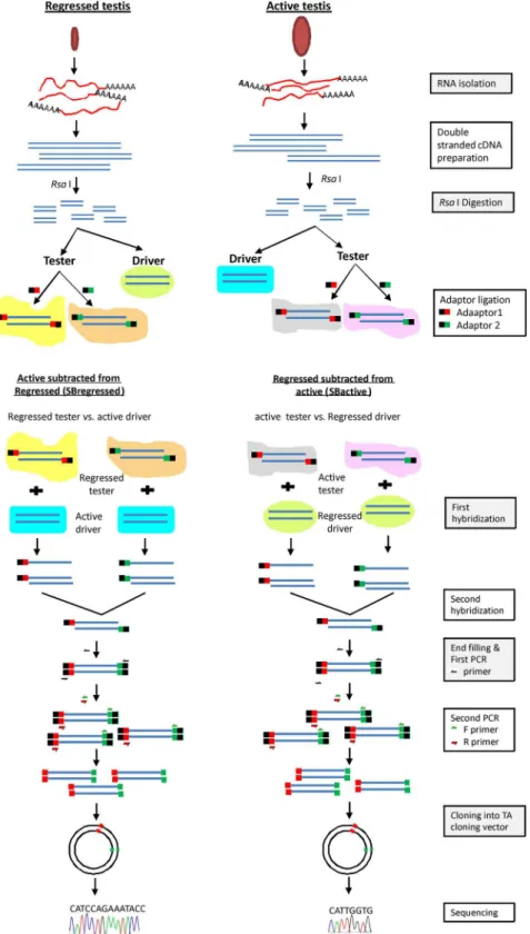
Subtractive hybridization was done according to the manufacturer’s protocol (Clontech,USA: cat.No–637401; protocol no- PT1117-1). In brief, 2μg of RNA isolated from testes in active
and regressed phases were used to generate double-stranded cDNAs, which were digested with RsaI enzyme, to generate blunt–end partial cDNAs. The digested resultant of each phase was divided in two portions named tester and driver. Testers from the active and regressed phases were again divided in two halves; the one half was ligated with adaptor1 and to another adap-tor2 (provided with the kit). The efficiency of adaptor ligation was determined as suggested in the kit protocol. Hybridization was performed between the active tester and the regressed driver, where regressed is subtracted from active (hereafter will be referred to SBactive) and the regressed tester and the active driver, where active is subtracted from regressed (hereafter will be referred to SBregressed). The hybridized samples were used for primary and secondary sub-traction PCRs with primers provided with the kit. Subsub-traction efficiency was evaluated between the subtracted and un-subtracted samples of the active phase using primers specific toH. flavi-viridis. PCR amplification was terminated in different cycles (28, 33, 38 and 42) and all PCR products were run on gel to validate the subtraction efficiency. The secondary PCR products, contained partial cDNAs, were ligated to a TA cloning vector in multiple cloning site (Promega Corp, USA), and were transformed intoE.coli. From each SBactive and SBregressed sample, randomly130 positive colonies were selected to perform colony-PCR to verify the insert size in TA cloning vector. Colonies showing higher molecular mass with a single amplification prod-uct in the colony-PCRs were considered; 48 different colonies, of each SBactive and SBre-gressed sample were selected. pDNA from those selected colonies were isolated with GenElute plasmid miniprep kit (Sigma, USA). The region of pDNA having partial cDNAs were
sequenced (Amnion Biosciences, India). A flowchart regarding the above-described subtractive hybridization process is illustrated inFig 1.
Sequence analysis and phylogenetic tree preparation
To find the identity of the partial cDNAs, the Basic Local Alignment Search Tool (BLAST) was used to align with nucleotide sequences deposited in the NCBI databases. Identities of partial cDNAs were assigned from the best aligned sequence having highest identity score and query coverage with least E value.
A phylogenetic tree was constructed with the partial cDNAs and their corresponding ortho-log sequences in other species. MEGA5 software was used for generation of the phyortho-logenetic tree by neighbor-joining statistical method and the bootstrap phylogenetic tree construction method, having a bootstrap replication of 1000 [14].
Reverse Transcription and Real-Time PCR
1μg of RNA, isolated from the testes samples of lizards and mice, was treated with DNaseI
according to the manufacturer’s protocol (Thermo Scientific, USA). To generate cDNAs form the treated RNAs, M-MLV reverse transcriptase (Promega Corp, USA) were used with oligo (dT)15as primer in a final volume of 25μl. cDNA was diluted 3 times with nuclease-free water,
and subsequent PCRs were carried out using 1μl of the diluted cDNA as template.
Fig 1. Flowchart of subtractive hybridization protocol for lizard testicular samples.Schematic representation depicting method of subtractive hybridization between RNA isolated from testes of two different reproductive phases (active and regressed phase) of wall lizard.
Real-time PCR was carried out with syber green using Mesa-green master mix kit (Eurogen-tech, Belgium) in Eppendorf Realplex mastercycler machine (Eppendorf, Germany). Reaction setup and PCR primer efficiency calculations were done by previously described procedure by us [15,16]. Primers with an efficiency of 1±0.2 were considered for further use. To evaluate the gene expression pattern in the testis of three reproductive phases of wall lizards and the three developmental phases of mice, three biological replicates were run for every phase of testicular sample, and for each biological sample, there were two technical replicates along with one con-trol for reverse-transcriptase and one non-template concon-trol. The expression ofGapdh (Glycer-aldehyde-3-Phosphate Dehydrogenase) for wall lizard andPpia(Peptidylprolyl Isomerase A) for mice was used as reference gene for normalizing the target genes expression. Calculation for relative fold change with respect to the active phase in wall lizards and 60 days in mice was performed by 2-ΔΔctmethod as previously described by us [17].
A clustered image heat-map was generated with the relative fold change values, using online tool CIMminer (http://discover.nci.nih.gov/cimminer/home.doas on date 22 July, 2015) to represent the gene expression profile between wall lizards and mice contemporary testis. For the generation of heat map methods like euclidean, average linkage, and quantile were used as distance method, cluster algorithm, and binning method, respectively.
Correlation between testis weight and gene expression
Testes weights (mg) of nine testicular samples from wall lizards (three biological replicates for each of three phases) were assigned percentage values by considering the highest value amongst all of them as 100. Here, the highest value came from one biological replicate of the active phase of wall lizard. In a similar manner, mice testicular weights from nine testicular samples of mouse (three biological replicates for each three developmental phases) were assigned per-centage values by considering the highest value amongst all of them as 100. A graph was Table 1. List of primers for real-time PCR.
Gene ID Species Primer sequences (5'-3') Product length NCBI Acc No
Forward Reverse
Hk1 Lizard GAATGACACGGTGGGAGTC TGTATGCCTCCTATGTTCTC 126 JZ822542
Mouse CAGTGTGAAGTCGGCCTGAT GGACCCATCATCCCCAAAGG 141 NM_001146100.1
Nme5 Lizard GGGATTATGTTCAATTAGCC CTATTCCTCAGCTGAAAGAG 199 JZ822546
Mouse CAGGTTCATGTTTCCAGCCG TCCTTGAAGTAGGGTTGGCG 100 NM_080637.3
Akap4 Lizard TGGAAAGTTGAGAGAGGAGG GGAAGTGGAACTGTACCTAG 105 JZ822547
Mouse AGGACAACAAGATCAGGACCG CAGCAGCACCCTTGGAATC 89 NM_001042542.2
Bco2 Lizard ACTATACAAAGCCCCTCATG GCTGCAGTAGGAATAGGTC 120 JZ822548
Mouse ATGTTGGGACCGAAGCAGAG GCAACGCCATTCCATCAAACC 196 NM_133217.3
Arih1 Lizard ATGTCTTGCAGCGAATCTTG AACCAGTCCATTATCTTCG 107 JZ822545
Mouse CAGTGTCGTGCCACACTCAT CGAAAGCACCTCTGTGGCAT 120 NM_019927.2
Rassf7 Lizard GGCTAGCCCTGACAAACGT AGCCAAGTGTGGACAGTATG 128 JZ822549
Mouse ACAGGTCGATTTGTCCTTGT GTATCGCCGATGGCTTTGG 229 NM_025886.3
Hmgb1 Lizard GTAGGGTATGCAGAACGAAG GACTTGTCCTGTGTTATACC 100 JZ822550
Mouse GGACTCTCCTTTAACCGCCA CCTTCGCTGGGACTAAGGTC 148 NM_010439.3
Tubb4b Lizard TACAGCTGAGGAGGAAGGAG GATGACACCAGGAAGAAAGG 170 JZ822551
Mouse CACTTACCACGGAGATAGCGA ACCTTCTGTGTAGTGCCCCTT 229 NM_146116.2
Gapdh Lizard ACACAGTCCATGCCATCACA GACCTTGCCAACAGCCTTA 134 JF303078.1
Ppia Mouse GTCGCTTTTCGCCGCTTGC TCTGCTGTCTTTGGAACT 127 NM_008907.1
generated for each gene, where y axis was assigned to the relative fold change values (obtained by 2-ΔΔctmethod) of testicular samples against their corresponding testis-weight in percentage on x axis. A linear regression line was drawn by connecting the expression values plotted on the graph. In the graph, independent linear regression line was generated for wall lizards and mice.
Statistical analysis
The relative fold change in gene expression data were represented as ±SEM (n = 3). Statistically significant difference among the samples were analyzed by one-way analyses of variance (ANOVA) followed by Newman-Keuls multiple comparison test. Difference in gene expression was considered significant when the p value was found to be less than 0.05. Correlations were determined between testis wt. and relative gene expression by Pearson's correlation coefficient analysis. p<0.05 was considered statistically significant correlation. All statistics and graph
preparation were done by GraphPad prism software (USA) version 5.0.
Results
Comparison of seasonal testicular growth in wall lizards with the
testicular development in mice
Cross section of testis from the regressed phase of a wall lizard showed small seminiferous tubules having small lumen, consisting mostly Sertoli cells and very few early germ cells (Fig 2A). The 5-day old mouse testis (Fig 2D) also had similar structure having mostly Sertoli cells and few early germ cells, except that it did not have any lumen in the seminiferous tubule.Fig 2B and 2Eshow cross sections of the recrudescent phase of wall lizards and 20-day old mouse testis, respectively. Testis from both species consisted of mostly pre-meiotic and meiotic stages of germ cells. Testis from active phase of wall lizards (Fig 2C) and adult 60-day old mice (Fig 2F) showed presence of all stages of germ cells along with spermatids. In the both testis, tubular diameter and testicular weight increased (Fig 2G, 2H, 2J and 2K) from regressed to active (in wall lizards) and from 5 days towards 60 days testis (in mice). The Body weight of wall lizards did not differ significantly among various reproductive phases (Fig 2I); whereas, body weight of mouse increased with age (Fig 2L).
Subtractive hybridization between active and regressed phase of
testicular RNA from wall lizards
Subtractive hybridization was done to identify genes that are differentially expressed between active and regressed phases of the testis. To get transcripts that were highly expressed in the active phase in comparison to the regressed phase, the testicular RNA from regressed phase was subtracted from the testicular RNA of active phase (SBactive). For identification of tran-scripts expressed more in the regressed phase compared with the active phase, the active phase testicular RNA was subtracted from the testicular RNA of regressed phase (SBregressed).
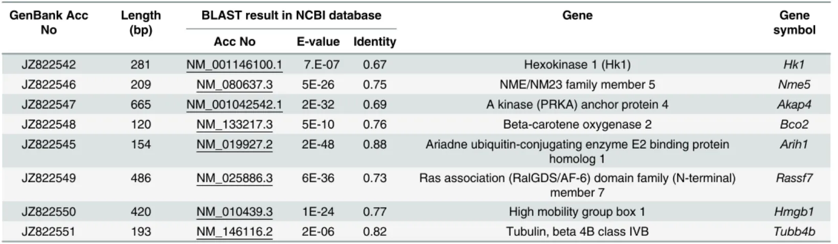
Sequencing of the fragments of transcripts, obtained from SBactive and SBregressed pro-vided 35 unique sequences (NCBI Acc No- JZ822542–JZ822576). Out of these, 27 sequences were from the active phase (SBactive) and 8 were from the regressed phase (SBregressed). Out of 27 sequences in the SBactive sample, 10 sequences were found to have similarity with known gene sequences from different species (Fig 3). Out of these 10, 8 partial gene sequences (Hk1,
Fig 2. Comparative analysis of testicular development in wall lizard and mice at different reproductive phase of testes.(A), (B) and (C) represent the cross section of testis isolated from regressed, recrudescent and active phase of reproductive cycle in wall lizard, respectively. Figure (D), (E) and (F) represent testis isolated from 5 day, 20 day and 60 day old mouse respectively. In the figure (C & F), as for representation, Leydig cell (blue), peritubular cell (red), Sertoli cell (black), germ cell (green), and spermatids (yellow) are shown with respective arrowhead. Figure (G) & (J) shows comparison of tubular diameter (n = 6), (H) & (K) shows comparison of testis weight (n = 6), (I) & (L) depicts the comparative analysis body weight (n = 3) between wall lizard and mice, respectively. One way analysis of variance followed by newman-keuls multiple comparison test was performed to get P value as significance level p<0.05. In a graph, different letters above the bars denotes significant difference in values (p<0.05), Bars refers to S.D. between replicates (n = 3). Scale bar on the figure A-F indicates 0.01mm. Rg-T, Rc-T, Ac-T are testes isolated from regressed, recrudescent, and active phase of wall lizard, respectively. 5d-T, 20d-T, and 60d-Tdenotes testes isolated from 5 days, 20 days and 60 days old mouse, respectively.
A phylogenetic tree was constructed using the bootstarp method, with known homolog sequences found in NCBI databases (S1 Fig). The sequence ofArih1(JZ822545) fromH. flavi-viridiswas found to be nearest toAnoliswith the highest homology, and formed an altogether different node. The sequences ofNme5(JZ822546) andTubb4b(JZ822551) were in greater homology withZ.albicollis. SequencesBco2(JZ822548),Rassf7(JZ822549) andHmgb1
(JZ822550) were in greater homology withXenopus. The sequence similarities ofHk1
(JZ822542) andAkap4(JZ822547) found to be higher withDrosophila.
Fig 3. Summarized result of subtractive hybridization analysis.Distribution of total partial sequences, obtained from subtractive hybridizations analysis between testicular RNA isolated from regressed and active phase of wall lizard.
doi:10.1371/journal.pone.0151150.g003
Table 2. List of partial transcript sequences of lizard and their sequence similarity with homolog-genes of mice.
GenBank Acc No
Length (bp)
BLAST result in NCBI database Gene Gene
symbol Acc No E-value Identity
JZ822542 281 NM_001146100.1 7.E-07 0.67 Hexokinase 1 (Hk1) Hk1
JZ822546 209 NM_080637.3 5E-26 0.75 NME/NM23 family member 5 Nme5
JZ822547 665 NM_001042542.1 2E-32 0.69 A kinase (PRKA) anchor protein 4 Akap4
JZ822548 120 NM_133217.3 5E-10 0.76 Beta-carotene oxygenase 2 Bco2
JZ822545 154 NM_019927.2 2E-48 0.88 Ariadne ubiquitin-conjugating enzyme E2 binding protein homolog 1
Arih1
JZ822549 486 NM_025886.3 6E-36 0.73 Ras association (RalGDS/AF-6) domain family (N-terminal) member 7
Rassf7
JZ822550 420 NM_010439.3 1E-24 0.77 High mobility group box 1 Hmgb1
JZ822551 193 NM_146116.2 2E-06 0.82 Tubulin, beta 4B class IVB Tubb4b
Comparative gene expression in the testis of wall lizards and mice by
real-time PCR analysis
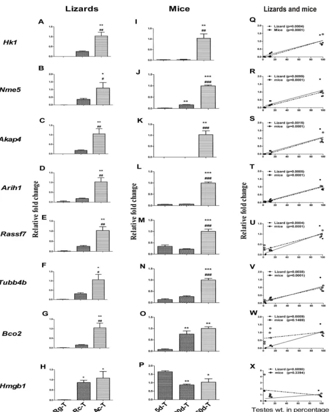
In order to analyze the mRNA expression pattern of 8 genesHk1,Nme5,Akap4,Arih1,Rassf7,
Tubb4b,Bco2andHmgb1in different testicular phases of wall lizard and mice, real-time PCR was performed using testicular RNA. ExceptHmgb1, rest other 7 genes were significantly high in the active phase of testis as compared to the regressed and recrudescent phases of wall liz-ards’testis (Fig 4A–4G). Significantly higher expression of six genesHk1,Nme5,Akap4,Arih1,
Rassf7andTubb4bwere found in the 60 days old mouse testes as compared to 5 days and 20 days old testis (Fig 4I–4N). Interestingly, the expression ofNme5andBco2were significantly higher in the 20-day old testis as compared to 5-day old testis in mice (Fig 4J and 4O). In wall lizards as well as in mice,Hk1,Nme5,Akap4,Arih1,Rassf7andTubb4bexpression were found to have significant correlation with testicular weight (Fig 4Q–4V). The increase in expression ofBco2in 20 days old mice did not have any significant correlation with the testicular weight, although the increase in expression in the wall lizards’testis was found to have significant posi-tive correlation with increase in testicular weight of wall lizards (Fig 4W). The relative expres-sion ofHmgb1was found to be significantly higher in the recrudescence phase compared with the regressed phase of wall lizards’testis. Comparable level of expressions was found between the recrudescence and the active phase of the wall lizards’testis (Fig 4H). In the mice testis, the expression level ofHmgb1was found to be similar in day 20 and day 60, however, there was significantly higher expression observed in the 5-days as compared to that at 20-day and 60-day testes (Fig 4P). Neither in wall lizards nor in mice, was the expression level ofHmgb1
found to be correlated with the increased testicular weight (Fig 4X).
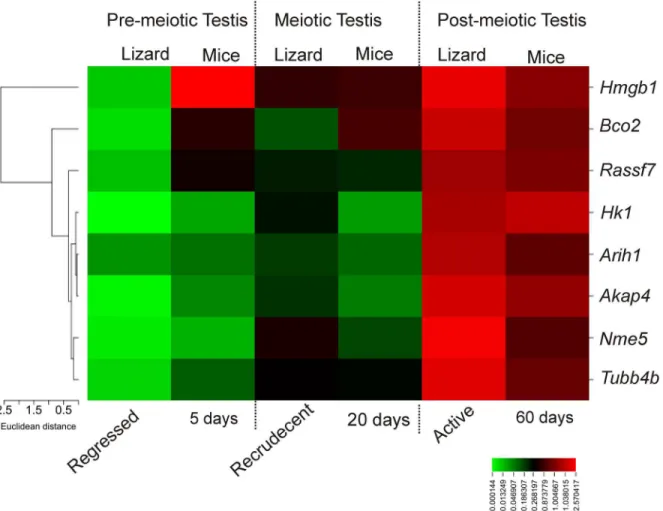
Relative fold change in gene expression values of all 8 genes were used to generate a heat map to compare the expression status between wall lizards and mice on similar testicular phases like pre-meiotic, meiotic and post-meiotic (Fig 5). The expression ofHmgb1was found to be high in pre-meiotic testis of mice, but not in the testis of wall lizards. The expression of
Hmgb1in meiotic and post-meiotic testis in wall lizards and mice were found to be compara-ble. We found thatHk1,Nme5,Akap4,Arih1,Rassf7,and Tubb4b and Bco2had a similar high expression in the testis of wall lizards during active phase and at 60days in the mice.Hk1,
Arih1,Akap4andNme5had comparable expressions in the regressed phase in wall lizards and 5 days old mice testis.Bco2,Rassf7andTubb4bhad higher expression in testis of 5 days old mice compared to testis in regressed phase in the wall lizard.Rassf7,Aih1andTubb4bhad a comparable expression in the testis of recrudescent phase of wall lizard and in 20-day old mice testis. The expression ofBco2was found to be high in testis of 20days old mice than in the testis of recrudescent phase of the wall lizard. During recrudescent phase of the wall lizard, expres-sion status ofHk1andAkap4were higher as compared to the testis of 20 days old mice. There was higher abundance ofNme5in the testis from recrudescent phase of lizards although it was not in testis of 20 days old mice (Fig 5) which is a comparable testicular phase with recrudes-cent of lizards.
Probable protein-protein interactions of differentially expression genes in
testicular phases of wall lizards and mice
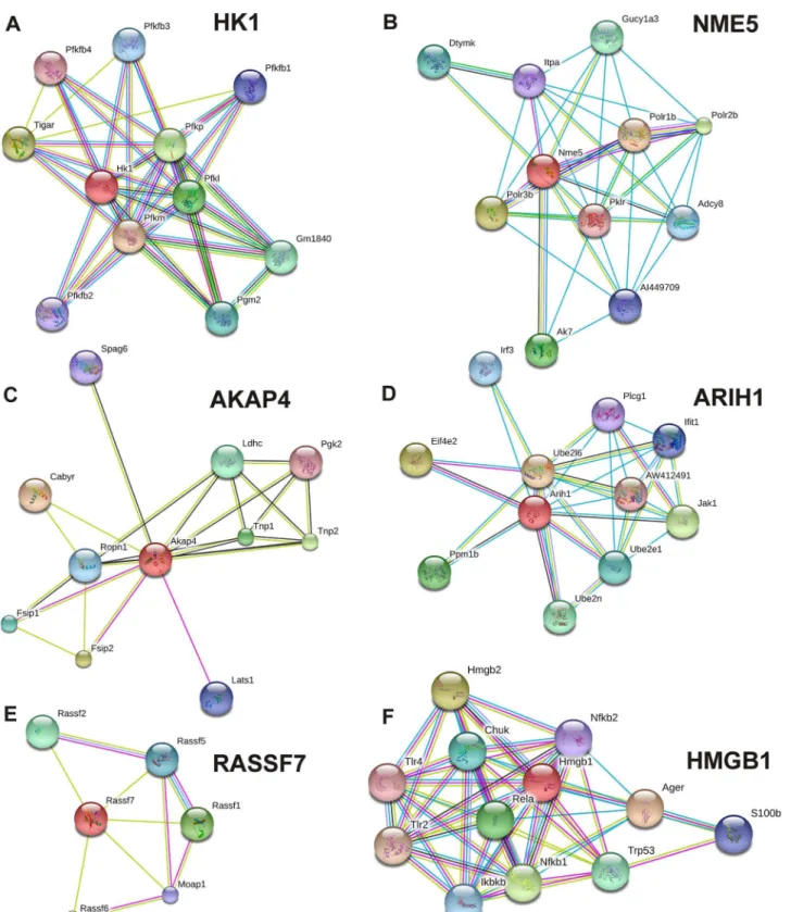
In the STRING database, there were known and predicted protein-protein interactions ofHk1,
PLCG1. RASSF7 interacted with RASSF6, RASSF1, RASSF2, RASSF5, and MOAP1. HMGB1 interacted with AGER, HMGB2, NFKB1, RELA, TRP53, CHUK, IKBKB, S100B, NFKB2, TLR4, and TLR2. The interaction score and name of interacting proteins are mentioned inS1 Table. Interactome ofBco2andTubb4bwere not found in the STRING database.
Discussion
In this study, we have compared the gene expression profiles in the testis of wall lizards and mice during various phases of testicular activity (pre-meiotic, meiotic and post-meiotic). Ear-lier we have compared the differential transcriptome of three reproductive phases of testis from wall lizard [12]. Since lizard array chip was not available, wall lizard testicular RNA was hybridized on known mouse array chip. As gene sequences of wall lizard and mouse vary widely, the expression signals due to hybridization was low in confidence. Additionally as liz-ards gene sequences are not available, the validation of microarray data and further expression
Rc-T, Ac-T are testes from regressed, recrudescent, and active phase of wall lizard. 5d-T, 20d-T, and 60d-T are testes from 5 days, 20 days and 60 days old mice.*p<0.05,**p<0.01 and***p<0.001 represents the comparison of Rg-T with Rc-T and Ac-T in wall lizards and 5d-T with 20d-T and 60d-T in mice.
#p<0.05,##p<0.01 and###p<0.001 represents the comparison of Rc-T with Ac-T in wall lizards and 20d-T with 60d-T in mice.
doi:10.1371/journal.pone.0151150.g004
Fig 5. Heat map analysis of gene expression between lizards and mice.Heat map analysis showing differential gene expression pattern in pre-meiotic, meiotic and post-meiotic testes of wall lizard and mice. The relative fold change in gene expression for genesHk1,Nme5,Akap4,Arih1,Tubb4b,Rassf7, Bco2, andHmgb1were compared in corresponding testes of wall lizards and mice. Color from red to green indicates high to low expression.
Fig 6. Predicted interaction among proteins.Probable interactome of different proteins, of which mRNA were differentially expressed in different testicular phase of wall lizard mice. Probable interacting partner of gene products,Hk1(A),Nme5(B),Akap4(C),Arih1(D),Rassf7(E), andHmgb1(F) with other proteins as per STRING database.
studies with gene specific primers are not efficient. To overcome these problems, we have com-pared the testicular RNA expression profile between regressed and active phase of spermato-genesis in lizard by subtractive hybridization.
Thirty five unique partial RNA sequences were identified in testis of wall lizard. The 17 and 8 partial sequences from active and regressed phase, respectively, showed no significant simi-larity with known mRNA sequences in the database. The reason for the lack of simisimi-larity of these partial sequences with known mRNA sequences may reside in the lack of longer sequence reads (eg. JZ822572 and JZ822543), which is necessary to attain a significant match. There were other sequences, which were found to be relatively longer and showed partial similarity to short region of more than one mRNAs (eg. JZ822573, JZ822574, JZ822575); the query coverage for these sequence alignments were also very low (below 40%). Therefore, these 17 and 8 sequences could not be reliably identified as mRNA sequences of known genes. Hence, in the present study we could not consider these partial sequences for further experiments.
Ten partial RNAs among 35, showed sequence homology with mRNA sequences of ortholo-gus genes from other species and found to be abundant in the testis during active phase com-pared to regressed phase of wall lizards. Out of 10 sequences, 8 showed sequence similarity with corresponding mice gene sequences. In order to evaluate testicular phase related similarity in the expression pattern between two species, we analyzed the expression profiles of these 8 genes using real-time PCR in different phases of mice testicular development and lizard repro-ductive cycle. 7 genes,Hk1,Nme5,Akap4,Arih1,Rassf7,Bco2andTubb4bwere found to be uniformly associated with testis with active spermatogenesis in both the species. Expression of 6 genesHk1,Nme5,Akap4,Arih1,Rassf7andTubb4bshowed positive correlations with increased testicular weight in both wall lizards and mice, that represents their conserved pat-tern of expression between these two species. Specifically higher expression ofHmgb1in testis of 5 days old mice suggested that the regressed testis cannot be considered as neonatal testis in terms of developmental status.
found in adult testis ofMacropus eugenii, located in the cytoplasm of round and elongated spermatids, and in the sperm tail [26]. In the interactome database, it interacted with FSIP1 and FSIP2, which are fibrous sheath interacting proteins, and with germ-cell-specific proteins like SPAG6 and PGK2, and TNP1 and TNP2, which are nucleosome compaction proteins. BCO2enzyme is a mitochondrial carotenoid-oxygenase, which catalyses asymmetric cleavage of carotenoids and protects the cells from carotenoid induced oxidative stress [27]. We found a higher expression ofBco2at 20 days (meiotic) and 60 days (post-meiotic) in comparison to that in 5 days old mice testis. This higher expression ofBco2in testis showing meiosis can be correlated with the requirement of retinoic acid (RA) for the meiotic initiation of germ cells. When germ cells undergo meiosis, they require retinoic acid [28] which is product of beta-car-otene. Hence, there is a possibility that during meiosis, germ cells may undergo oxidative stress due to higher amount of beta-carotene; therefore,Bco2may create a balance for maintaining the right amount of beta-carotenoid in the germ cells.ARIH1can induce aggresome formation and helps in the ubiqutinilation of target proteins [29,30]. It is also found to be the marker for proliferation [31]. As the active testis of wall lizards and adult testis of mice are highly prolifer-ative, a higher expression ofArih1in those phases of testis in wall lizards and mice can be justi-fied. In the predicted interactome database, this protein interacts with the JAK1, which is a protein-tyrosine kinase found in human spermatozoa [32] and helps in germ cell differentia-tion inXenopus[33].RASSF7is a ras association domain-containing protein 7, which nega-tively regulates stress-induced JNK activation [34]. It is also a regulator of microtubule dynamics in a cell and is required for mitosis [35]. The important paralog of this gene isRassf8, found to be essential for maintaining adherent junctions (AJ) in epithelial cells and has a role in epithelial cell migration [36]. We observed a higher expressionRassf7in the testis of active phase and adult testis of wall lizards and mice, respectively. In the interactome data, it showed interaction with other ras-associated proteins.TUBB4Bis a tubulin protein. We found its expression in the adult testis of mice and active testis of lizards, from which we can infer that the requirement of this component of microtubules may be higher in mature testis for provid-ing structural support to large number of germ cells.HMGB1is a high-mobility group binding protein. The expression ofHmgb1was found to be comparable during recrudescence and active phase of lizards. This was also comparable in 20 and 60 days old testis of mice. However, this was highest in neonatal testis (5 days) of mice. Interactome data of HMGB1, suggested its interaction with several proteins related to PI3K-Akt signaling pathway, like NFKB1, RELA1, CHUK, and IKBKB, which are known to play important role in cellular growth and differentia-tion. TheHmgb1gene was found to be one of the 159 transcripts from testicular biopsy where the expression was found to be high in idiopathic infertile patients of AZFc microdeletion [37]. In many patients the cause of infertility was due to severe hypo spermatogenesis, this can be because the testis remains in state of immaturity which fails to develop sufficient functional sperm. The higher expression ofHmgb1in neonatal (immature) testis of mice and testis of infertile patients, led us to hypothesize that higher expression of HMGB1can keep the testis in immature state whereas moderate expression is seen during spermatogenesis.
Supporting Information
S1 Fig. Phylogenetic tree with partial sequences of lizards obtained from subtractive hybridization of testicular samples.Phylogenetic tree for the genes,Hk1(A),Nme5(B),
Akap4(C), Bco2 (D),Arih1(E),Rassf7(F),Hmgb1(G), andTubb4b(H). (TIF)
S1 Table. Predicted interacting proteins and their score of interaction-confidence. (DOC)
Acknowledgments
We are thankful to Dr. Indrashis Bhattacharya, of HNB Garhwal University, Uttarakhand for critical analysis of the paper and valuable suggestions.
Author Contributions
Conceived and designed the experiments: HS UR SSM. Performed the experiments: HS. Ana-lyzed the data: HS SA UR SSM. Wrote the paper: HS UR SSM.
References
1. Fawcett DW, Neaves WB, Flores MN. Comparative observations on intertubular lymphatics and the organization of the interstitial tissue of the mammalian testis. Biol Reprod.1973; 9(5):500–532. PMID: 4203289
2. Russel LD, Ettlin R., Hikim AP, Clegg ED. Mammalian spermatogenesis. Histological and Histopatho-logical Evaluation of the Testis. 1st ed. Cache river press, Bolesta, USA; 1990.
3. Clermont Y. Contractile elements in the limiting membrane of the seminiferous tubules of the rat. Exp Cell Res.1958; 15(2):438–440. doi:10.1016/0014-4827(58)90052-1PMID:13597909
4. De Rooij DG, Russell LD.All you wanted to know about spermatogonia but were afraid to ask. J Androl. 2000; 21(6):776–798. PMID:11105904
5. Kerr JB, Loveland KL, O’Bryan MK, de Kretser DM. Cytology of the Testis and Intrinsic Control Mecha-nisms. In: Neill DJ, editor. Knobil and Neill’s physiologyof reproduction. 3rd ed. Elsevier,
London.2006. pp. 827–948.
6. Nebel BR, Amarose AP, Hacket EM. Calendar of gametogenic development in the prepuberal male mouse. Science.1961; 134(3482):832–833. doi:10.1126/science.134.3482.832PMID:13728067
7. Kumar S, Roy B, Rai U. Hormonal Regulation of Testicular Functions in Reptiles. In: Norris OD and Lopez HK,editors. Hormonesand reproduction of vertebrates. 3rd Vol: Elsevier, London.2011. pp. 63–
88.
8. Sanyal MK, Prasad MRN. Reproductive cycle of the Indian House Lizard, Hemidactylus flaviviridis Rup-pell. Copeia.1967; 3:627–633. doi:10.2307/1442242
9. Khan UW, Rai U. Interrelationship among testicular cells in wall lizard Hemidactylus flaviviridis (Rup-pell): An ultrastructural seasonal and experimental study. Indian J Exp Biol.2004; 42(4):378–388. PMID:15088688
10. Stukenborg J-B, Kjartansdóttir KR, Reda A, Colon E, Albersmeier JP, Söder O. Male germ cell develop-ment in humans. Horm Res Paediatr.2014; 81(1):2–12. doi:10.1159/000355599PMID:24356336
11. Khan UW, Rai U. Paracrine role of testicular macrophages in control of Leydig cell activities in the wall lizard, Hemidactylus flaviviridis. Gen Comp Endocrinol.2008; 156(1):44–50. doi:10.1016/j.ygcen.2007. 10.006PMID:18086472
12. Gautam M, Mathur A, Khan MA, Majumdar SS, Rai U. Transcriptome Analysis of Spermatogenically Regressed, Recrudescent and Active Phase Testis of Seasonally Breeding Wall Lizards Hemidactylus flaviviridis. PLoS One; 2013; 8(3):e58276. doi:10.1371/journal.pone.0058276PMID:23536792
14. Tamura K, Peterson D, Peterson N, Stecher G, Nei M, Kumar S. MEGA5: Molecular evolutionary genet-ics analysis using maximum likelihood, evolutionary distance, and maximum parsimony methods. Mol Biol Evol.2011; 28(10):2731–2739. doi:10.1093/molbev/msr121PMID:21546353
15. Bhattacharya I, Pradhan BS, Sarda K, Gautam M, Basu S, Majumdar SS. A switch in Sertoli cell responsiveness to FSH may be responsible for robust onset of germ cell differentiation during prepu-bartal testicular maturation in rats. Am J Physiol Endocrinol Metab.2012; 303(7):E886–E898. doi:10. 1152/ajpendo.00293.2012PMID:22850685
16. Majumdar SS, Sarda K, Bhattacharya I, Plant TM.Insufficient androgen and FSH signaling may be responsible for the azoospermia of the infantile primate testes despite exposure to an adult-like hor-monal milieu. Hum Reprod. 2012; 27(8):2515–2525. doi:10.1093/humrep/des184PMID:22669085
17. Bhattacharya I, Basu S, Sarda K, Gautam M, Nagarajan P, Pradhan BS, et al. Low levels of Gαs and
Ric8b in testicular sertoli cells may underlie restricted FSH action during infancy in primates. Endocri-nology.2015; 156(3):1143–1155. doi:10.1210/en.2014-1746PMID:25549048
18. Schindler A, Foley E. Hexokinase 1 blocks apoptotic signals at the mitochondria. Cell Signal.2013; 25 (12):2685–2692. doi:10.1016/j.cellsig.2013.08.035PMID:24018046
19. Nakamura N, Shibata H, O’Brien DA, Mori C, Eddy EM.Spermatogenic cell-specific type 1 hexokinase is the predominant hexokinase in sperm. Mol Reprod Dev.2008; 75(4):632–640. doi:10.1002/mrd. 20791PMID:17924400
20. Nakamura N, Miranda-Vizuete A, Miki K, Mori C, Eddy EM. Cleavage of disulfide bonds in mouse sper-matogenic cell-specific type 1 hexokinase isozyme is associated with increased hexokinase activity and initiation of sperm motility. Biol Reprod.2008; 79(3):537–545. doi:10.1095/biolreprod.108.067561 PMID:18509164
21. Hwang KC, Ok DW, Hong JC, Kim MO, Kim JH. Cloning, sequencing, and characterization of the murine nm23-M5 gene during mouse spermatogenesis and spermiogenesis. Biochem Biophys Res Commun.2003; 306(1):198–207. doi:10.1016/S0006-291X(03)00916-1PMID:12788088
22. Munier A, Feral C, Milon L, Pinon VPB, Gyapay G, Capeau J, et al.A new human nm23 homologue (nm23-H5) specifically expressed in testis germinal cells. FEBS Lett.1998; 434(3):289–294. doi:10. 1016/S0014-5793(98)00996-XPMID:9742940
23. Munier A, Serres C, Kann ML, Boissan M, Lesaffre C, Capeau J, et al. Nm23/NDP kinases in human male germ cells: role in spermiogenesis and sperm motility? Exp Cell Res. 2003; 289(2):295–306. doi: 10.1016/S0014-4827(03)00268-4PMID:14499630
24. Brown PR, Miki K, Harper DB, Eddy EM.A-kinase anchoring protein 4 binding proteins in the fibrous sheath of the sperm flagellum. Biol Reprod. 2003; 68(6):2241–2248. doi:10.1095/biolreprod.102. 013466PMID:12606363
25. Miki K, Willis WD, Brown PR, Goulding EH, Fulcher KD, Eddy EM.Targeted disruption of the Akap4 gene causes defects in sperm flagellum and motility. Dev Biol. 2002; 248(2):331–342. doi:10.1006/ dbio.2002.0728PMID:12167408
26. Hu Y, Hu H, Park AJ, O’Brien DA, Shaw G, Renfree MB. A-kinase anchoring protein 4 has a conserved role in mammalian spermatogenesis. Reproduction. 2009; 137(4):645–653. doi:10.1530/REP-08-0337 PMID:19139142
27. Amengual J, Lobo GP, Golczak M, Li HNM, Klimova T, Hoppel CL, et al.A mitochondrial enzyme degrades carotenoids and protects against oxidative stress. FASEB J. 2011; 25(3):948–959. doi:10. 1096/fj.10-173906PMID:21106934
28. Hogarth CA, Griswold MD. Review series The key role of vitamin A in spermatogenesis. J Clin Invest. 2010; 120(4):956–962.
29. Parelkar SS, Cadena JG, Kim C, Wang Z, Sugal R, Bentley B, et al. The parkin-like human homolog of Drosophila ariadne-1 (HHARI) can induce aggresome formation in mammalian cells and is immunologi-cally detectable in Lewy bodies. J Mol Neurosci. 2012; 46(1):109–121. doi: 10.1007/s12031-011-9535-1PMID:21590270
30. Ardley HC, Tan NG, Rose SA, Markham AF, Robinson PA. Features of the parkin/ariadne-like ubiquitin ligase, HHARI, that regulate its interaction with the ubiquitin-conjugating enzyme, Ubch7. J Biol Chem. 2001; 276(22):19640–19647. doi:10.1074/jbc.M011028200PMID:11278816
31. Elmehdawi F, Wheway G, Szymanska K, Adams M, High AS, Johnson CA, et al.Human Homolog of Drosophila Ariadne (HHARI) is a marker of cellular proliferation associated with nuclear bodies. Exp Cell Res. 2013; 319(3):161–172. doi:10.1016/j.yexcr.2012.10.002PMID:23059369
33. Hyakutake K, Kawasaki T, Zhang J, Kubota H, Abe S-I, Takamune K. Asymmetrical allocation of JAK1 mRNA during spermatogonial stem cell division in Xenopus laevis. Dev Growth Differ.2015; 57:389–
399. doi:10.1111/dgd.12219
34. Takahashi S, Ebihara A, Kajiho H, Kontani K, Nishina H, Katada T. RASSF7 negatively regulates pro-apoptotic JNK signaling by inhibiting the activity of phosphorylated-MKK7. Cell Death Differ. 2011; 18 (4):645–655. doi:10.1038/cdd.2010.137PMID:21278800
35. Recino A, Sherwood V, Flaxman A, Cooper WN, Latif F, Ward A, et al. Human RASSF7 regulates the microtubule cytoskeleton and is required for spindle formation, Aurora B activation and chromosomal congression during mitosis. Biochem J. 2010; 430(2):207–213. doi:10.1042/BJ20100883PMID: 20629633
36. Lock FE, Underhill-Day N, Dunwell T, Matallanas D, Cooper W, Hesson L, et al.The RASSF8 candidate tumor suppressor inhibits cell growth and regulates the Wnt and NF-kappaB signaling pathways. Onco-gene. 2010; 29(30):4307–4316. doi:10.1038/onc.2010.192PMID:20514026