STUDIES ON PICOPHYTOPLANKTON IN THE SOUTHERN GULF OF MEXICO:
RECOGNITION OF PICOPROKARYOTES AND ABUNDANCES OF
PICOPHYTOPLANKTON DURING "DRY SEASON"
Aldo Aquino-Cruz1, David Uriel Hernández-Becerril1,*, Martha Signoret-Poillon2, David Alberto Salas-de-León1 and María Adela Monreal-Gómez1
1Universidad Nacional Autónoma de México
(Circuito Exterior, S/N, Cd. Universitaria, Apartado Postal 70-305, C.P. 04510, México, D.F., México)
2Universidad Autónoma Metropolitana-Xochimilco- Departamento El Hombre y su Ambiente
(Calz. Del Hueso 1100, Col. Villa Quietud, 04960, México, D.F., México)
*Corresponding author: [email protected]
A
B S T R A C TThe abundance and distribution of total autotrophic picophytoplankton (PFP), temperature, salinity, PAR, and chlorophyll a were determined in two presumably contrasting environments: (1) two coastal areas (close to the mouths of three rivers), and (2) one oceanic area (Campeche Canyon), of the southern Gulf of Mexico, during the "dry season" (June-July, 2004). The picoprokaryotes Prochlorococcus and Synechococcus were identified by TEM, whereas Synechococcus and picoeukaryotes populations were also recognized by flow cytometry. The highest PFP abundance (1.67×10⁵ cells ml-1) was found in shallow waters (~10 m depth) around the Grijalva-Usumacinta river mouth, followed by that found at a station close to the Coatzacoalcos River (1.19×10⁵ cells ml-1); PFP abundances in the Campeche Canyon were usually lower (maximum 1.53×10⁴ cells ml-1). Greater variability in PFP abundances was found in coastal stations than in oceanic waters, and weak relationships appeared between the patterns of chlorophyll a and PFP abundance. Peaks of PFP were detected in both coastal and more oceanic areas, but in the Campeche Canyon they were located deeper (60 m), relatively closer to the deep maximum of chlorophyll (located at about 75 m). Results suggest that PFP populations include a substantial photosynthetic component in both coastal and oceanic waters of the southern Gulf of Mexico.
R
E S U M OAbundância e distribuição do picofitoplâncton autotrófico total (PFP), temperatura, salinidade, PAR e clorofila-a, foram determinados em dois ambientes presumivelmente diferentes: (1) duas áreas costeiras (perto da foz de três rios) e (2) uma área oceânica (Campeche Canyon), ambas situadas ao sul do Golfo do México, durante a "estação seca" (Junho-Julho, 2004). Os picoprocariontes Prochlorococcus e Synechococcus foram identificados por TEM, e as populações de Synechococcus e de picoeucariontes também foram reconhecidas por citometria de fluxo. A maior abundância de PFP (1,67 × 10⁵ células ml-1) foi encontrada em águas rasas (~ 10 m de profundidade) em torno dos rios Grijalva Usumacinta, seguida de uma estação perto do Rio Coatzacoalcos (1,19 × 10⁵ células ml-1). As abundâncias de PFP em Campeche Canyon foram geralmente menores (máximo 1,53 × 10⁴ células ml-1). A maior variabilidade em abundâncias de PFP foi encontrada em estações costeiras quando comparado às águas oceânicas, e quase não houve correlação entre os padrões de clorofila-a e abundância de PFP. Picos de PFP foram detectados nas áreas costeiras e oceânicas, mas em Campeche Canyon localizaram-se em maior profundidade (60 m), relativamente mais perto do local onde se registrou o máximo de clorofila (cerca de 75 m). Os resultados sugerem que as populações de PFP englobam um componente fotossintético substancial em ambas as águas costeiras e oceânicas do sul do Golfo do México.
Descriptors: Abundance, Distribution, Picophytoplankton, Prochlorococcus, southern Gulf of Mexico, Synechococcus.
I
NTRODUCTIONThe term autotrophic picoplankton or picophytoplankton (PFP) refers to the smallest photosynthetic prokaryote and eukaryote organisms of aquatic ecosystems with a cell size between 0.5 and 3µm, of worldwide distribution in marine waters (STOCKNER, 1988; PARTENSKY et al., 1999; MAN-AHARONOVICH et al., 2010; CERINO et al.,
2012). This photosynthetic community of
microorganisms has shown a great and unexpected species diversity (LE GALL et al., 2008; NOT et al., 2009; MOON-VAN DER STAAY et al., 2001; MAN-AHARONOVICH et al., 2010). Picoprokaryotes,
basically composed of the cyanobacteria
Prochlorococcus and Synechococcus are generally a
dominant photosynthetic component in oligotrophic
waters (AGAWIN and AGUSTÍ, 2005;
BERTILSSON et al., 2005; GROBet al., 2007), but may also be important in more eutrophic waters in coastal areas (CHEN et al., 2011; MITBAVKAR et al., 2012). Eukaryotic picoplankton include several
classes of phytoplankton (Haptophytes,
Pelagophyceae, Prasinophyceae, etc.), they are of worldwide distribution in coastal waters and their dynamic has been linked to the large amounts of biomass found in phytoplankton communities (NOT et al., 2004; MARIE et al., 2010).
The southern Gulf of Mexico is a dynamic marine ecosystem where many physical and biological processes occur in both coastal and oceanic waters (SALAS-DE-LEÓN et al., 2004, 2008; SIGNORET et al., 2006). One of the most important hydrological aspects of this area is the influence of both the Coatzacoalcos and Grijalva-Usumacinta Rivers, this latter discharging the second-largest volume of freshwater into the Gulf of Mexico (YÁÑEZ-ARANCIBIA and DAY, 2004, MÉXICO, 2008). The climate in this tropical environment presents a dry and a rainy season that modify the hydrodynamic and freshwater influence on the continental shelf (TORRES-BEJARANO et al., 2012). The dry season in the southern Gulf is normally associated with both tide penetration up the river (TORRES-BEJARANO et al., 2012) and the slight influence of terrigenous material (nutrient load) on the continental shelf, which together result in decreasing concentrations of chlorophyll a in the water column (SIGNORET et al.,
2006). During the rainy season, a considerable amount of continental water flows downstream to the mouth on the Gulf of Mexico and induces thermal and haline fronts associated with river plumes in the region of the Coatzacoalcos and Grijalva-Usumacinta Rivers (SIGNORET et al., 2006; HERNÁNDEZ-BECERRIL et al., 2008). Average flow rates in the Coatzacoalcos River have attained 405 and 1104.9 m3 s-1,
respectively, during the dry and rainy season (TORRES-BEJARANO et al., 2012), while the Grijalva-Usumacinta River has reached up to 2154 m3
s-1 (ALVÁREZ-GÓNGORA et al., 2012). Total mean
natural surface runoff recorded 39482 and 117546 hm3/year in the Coatzacoalcos and
Grijalva-Usumacinta Rivers, respectively (CONAGUA, 2008). Nutrient inputs onto the continental shelf in the southern Gulf are known to control biodiversity and primary productivity in oligotrophic and coastal regions (ALVÁREZ-GÓNGORA et al., 2012). Due to the fact that the Coatzacoalcos River has been drastically contaminated by many anthropogenic sources over the years (mainly because of land use change, urban pollution and petrochemical production activities), water discharges in the Coatzacoalcos region have damaged extensive coastal ecosystems and represent a threat to aquatic marine life as well as to humans through the consumption of contaminated seafood (RUIZ-FERNÁNDEZ et al., 2012).
The coastal waters of the southern Gulf exhibit a remarkable temporal and spatial variability in the distribution of chlorophyll a, which is dependent
on the interaction of temperature, salinity, irradiance, nutrient availability, and water circulation in the water column (SIGNORET et al., 2006). The Campeche Canyon is a geomorphological feature encountered in the oceanic waters of the southern Gulf of Mexico (~2400 m depth),in the region of which currents, gyres, and atmospheric conditions impact various biological processes throughout the year (SALAS-DE-LEÓN et al., 2004). In this region autotrophic-heterotrophic biogeochemical processes are met with which lead to a deepening of the chlorophyll maximum (to between 78 and 89 m) and the depth of the euphotic zone as a result of the influence of an anticyclonic eddy (SALAS-DE-LEÓN et al., 2004).
There are few studies of PFP in this region. Hernández-Becerril et al. (2012) studied accessory pigments associated with PFP and the picoeukaryote fraction, and found that Prorochlorococcus, Synechococcus, and some picoeukaryotes
(Micromonas pusilla) were numerically important and
distributed throughout the euphotic zone of the southern Gulf of Mexico.
M
ATERIAL ANDM
ETHODS Study AreaThis investigation was conducted in the southern Gulf of Mexico, located between 18-21º N and 92-95ºW (Fig. 1). This region pertains to a tropical marine ecosystem with atmospheric and surface water temperatures above 29°C during summer (TOLEDO, 1996). The Coatzacoalcos and Grijalva-Usumacinta Rivers are two hydrological components that transport large amounts of nutrients of terrigenous origin onto the continental shelf. The average annual discharge of the Grijalva-Usumacinta and Coatzacoalcos river is 115,536 hm3 and 32,752
hm3, respectively (MÉXICO, 2008). Mesotrophic and
eutrophic waters have been encountered in association with the continental shelf while oligotrophic waters dominate the oceanic environment (SIGNORET et al., 2006). Chlorophyll-a fluorescence has indicated
significant amounts of photosynthetic biomass associated with the thermocline depth and low-light conditions (SALAS-DE-LEÓN et al., 2004; SIGNORETet al., 2006). In summer (the dry season) stratified waters occur in the southern Gulf of Mexico, with a thermocline at depths of more than 70 m, generally associated with the Campeche Canyon (ESPINOSA-FUENTES and FLORES-COTO, 2004). A cyclonic gyre develops in autumn (<150 km diameter) when colder water masses rise to the surface and reduce the thermocline's depth (MONREAL-GÓMEZ and SALAS-DE-LEÓN, 1997). In addition, the influence of energetic and warm water masses from the Caribbean Sea, brought by the Yucatan current, lead to the formation of anticyclonic and cyclonic gyres that carry planktonic organisms into the southern Gulf of Mexico (SANVICENTE-AÑORVE et al., 2000). The continental shelf receives large amounts of nutrients from the water discharges of the Coatzacoalcos and Grijalva-Usumacinta Rivers (Fig. 1), the latter of which is considered the second greatest provider of continental water for the Gulf of Mexico (YÁÑEZ-ARANCIBIA and DAY, 2004).
Collection of Water Samples
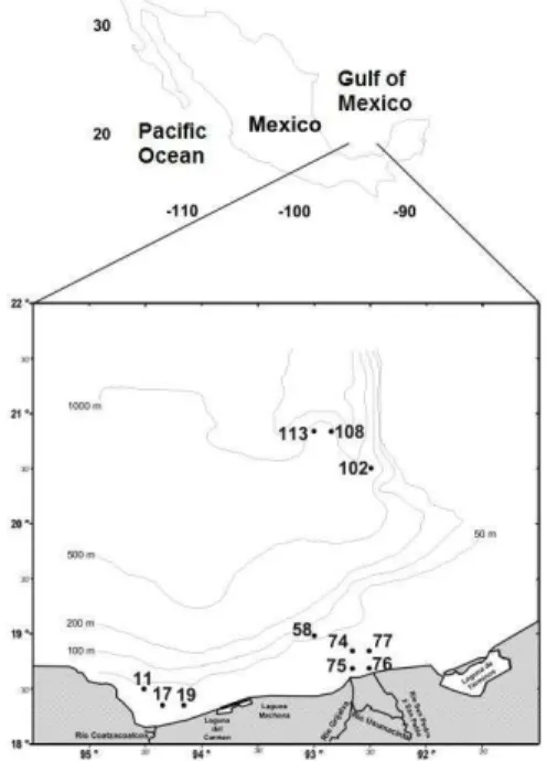
Sampling was carried out on board the R/V “Justo Sierra”, on the cruise PROMEBIO IX, during the “dry season” (June-July, 2004). Sampling stations in the southern Gulf of Mexico were located in two coastal areas (eight stations in all: three in the vicinity of the Coatzacoalcos River and five around the Grijalva-Usumacinta Rivers) and in an oceanic area (three stations at the Campeche Canyon) (Fig. 1). Water samples for studying picoplankton were collected from the water column with Niskin bottles (8 L) attached to a CTD-Rosette system (Neil Brown Mark III) every 4-10 m over the continental shelf where depths were less than 40 m, otherwise samples
were collected at 10, 20, 40, 60, 80, 100, 120, 150 and 180 m. At each sampling station, CTD casts recorded temperature and salinity throughout the water column. Vertical profiles of both chlorophyll a (deep
chlorophyll maximum, DCM) and PAR0
(photosynthetic active radiation) were measured with a passive fluorometer (Biospherical Instruments, model PNF-300) based on in vivo fluorescence and an
algorithm provided by the manufacturer.
Identification of Picoprokaryote Cells by TEM
Picoprokaryote organisms were identified by transmission electron microscopy (TEM) (JEOL 1200 CX). A 300 ml water sample with a high concentration of phytoplankton was centrifuged at 10,500 g for 15 min. Cell pellets were separated from the supernatant by decantation and fixed in 5 ml of filtered seawater (0.2 µm) with glutaraldehyde (3% final concentration) for 24 h at 4 °C. Cells were post-fixed with osmium tetroxide (2%) and phosphate buffer (100 mM) for 2 h. Samples were dehydrated through an ethanol series (10, 30, 50, 70, 90, 100%). Cells were then embedded in epoxy resin, with posterior polymerization in an oven (60°C) for 48 h. Thin sections of the embedded cells were made every 100 nm using an ultramicrotome Reichert-Jung. The cells were then mounted on grids and treated conventionally for contrast (ammonium acetate and lead citrate) before observation by TEM.
Cell Counts by Epifluorescence Microscopy
150 ml of sea water were collected at each depth, fixed with paraformaldehyde (1% final concentration) in dark glass bottles and stored at –15° C. Samples were maintained at this temperature until analysis. Samples were thawed out in water (~37° C) and an aliquot of 15-25 ml of sea water was filtered by hand through a nitrocellulose black membrane (0.22µm) using a filter unit attached to a 60 ml syringe. Filters with the biological material were mounted on microscope slides with a drop of immersion oil (Zeiss) on the top and bottom of the filters before they were secured with a cover slide. Picoplankton cells were identified and quantified by natural fluorescence under an epifluorescence microscope (Olympus BX-40) equipped with an excitation filter with a wavelength at 470 nm (blue light), a dichroic filter (495 nm) and an emission filter (515 nm) manufactured by Chroma Technology. A total of 300-350 cells were counted at each sampling depth and cell concentration was inferred in accordance with the equation:
where N= total number of PFP cells per ml-1; n= total
number of cells counted on filter, V= volume of
sample filtered; S= total filter area (cm2); P= area
where cells were counted (cm2). This study was
limited to distinguishing between prokaryote and eukaryote cells since natural fluorescence was too dim to distinguish red from orange. Therefore, the cell counts shown in this study refer to the total number of PFP (prokaryote and eukaryote) cells.
Cells Analysis by Flow Citometry
In general, procedures for studying picophytoplankton samples by flow cytometry
(sampling, identifying and counting
picophytoplankton) followed recommendations by Marie et al. (1999). Aliquots of 5 ml from selected bottle samples were placed into cryovials and kept frozen in liquid nitrogen until analysis by flow cytometry. Approximately 1-2 ml of thawed samples were analyzed by flow cytometer (FACScalibur, Becton Dickinson). Three fluorescences were used: FL1 (green fluorescence), FL2 (red fluorescence) and FL3 (orange fluorescence), and Polyscience beads of 1 µm (5-10 µl at bead concentrations of 105 beads ml-1)
were also used for calibration. Acquisitions varied from 50 to 100 X 103 events, depending on the cells
density. The cell quest program was used to analyze data.
R
ESULTSHydrographic Conditions and Chlorophyll a
Table 1 shows data of total depth, thermocline depth, PAR0, total picophytoplankton
abundance, and chlorophyll a found in the stations
sampled in the southern Gulf of Mexico. Coastal Stations (11, 17 and 19) in the vicinity of the Coatzacoalcos River were relatively shallow, with depths of less than 65 m. The mixing layer depth at those stations was ~15 m with water temperatures between 25 and 27°C (Fig. 2, Table 1). The Coatzacoalcos River's discharge had no influence on the water of the continental shelf since salinity was never below 36. The depth of the photic zone (1% PAR0) was detected at 57 m (Sta. 11) and 37 m (Sta
17), whereas 46% of PAR0 reached the bottom at
Station 19 (Fig. 2, Table 1). The chlorophyll maximum (CM) was very weak, except at Station 19 (Table 1), and was detected below the mixed layer, at 33 m (Sta. 11), 26 m (Sta. 17), and 19 m (Sta. 19) (Fig. 2).
Coastal stations distributed in the proximity of the Grijalva-Usumacinta Rivers were rather shallow, between 10 and 25 m (Sta. 74-77) (Fig. 2), whereas towards the northwest, linked to the narrow end of the continental shelf, the depth reached 120 m (Sta. 58) (Fig. 2). Water temperature did not vary substantially in the water column, except at Stations 77 and 58, where the mixing layer reached depths of 17 m and 33.5 m, respectively (Fig. 2, Table 1); a well-defined thermocline was found at Station 58 (Fig. 2, Table 1). All stations showed salinities >36, with DCM towards the bottom and irradiances spanning the entire water column, except at Station 58 (1% PAR0 at 109 m) (Fig. 2, Table 1). The vertical
distribution of chlorophyll showed no clearly defined pattern, and the layers of CM were not apparent, except at Station 58, where a CM was located at 66 m (Fig. 2, Table 1), and at Station 76 (close to the surface, 5.5 m, Fig. 2) where the chlorophyll concentration attained the highest value found in this study (2.81 mg m-3) (Table 1).
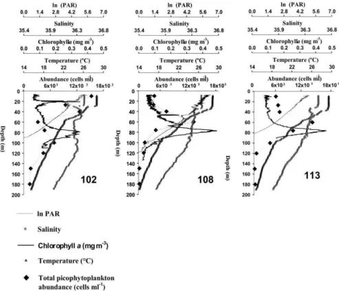
Stations located over the Campeche Canyon had maximum depths ~2379 m (Table 1). The mixing layer depth was found between 30-42.5 m, with warm temperatures ~28.5° C (Fig. 3); well-defined thermoclines were found at all three Stations. In this area (Sta. 102-113) the photic zone (1% PAR0) varied
between 81.5-118.5 m, with salinities of from 35.5 to 36.6 (Fig. 3). Chlorophyll a concentrations were rather
low, and the chlorophyll maximum reached 0.35 mg m-3 at both 21 and 78 m depths at Station 102, whereas
concentrations of 0.49 and 0.45 mg m-3 were found at
Table 1. Total depth, thermocline depth, depth of 1% PAR, maximum abundance of picoplankton, and deep chlorophyll maximum encountered at coastal and oceanic stations in the southern Gulf of Mexico. NP: not present; *max. indicates maximum percentage of irradiance detected at the bottom.
Station Depth
(m)
Thermocline depth (m)
Depth of 1% PAR (m)
Maximum abundance (cells ml-1)
Maximum chlorophyll a
(mg m-3)
11 68 14 57 8.12×10⁴ at 32 m 0.44 at 33 m
17 42 13.5 31 1.19×10⁵ at 5 m 0.46 at 26 m
19 34 14.5 *max. 46% 8.72×10⁴ at 12 m 1.98 at 19 m
58 120 33.5 109 2.78×10⁴ at 20 m 0.91 at 66 m
74 25 NP *max. 10% 9.54×10⁴ at 21 m 0.60 at 21.6 m
75 14 11.4 *max. 20% 1.16×10⁵ at 5 m 1.20 at 12.5 m
76 10 NP *max. 24.5% 1.67×10⁵ at 9 m 2.81 at 5.5 m
77 21 16.5 *max. 58% 9.68×10⁴ at 10 m 1.49 at 17 m
102 2371 30 90 1.53×10⁴ at 10 m 0.35 at 21 m
0.35 at 78 m
108 2219 40 118.5 1.25×10⁴ at 60 m 0.49 at 77 m
113 2196 42 81.5 1.32×10⁴ at 60 m 0.45 at 76 m
Fig. 3. Vertical profiles of some environmental variables, chlorophyll a and picophytoplankton in one oceanic area: Sta. 102-113 (Campeche Canyon).
Identification of Picophytoplankton Cells
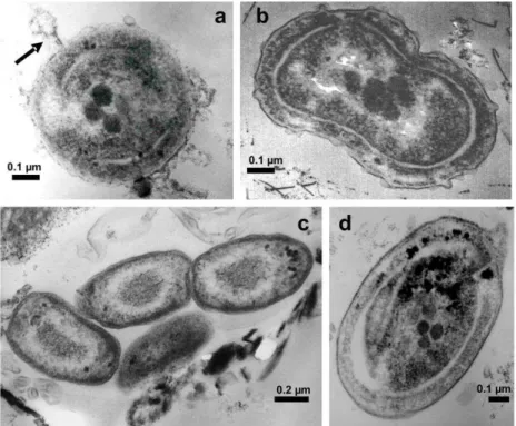
Picoprokaryote organisms were successfully recognized for the first time in the southern Gulf of Mexico by TEM, in terms of shape, size and ultrastructure. Both Prochlorococcus (Fig. 4a) and Synechococcus (Figs 4b-d) cells were identified: the Prochlorococcus cells presented a spherical to
subspherical shape and sizes varying between 0.58 and 0.65 µm diameter, some of them with virus attached (Fig. 4a, arrow), whereas Synechococcus cells were
more elongated (Figs 4c, d), solitary or in clumps (up to three cells) (Fig. 4 c) and varied between 0.72 and 0.98 x 0.41 and 0.73 µm in size, with some cells found in division (Fig. 4b). They were present in all the samples obtained and analyzed throughout this study.
Additionally, picoeukariote populations (even two populations were observed, Fig. 5, 26 m) could be recognized by flow cytometry by showing high red fluorescence (due to chlorophyll a) and very
low orange fluorescence, whereas Synechococcus
populations were relatively easily identified by the combination of red and orange fluorescences (Figs 5, 6); Prochlorococcus populations were probably
masked by the electronic noise and could not be recognized positively (Figs 5, 6). However, unfortunately, cell counting by flow cytometry provided an overestimation of cells when the calculations were made and the data proved to be unreliable for corrections.
Abundance and Distribution of Picophytoplankton
Fig. 4. Picoprokaryotes cells, TEM. Fig. 4a.Prochlorococcuscell showing some ultrastructural characteristics and one virus attached (arrow) (0.64 µm diameter). Fig. 4b.Synechococcus cell in division (1.12 µm, longest axis). Fig. 4c.Three Synechococcus cells (0.72 x 0.41 µm).Fig. 4d. Detail of one Synechococcus cell (0.98 x 0.73 µm).
Fig. 6. Same legend as Fig. 5, for samples in Station 113 (Campeche Canyon).Syn = Synechococcus, Euk = Unidentified picoeukaryote population.
The highest picophytoplankton abundance (1.67×10⁵ cells ml-1) was detected in a coastal area, in
shallow waters in the vicinity of the Grijalva-Usumacinta Rivers (Sta. 76, 9 m), followed by the abundance found in another coastal area, near the mouth of the Coatzacoalcos River (1.19×10⁵ cells ml-1,
Sta. 17, 5 m), whereas in the Campeche Canyon picophytoplankton abundances reached a maximum of 1.53×10⁴ cells ml-1 (Sta. 102, 10 m) (Table 1).
Additionally, the vertical distributions of picophytoplankton showed a weak correspondence with those of the chlorophyll a. Only at Station 11 was
there a close association (1 m difference) between the CM and maximum cell PFP abundance (Fig. 3), and although the distribution patterns of both vertical distributions (picophytoplankton and chlorophyll a)
were similar at Stations 108 and 113 (showing a deep maximum concentration), these peaks did not coincide and were 20 m apart, with the PFP maximum concentration above the CM (Fig. 3).
Close to the CoatzacoalcosRiver,
picophytoplankton varied in the water column from 5.82×10⁴ to 1.19×10⁵cells ml-1 peaking between 5 and
32 m depth (Fig. 3, Table 1). In this area, maximum cell abundance occurred in the mixed layer, except at Station 11.
Over the continental shelf near the Grijalva-Usumacinta Rivers PFP concentrations ranged between 9.61×10³ and 1.67×10⁵ cells ml-1. Highest cell
abundance occurred in subsurface waters between 5 and 21 m depth (24-78% PAR0) within the mixed
layer. Stations had shallow waters where higher
picophytoplankton abundance occurred towards the bottom with no relation to the chlorophyll a pattern,
except at Station 74.
Picophytoplankton abundances in the oceanic region (Campeche Canyon) ranged between 275 cells ml-1 and 1.53×10⁴ cells ml-1. Maximum cell
abundances differed between Stations 108-113 in terms of cell concentration and depth. Station 102 attained its highest cell abundance at 10 m depth, above the thermocline, whereas Stations 108 and 113 attained up to 1.25×10⁴ cells ml-1 (68% PAR
0) and
1.32×10⁴ (31% PAR
0), respectively, at 60 m depth,
below the mixing layer (Fig. 3, Table 1).
D
ISCUSSIONwaters (above 100 m, photic zone) and reached a maximum cell concentration of 1.67×10⁵ cell ml-1 at
salinities >35.5. Schapira et al. (2010) found a higher concentration of PFP (1.3-1.4×10⁶ cell ml-1) at lower
salinities (8-11%) associated with brackish-marine waters in a coastal lagoon in Australia. Since river runoff did not cause a detectable thermal and salinity gradient (freshwater influence) in coastal waters - as salinity values never decreased below 36 throughout the water column - possibly insufficient nutrient availability in the water for the growth of PFP populations occurred over the continental shelf during the dry season.
Temporal and spatial variations of PFP have been previously reported in diverse marine environments worldwide (ALONSO-LAITA et al., 2005). In this study, the distribution of PFP showed higher fluctuations between shallow coastal Stations within the mixed layer than in more oceanic waters of the Campeche Canyon. Rapid physicochemical changes are known to occur at different scales in coastal water (ÁLVAREZ-GÓNGORA et al., 2012) and changes in the dynamic of PFP have been suggested (MOORE and CHISHOLM, 1999; VELDHUIS et al., 2005), as a result of nutrient concentration (RAVEN, 1998), water circulation, seasons, stratification (BOUMAN et al., 2011),
grazing (PERNTHALER, 2005), salinity
(MITBAVKAR et al., 2012), and light availability in the water column (CHEN et al., 2011). As for the southern Gulf of Mexico, the vertical structure of chlorophyll a (pico, nano, and microphytoplankton)
has been postulated as a function of thermal fluxes, haline fronts, irradiance, nutrient uptake, and regional circulation patterns (SIGNORET et al., 2006). In addition, in situ profiles of natural chlorophyll-fluorescence in the southern Gulf have revealed that
autotrophic communities had maximum growth yield related to both the thermocline and the limit of the euphotic layer (SIGNORET et al., 2006). In general, this study found that total PFP abundance had a weak correspondence with the depth of chlorophyll a
maximum (DCM) which suggested that higher photosynthetic cells other than PFP contributed significantly to the DCM.
The transition from oligotrophic waters to nutrient-rich and well-mixed waters in coastal areas has been linked to an increasing concentration of
Synechococcus and picoeukaryotes (ANSOTEGUI et
al., 2003; GUILLOU et al., 2004; NOT et al., 2004). This has been also reported in the southern Gulf of
Mexico, where the photosynthetic pigments
zeaxanthin (representing Synechococcus) and
prasinoxanthin (representing picoprasinophytes) reached high concentrations towards coastal waters influenced by nutrient loads (HERNÁNDEZ-BECERRIL et al., 2012). Therefore, this suggests that
the major abundance of coastal PFP reported here may be assigned to the presence of Synechococcus and
picoeukaryote populations. However, it is not clear what the PFP contribution to biomass is, since chlorophyll a concentrations (CM) had little
relationship to PFP abundance in the water column at most Stations.
Some PFP species (picoprokaryotes) have developed physiological adaptations that link them to certain ecological niches in the environment. For example, Prochlorococcus is known by at least two
physiologically and genetically different ecotypes in the water column: one adapted to high light conditions and another to low light conditions (BOUMAN et al., 2011). As for the coastal and oceanic waters of the Gulf of Mexico, the specific divinyl-chlorophyll a
marker has suggested high cell concentrations of
Prochlorococcus (above and below 1% PAR0) in
winter (HERNÁNDEZ-BECERRIL et al., 2012),
although little is yet known of PFP ecotypes in terms of function, distribution, and contribution to total biomass in Mexican waters. Physiological adaptations of PFP in the water column to light conditions, nutrient availability, and temperature have been discussed in some studies and their advantages have been associated with the distribution of these organisms in diverse environments (ANDERSSON et al., 1994; RAVEN, 1998; MOORE and CHISHOLM, 1999; VELDHUIS et al., 2005).
Oligotrophic waters have been regarded as a suitable environment for the growth and numerical dominance of PFP within the phytoplankton
community (HALL and VINCENT, 1990;
GOERICKE and REPETA, 1992; CROSBIE and FURNAS, 2001). For instance, in offshore waters of the southern Adriatic Sea, Cerino et al. (2012)
determined that PFP organisms, mainly
picoprokaryotes, were the most important
phototrophic fraction with 96% of total abundance and up to 49% of total biomass. In oligotrophic waters of the Campeche Canyon, PFP showed still moderate densities (1.5×10³ cell ml-1) at Station 102, at 180 m
(Fig. 3), as previously reported in other regions (PARTENSKY et al., 1999; LANDRY, 2002; CHEN et al., 2011), although concentrations were up to 3 orders of magnitude lower than those recorded in coastal areas. Moreover, the peak of PFP in oligotrophic waters was above the CM (~20 m), which also suggested the potential dominance of larger
photosynthetic forms (e.g. nano or
remarkable occurrence of picoeukaryotes linked to well-mixed waters whereas Prochlorococcus was
substantially predominant along stratified oligotrophic oceanic systems.
Regarding the natural mechanisms of productivity in oligotrophic waters of the southern Gulf of Mexico, Salas-de-León et al.(2004) suggested that the development of the upwelling brings nutrient loads from mid-waters that stimulate the growth of autotrophic organisms triggering unimodal DCM (up to 0.32 mg m-3) at depths ~1% PAR
0 (78-89 m depth).
Although the structure of DCM reported in this study coincided with that given in previous studies, surprisingly higher concentrations at the DCM (up to 0.49 mg m-3) were detected in oligotrophic waters than
in either coastal areas or in other studies of the Gulf. Furthermore, a bimodal chlorophyll a distribution
(both with 0.35 mg m-3) occurred in the Campeche
Canyon (Station 102), within the mixed layer and ~1% PAR0. This study was, however, limited to
determining both the physical mechanisms and autotrophic components involved in the DCM in the Campeche Canyon.
The picoprokaryotes Prochlorococcus and Synechococcus were identified in the study area by
TEM, but cell counts made no distinction between the abundances and contributions of picoprokaryotes and picoeukaryotes to total PFP abundance. Picoeukaryote populations (even two different populations were detected, Fig. 5) were also detected by flow cytometry in the southern Gulf of Mexico (Figs 5, 6), which agrees with the report of Hernández-Becerril et al. (2012), though we were unable to identify them by TEM.
PFP populations have been poorly investigated in the southern Gulf of Mexico and this
study suggests that the picoprokaryotes
Prochlorococcus and Synechococcus may be
important photosynthetic components for organic carbon production within trophic webs in both coastal and oceanic waters. In other regions elsewhere in the world, Prochlorococcus and Synechococcus usually
contribute above 70% of the total phytoplankton community (CROSBIE and FURNAS, 2001; CERINO et al., 2012), with cell abundances ranging between
104 -105 (CHISHOLM et al., 1988) and 103-105 cells
ml-1 (GROB et al., 2007), respectively.
In conclusion, PFP populations in the southern Gulf of Mexico showed high variability in coastal waters, possibly as a result of the influence of the mixed layer, irradiance, and nutrient availability. Although peaks of PFP abundances were detected in both coastal and more oceanic areas, those found in the oceanic areas were located deeper (60 m) in the water column, relatively closer to the DCM (located at about 75 m). During the dry season the abundances of PFP yielded higher concentrations in coastal areas
than in oligotrophic ones. However, quantitative results of total PFP had little or no relationship with either maximum cell abundance or maximum
chlorophyll a which suggests that larger
photosynthetic forms may be an important biological component in the southern Gulf of Mexico during the dry season. Prochlocoroccus and Synechococcus were
identified and illustrated by TEM, and at least
Synechococcus and two unidentified picoeukariote populations were also recognized by flow cytometry. Further studies are required to establish the importance and contribution of PFP (picoprokaryotes and picoeukaryotes) to the total phytoplankton community in different seasons and environments in the southern Gulf of Mexico.
A
CKNOWLEDGEMENTSWe wish to thank the Coordinación de la Investigación Científica (CTIC, UNAM) for their support for our use of the R/V “Justo Sierra” for the PROMEBIO IX cruise. This research project was funded by the Mexican Consejo Nacional de Ciencia y Tecnología (CONACyT) through grant G27777-B.
R
EFERENCESAGAWIN, N. S. R.; AGUSTÍ, S. Prochlorococcus and Synechococcus cells in the central Atlantic Ocean: distribution, growth and mortality (grazing) rates. Vie Milieu, v.55, n. 3/4, p. 165-175, 2005.
ALONSO-LAITA, P.; NAVARRO, N.; DUARTE, C. M. ; AGUSTÍ, S. Seasonality of pico-phytoplankton abundance and cell death in a Mediterranean Bay (Bay of Palma, Majorca Island). Vie Milieu, v.55, n. 3/4, p. 177-184, 2005.
ÁLVAREZ-GÓNGORA, C. C.; LICEAGA-CORREA, M. D.; HERRERA-SILVEIRA, J. A. Seasonal variations of community structures phytoplankton in groundwater discharge areas along the Northern Yucatan Peninsula coast. Rev. Biol.Trop., v. 60, n. 1, p. 157-72, 2012. ANDERSSON, A.; HAECKY, P.; HAGSTRÖM, Å. Effect
of temperature and light on the growth of micro-plankton nano-plankton and pico-plankton: impact on algal succession. Mar. Biol., v. 120, n. 4, p. 511-520, 1994. ANSOTEGUI, A.; SAROBE, A.; TRIGUEROS, J. M.;
URRUTXURTU, I.; ORIVE, E. Size distribution of algal pigments and phytoplankton assemblages in a coastal-estuarine environment: contribution of small eukaryotic algae. J. Plankton Res., v. 25, n. 4, p. 341-355, 2003. BERTILSSON, S.; BERGLUND, O.; PULLIN, M. J.;
CHISHOLM, S. W. Release of dissolved organic matter by Prochlorococcus. Vie Milieu, v. 55, n. 3/4, p.
225-331, 2005.
BOUMAN, H. A.; ULLOA, O.; BARLOW, R.; LI, W. K. W.; PLATT, T.; ZWIRGLMAIER, K.; SCANLAN, D. J.; SATHYENDRANATH, S. Water column stratification governs the community structure of subtropical marine picophytoplankton. Environ.
CERINO, F.; AUBRY, F. B.; COPPOLA, J.; FERLA, R. L.; MAIMONE, G.; SOCAL, G.; TOTTI, C. Spatial and temporal variability of pico-, nano- and microphytoplankton in the offshore waters of the southern Adriatic Sea (Mediterranean Sea). Cont. Shelf
Res., v. 44, sp. iss., p. 94-105, 2012.
CHEN, B. Z.; WANG, L.; SONG, S. Q.; HUANG, B. Q.; SUN, J.; LIU, H. B. Comparisons of picophytoplankton abundance, size, and fluorescence between summer and winter in northern South China Sea. Cont. Shelf Res., v. 31, n. 4, p. 1527-1540, 2011.
CHISHOLM, S. W.; OLSON, R. J.; ZETTLER, E. R.; GOERICKE, R.; WATERBURY, J. B.; WELSCHMEYER, N. A. A novel free-living prochlorophyte abundant in the oceanic euphotic zone.
Nature, v. 334, n. 6180, p. 340-343, 1988.
CONAGUA, SEMARNAT. Gobierno Federal. Secretaría de Medio Ambiente y Recursos Naturales. Programa Nacional Hídrico: 2007-2012. México: CONAGUA, SEMARNAT, 2008. 159 p.
CROSBIE, N. D.; FURNAS, M. J. Abundance, distribution and flow-cytometric characterization of picophytoprokaryote populations in central (17°S) and southern (20°S) shelf waters of the Great Barrier Reef. J.
Plankton Res., v. 23, n. 8, p. 809-828, 2001.
ESPINOSA-FUENTES, M. L.; FLORES-COTO, C. Cross-shelf and vertical structure of ichthyoplankton assemblages in continental shelf waters of the Southern Gulf of Mexico. Estuarine, Coastal Shelf Sci., v. 59, n. 2, p. 333-352, 2004.
GOERICKE, R.; REPETA, D. J. The pigments of Prochlorococcus marinus: the presence of divinyl chlorophyll-a and chlorophyll-B in a marine prokaryote.
Limnol. Oceanogr., v. 37, n. 2, p. 425-433, 1992. GROB, C.; ULLOA, O.; CLAUSTRE, H.; HUOT, Y.;
ALARCÓN, G.; MARIE, D. Contribution of picoplankton to the total particulate organic carbon concentration in the eastern South Pacific.
Biogeosciences,v. 4, n. 5, p. 837-852, 2007.
GUILLOU, L.; EIKREM, W.; CHRETIENNOT-DINET, M. J.; LE GALL, F.; MASSANA, R.; ROMARI, K.; PEDROS-ALIÓ, C.; VAULOT, D. Diversity of picoplanktonic prasinophytes assessed by direct nuclear SSU rDNA sequencing of environmental samples and novel isolates retrieved from oceanic and coastal marine ecosystems. Protist, v. 155, n. 2, p. 193-214, 2004.
HALL, J. A.; VINCENT, W. F. Vertical and horizontal structure in the picoplankton communities of a coastal upwelling system. Mar. Biol., v. 106, n. 3, p. 465-471, 1990.
HERNÁNDEZ-BECERRIL, D. U.; GARCÍA-RESÉNDIZ, J. A.; SALAS-DE-LEÓN, D. A.; MONREAL-GÓMEZ, M. A.; SIGNORET-POILLON, M.; ALDECO-RAMÍREZ, J. Nanoplankton fraction in phytoplankton structure from the southern Gulf of Mexico (April 2000).
Cienc. Mar., v. 34, n. 1, p. 77-90, 2008.
HERNÁNDEZ-BECERRIL, D. U.; AQUINO-CRUZ, A.; SALAS-DE-LEÓN, D. A.; SIGNORET-POILLON, M.; MONREAL-GÓMEZ, M. A. Studies on picophytoplankton in the southern Gulf of Mexico: pigment analysis and potential importance of the picoeukariote Prasinophyte Micromonas pusilla. Mar.
Biol. Res., v. 8, n. 4, p. 331-340, 2012.
LANDRY, M. R. Integrating classical and microbial food web concepts: evolving views from the open-ocean tropical Pacific. Hydrobiologia, v. 480, n. 1/3, p. 29-39, 2002.
LE GALL, F.; RIGAUT-JALABERT, F.; MARIE, D.; GARCZAREK, L.; VIPREY, M.; GOBET, A.; VAULOT, D. Picoplankton diversity in the South-East Pacific Ocean from cultures. Biogeosciences, v. 5, n. 1, p. 203-214, 2008.
MAN-AHARONOVICH, D.; PHILOSOF, A.; KIRKUP, B. C.; LE GALL, F.; YOGEV, T.; BERMAN-FRANK, I.; POLZ, M. F.; VAULOT, D., BÉJÀ, O. Diversity of active marine picoeukaryotes in the Eastern Mediterranean Sea unveiled using photosystem-II psbA transcripts. ISME J., v. 4, n. 8, p. 1044-1052, 2010. MARIE, D.; SHI, X. L.; RIGAUT-JALABERT, F.;
VAULOT, D. Use of flow cytometric sorting to better assess the diversity of small photosynthetic eukaryotes in the English Channel. FEMS Microbiol. Ecol., v. 72, n. 2, p. 165-178, 2010.
MITBAVKAR, S.; RAGHU, C.; RAJANEESH, K. M.; PAVAN, D. Picophytoplankton community from tropical marine biofilms. J. Exp. Mar. Biol. Ecol., v. 426/427, p. 88-96, 2012.
MONREAL-GÓMEZ, M. A.; SALAS-DE-LEÓN, D. A. Circulación y estructura termohalina del Golfo de México, In: LAVÍN, M. F. (Ed.). Contribuciones a la oceanografía física de México. Ensenada: Unión Geofísica Mexicana, 1997. p. 183-199. (Monografía (Unión Geofísica Mexicana); n. 3).
MOON-VAN DER STAAY, S. Y.; DE WACHTER, R.; VAULOT, D. Oceanic 18S rDNA sequences from picoplankton reveal unsuspected eukaryotic diversity.
Nature, v. 409, n. 6820, p. 607-610, 2001.
MOORE, L. R.; CHISHOLM, S. W. Photophysiology of the marine cyanobacterium Prochlorococcus: ecotypic differences among cultured isolates. Limnol. Oceanogr., v. 44, n. 3, p. 628-638, 1999.
NOT, F.; DEL CAMPO, J.; BALAGUÉ, V.; DE VARGAS, C.; MASSANA, R. New insights into the diversity of marine picoeukaryotes. PLoS ONE, v. 4, n. 9, p. E7143,
2009.
NOT, F.; LATASA, M.; MARIE, D.; CARIOU, T.; VAULOT, D.; SIMON, N. A single species, Micromonas pusilla (Prasinophyceae), dominates the eukaryotic picoplankton in the western English channel.
Appl. Environ. Microbiol., v. 70, n. 7, p. 4064-4072, 2004.
PARTENSKY, F.; HESS, W. R.; VAULOT, D. Prochlorococcus, a marine photosynthetic prokaryote of global significance. Microbiol. Mol. Biol. Rev., v. 63, n. 1, p. 106-127, 1999.
PERNTHALER, J. Predation on prokaryotes in the water column and its ecological implications. Nat. Rev.Microbiol., v. 3, n. 7, p. 537-546, 2005.
RAVEN, J. A. The twelfth Tansley Lecture. Small is beautiful: the picophytoplankton. Funct.Ecol., v. 12, n. 4, p. 503-513, 1998.
mobilization on coastal contamination (Coatzacoalcos River, Mexico). Cont. Shelf Res., v. 37, p. 57-65, 2012. SALAS-DE-LEÓN, D. A.; MONREAL-GÓMEZ, M. A.;
SIGNORET, M.; ALDECO, J. Anticyclonic-cyclonic eddies and their impact on near-surface chlorophyll stocks and oxygen supersaturation over the Campeche Canyon, Gulf of Mexico. J. Geophys. Res.: Oceans, v. 109, n. C5, p. C05012, 2004.
SALAS-DE-LEÓN, D. A.; MONREAL-GÓMEZ, M. A.; DÍAZ-FLORES, M. A.; SALAS-MONREAL, D.; VELASCO-MENDOZA, H.; RIVERON-ENZASTIGA, M. L.; ORTIZ-ZAMORA, G. Role of near-bottom currents in the distribution of sediments within the Southern Bay of Campeche, Gulf of México. J. Coastal
Res., v. 24, n. 6, p. 1487-1494, 2008.
SANVICENTE-AÑORVE, L.; FLORES-COTO, C.; CHIAPPA-CARRARA, X. Temporal and spatial scales of ichthyoplankton distribution in the southern Gulf of Mexico. Estuarine, Coastal Shelf Sci., v. 51, n. 4, p. 463-475, 2000.
SCHAPIRA, M.; BUSCOT, M. -J.; POLLET, T.; LETERME, S. C.; SEURONT, L. Distribution of picophytoplankton communities from brackish to hypersaline waters in a South Australian coastal lagoon.
Saline Syst., v. 6, p.2, 2010.
SIGNORET, M.; MONREAL-GÓMEZ, M. A.; ALDECO, J.; SALAS-DE-LEÓN, D. A. Hydrography, oxygen saturation, suspended particulate matter, and chlorophyll-a fluorescence in chlorophyll-an ocechlorophyll-anic region under freshwchlorophyll-ater influence. Estuarine, Coastal Shelf Sci., v. 69, n. 1/2, p.
153-164, 2006.
STOCKNER, J. G. Phototrophic picoplankton: an overview from marine and freshwater ecosystems. Limnol.
Oceanogr., v. 33, n. 4, p. 765-775, 1988.
TOLEDO, O. A. Caracterización ambiental del Golfo de México. In: BOTELLO, A. V.; ROJAS-GALAVIZ, J. L.; BENÍTEZ, J. A.; ZÁRATE-LOMELÍ, D. (Eds.).
Golfo de México, contaminación e impacto ambiental: diagnóstico y tendencias. Campeche: Universidad Autónoma de Campeche, 1996. p. 9-28. (EPOMEX. Serie Científica; 5).
RR S- J R , F R R , R R ,
C. A study case of hydrodynamics and water quality modelling: Coatzacoalcos River, Mexico. In: SCHULZ, H. E.; SIMÕES, A. L. A.; LOBOSCO, R. J. (Eds.).
Hydrodynamics: natural water bodies. [S.l.]: InTech,
2012. cap. 3, p. 49-66. Available at: http://www.intechopen.com/books/hydrodynamics-natural-water-bodies/. Accessed in: Aug.22, 2012. VELDHUIS, M. J. W.; TIMMERMANS, K. R.; CROOT, P.;
VAN DER WAGT, B. Picophytoplankton: a comparative study of their biochemical composition and photosynthetic properties. J. Sea Res., v. 53, n. 1/2, p. 7-24. 2005.
YÁÑEZ-ARANCIBIA, A.; DAY, J. W. The Gulf of Mexico: towards an integration of coastal management with large marine ecosystem management. Ocean Coastal Manage., v. 47, n. 11/12, p. 537-563, 2004.