vol. 42, n. 2, abr./jun., 2006
Characterization of xylose reductase extracted by CTAB-reversed micelles
from
Candida guilliermondii
homogenate
Ely Vieira Cortez
1, Adalberto Pessoa-Jr
1, Maria das Graças de Almeida Felipe
2, Inês Conceição
Roberto
2, Michele Vitolo
1**1University of São Paulo, School of Pharmaceutical Sciences, Department of Biochemical and Pharmaceutical
Technology. 2Department of Biotechnology, FAENQUIL
Xylosereductase (XR) (E.C.1.1.1.21), produced by Candida guilliermondii, grown in sugar cane bagasse hydrolysate, was separated directly from the cell homogenate by reversed micelles of cetyl trimethyl ammonium bromide (CTAB), attaining a recovery yield of 100% and enrichment factor of 5.6 fold. The extraction conditions were: pH=7.0, electrical conductivity= 14 mS/cm, T=5 oC, 5% (w/w) of hexanol, 22% (w/w) of butanol and 0.15 M
CTAB. The XR after extraction was stable in pH interval of 6.0-6.5 and its heat inactivation constant was about 6.0-6.5 fold higher than that before extraction. The Vmax values against both xylose and NADPH for XR before and after extraction by reversed-micelles differed about 6%, whereas the difference on KM values were more pronounced. The (KM)xylose for XR after extraction was about 35% higher than before extraction, meanwhile (KM)NADPH was about 30% lower after than before extraction. As the KM variations indirectly signaled, the XR affinity simultaneously diminishes for xylose and increases for NADPH. This could probably explain why the Vmax values for XR before and after extraction were quite similar.
Uniterms
• Xylose reductase • Reversed micelles • Candida guilliermondii
INTRODUCTION
Liquid-liquid extraction by reversed micelles is a useful and very versatile tool for separating biomolecules, consisting of two steps. First, the so called forward extraction by which the target protein or the contaminants are transferred from an aqueous solution to a reversed micelles organic phase. Second, the so called backward extraction by which the biomolecules are released from the reversed micelles and transferred to a fresh aqueous phase (Kilikian et al., 2000).
This process shows a close similarity to the liquid-liquid traditional extraction process, because both are biphasic and consist in partitioning a targeted solute between an aqueous feed phase and an organic phase, with a subsequent back transfer to a second aqueous stripping phase (Pessoa-Jr, Vitolo, 1998). Reversed micelles systems have great potential for industrial application because they provide a favorable environment for the solubility of protein in the organic phase with preservation of biological activity (Rodrigues et al., 1999).
*Correspondence:
M. Vitolo
A good perspective should be to apply this technique directly in a crude microbial homogenate, aiming to purify a specific protein or enzyme. An example, should be the purification of xylose reductase (XR) from the Candida guilliermondii homogenate, which, after removing the cell debris and other contaminants, could be used in the “in vitro” conversion of xylose into xylitol. The xylitol production is in great demand, because it can be used successfully in food industry (for its sweetening power and insulin-independent metabolism), dentistry formulations (for its anticariogenicity, tooth hardening and mineralizing properties) and pharmaceutical formulations (for its capability of preventing eardrum infection and its possibility of being used as a sweetener in syrups, tonics and vitamin formulation) (Pessoa-Jr, Vitolo, 1997). Besides, the enzymatic xylose/xylitol conversion could become an alternative to the conventional process based on the reduction of xylose with inorganic catalyst (Ni or Pt).
This work deals with the effect of pH, temperature, concentration of xylose and NADPH on the activity of xylose reductase, which was attained from Candida guilliermondii (grown in sugar cane bagasse hydrolysate) and extracted from the cell homogenate by reversed micelles technique.
MATERIALS AND METHODS
Chemicals
Reduced nicotinamide adenine dinucleotide phosphate (NADPH), β-mercaptoethanol, and xylose were purchased from Sigma Chemical Company (St. Louis, MO, USA). All other chemicals were of analytical grade.
Microorganism
The cells were obtained from fermentations conducted with Candida guilliermondii FTI 20037 described by Barbosa et al (1988). The yeast was maintained on malt-extract agar slants at 4 oC.
Inoculum preparation, medium and fermentation conditions
A medium containing 3.0 g/L of xylose supplemented with 20.0 g/L of rice bran extract, 2.0 g/L of (NH4)2SO4 and 0.1 g/L of CaCl2.2H2O was used for growing the inoculum. Erlenmeyer flasks (125 mL), each containing 50 mL of medium with inoculum (initial pH 5.5), were incubated on a rotary shaker (200 rpm) at 30 oC for 24 h.
For the fermentation, concentrated bagasse hemicellulosic hydrolysate containing 42 g/L of xylose was employed. The hydrolysate was prepared according to Alves
et al. (1998). The hydrolysate was supplemented with the same nutrients used for the inoculum preparation. The cultivation was done by a batch process in a 1.25 L fermentor BIOFLO III (New Brunswick Scientific Co., Inc-Edison-New Jersey-USA), under agitation of 300 min-1 and aeration rate of
0.6 vvm (KLa 22.5 h-1), at 30 oC, initial pH 5.5.
Preparation of cell-free extracts
Cells were harvested by centrifugation at 800g, and washed in phosphate buffer (50 mM, pH 7.2). The cell pellets were stored in a freezer. For enzymatic analysis, cell extracts were thawed and disrupted by a sonic disruption technique using a Sonics & Materials disrupter. Cell homogenate was then centrifuged at 10,000 g (Jouan MR 1812) at 4 oC for 10 min, and the supernatant solution (CH)
was used for enzyme assays.
Liquid-liquid extraction
From the CH the enzyme was extracted by CTAB-reversed-micelles in isooctane, by a two-step procedure. In the first step (forward –extraction), 3.0 mL of the crude extract was mixed with an equal volume of micellar microemulsion (CTAB in isooctane/hexanol/water), under the following conditions: pH = 7.0; electrical conductivity = 14 mS/cm; T = 5 oC; hexanol = 5%; butanol = 22%; CTAB
(cetyl trimethyl ammonium bromide) = 0.15M. This mixture was agitated on the vortex for 1 minute, to obtain the equilibrium phase, and again separated into two phases (aqueous phase-I and micellar phase-I) by centrifugation at 657g for 10 min (Jouan Centrifuge Mod. 1812, Saint-Herblain, France). Afterwards, 2 mL of CTAB-micellar phase-I was mixed with 2.0 mL of fresh aqueous phase (acetate buffer 1.0 M at pH 5.5 with 1.0 M NaCl), in order to transfer the enzyme from the micelles to this fresh aqueous solution, called the aqueous phase-II (APII) (backward-extraction), which was finally collected by centrifugation (657g; 10 min). Both aqueous phases (first and second), and the homogenate were assayed to determine XR activity. So that, it was attained in APII an enzyme recovery yield of 100% and an enrichment factor of 5.6 fold in relation to the CH, which was taken as reference.
Enzyme assay
50 µL of 3.0 mM NADPH, 250 µL of enzyme extract and 600 µL of phosphate buffer (pH 6.2).
The reaction was carried out in 1 mL spectro-photometer cell at room temperature and the NADPH consumption followed by the variation of absorbance during 30 s at λ = 340 nm. The extinction coefficient used was equal to 6.22 x 10⁻³ M-1cm-1. Each activity
deter-mination was made in triplicate, being the variation coefficient not higher than 5%.
One XR (U) was defined as the amount of enzyme catalyzing the formation of 1 µmol of NADP per min. Throughout the work the XR activity was expressed as U per mL of reaction medium.
Effect of pH, temperature, xylose and NADPH concentration on the xylose reductase activity
The pH and temperature of the standard reaction test were changed one by one at intervals of 4.5-8.5 and 25-65 oC, respectively. The xylose and NADPH
concen-trations were varied from 5 to 100mM and from 0.02 to 0.20 mM, respectively.
While investigating the stability, the CH or the APII containing XR was incubated at room temperature for 30 min at the desired pH, or up to 150 min at the temperatures of 30, 35, 40,45 or 50 oC before the standard
assay was carried out.
To accomplish these experiments, it were always mixed in a volumetric proportion of 1:9 the media (CH or APII ) and an appropriate 0.1 M buffer solution (acetic acid-acetate buffer pH 4.5 and 5.5; phosphate buffer pH 6.0 and 6.5; and Tris-HCl pH 7.0, 7.5, 8.0 and 8.5). In all tests the pH was checked after homogenizing the mixture. The kinetic constants (KM and Vmax) for XR were determined through the conventional Lineweaver-Burk’s method.
RESULTS AND DISCUSSION
The optimum pH for xylose reductase (XR) activity before and after extraction with reversed micelles was about 6.0 (Figure 1). This result is in accordance with the literature, in so far as the optimum pH for XR activity was 5.5 for
Candida guilliermondii (Silva, et al., 1996), 6.0 for
Pachysolen tannophylus (Dietzelmuller et al., 1984) and Pichia stipitis (Verduyn et al., 1985). However, the maximum XR activity diminished about 44% after submission to the extraction (Figure 1), probably due to the direct effect of the organic solvent and CTAB on the structure of the enzyme (Kopp, Schede, 2004). The solvent could interfere with the most sensitive hydrophobic sites of
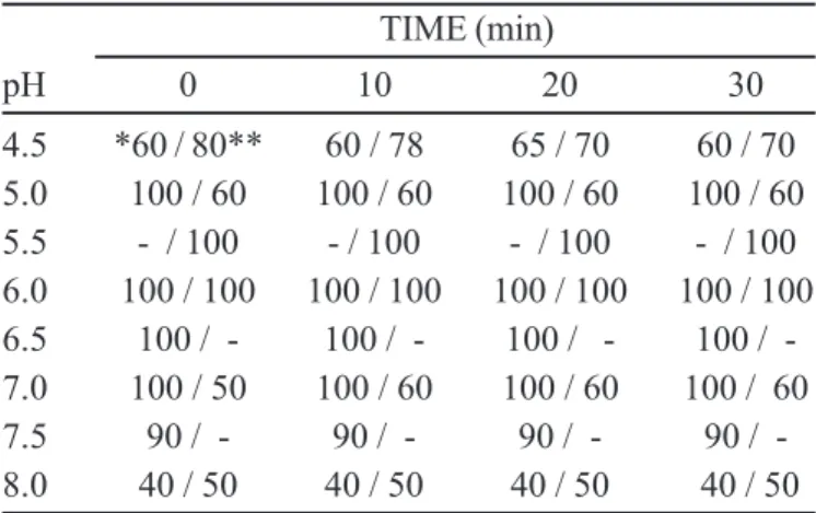
the macromolecule, causing their denaturation. Meanwhile CTAB, a cationic molecule, could interact with negatively charged groups located in the side chains of amino acids by electrostatic interaction, leading either to the alteration of the overall macromolecule charge or interfering on the proper ionization degree of such amino acid groups. Moreover, the pH variation itself could promote the formation of inadequate ionic groups at the active site and/or at another domain important to stabilize the protein structure. From Table I it can be seen that during a fixed interval of time (0-30 min), the activity of XR before and after extraction was stable (100% of catalytic activity retention) in the pH intervals of 5.5-7.0 and 6.0-6.5, respectively. The sharp diminution of XR stability (about 50%) for pH > 7.5, probably, was due to the denaturation of the protein (Table I and Figure 1).
TABLE I - Comparison of the stability against pH of the xylose reductase present in the cell-free homogenate (CH) and after extraction by CTAB-reversed-micelles (APII)
TIME (min)
pH 0 10 20 30
4.5 *60 / 80** 60 / 78 65 / 70 60 / 70 5.0 100 / 60 100 / 60 100 / 60 100 / 60 5.5 - / 100 - / 100 - / 100 - / 100 6.0 100 / 100 100 / 100 100 / 100 100 / 100 6.5 100 / - 100 / - 100 / - 100 / -7.0 100 / 50 100 / 60 100 / 60 100 / 60 7.5 90 / - 90 / - 90 / - 90 / -8.0 40 / 50 40 / 50 40 / 50 40 / 50
*Percent of the residual activity of xylose reductase in the CH; **Percent of the residual activity of xylose reductase in the APII.
From Table II it can be seen that the highest activity for XR present in the CH (0.611 U/mL) and APII (0.514 U/mL) occurred at 65 oC.
By applying the conventional Arrhenius’ method [Log v = f(1/T)] (Table II), the following equations were established:
(CH) Log v = 1.86 – 701.(1/T) (r = - 0.996) (1)
(APII) Log v = 1.85 – 725.(1/T) (r = - 0.996)(2)
where T is the temperature expressed as Kelvin degrees (K).
The activation energy (Ea) calculated from equations 1 and 2 for XR in the CH and APII were 13.4 kJ/mol and 13.9 kJ/mol, respectively. By using the conventional thermodynamic relations (Owusu, Makhzoum, 1992), the thermodynamic parameters related to the reaction catalyzed by XR and carried out at 303K were estimated (Table III). As can be concluded from Table III, the main thermodynamic parameters related to XR catalysis did not depend on the purity degree of the enzyme.
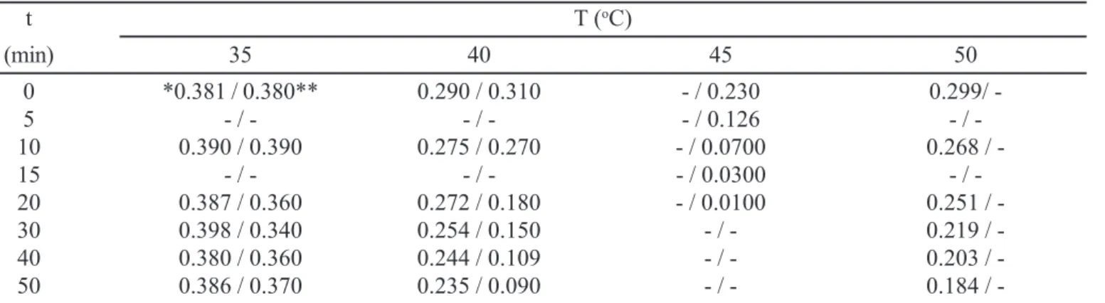
In regard to the stability against temperature, it is clear that the more purified XR (in APII) was less stable to the temperature than the crude extract (CH) (Table IV). Through plots of Log v versus time (Figure 2) and from the correspondent linear equations established (Equations 3-6)
it were possible to calculate the heat inactivation constant (k) for the more and less purified forms of XR. For instance, at 40 oC the XR present in CH and APII had k
values equal to 1.74x10⁻³ min-1 and 1.13x10⁻² min-1,
respectively. Such difference certainly resulted from the fact that in the cell-free homogenate (CH) there were compounds (residual xylose and/or NADPH, for instance) conferring some degree of heat stability to the enzyme. It is well demonstrated the fact that the presence of substrate/ cofactor in the reaction medium confer to the enzymes an increased stability against factors as pH, temperature and inhibitors (Vitolo, 2001).
(40 oC/CH) Log v = - 0.541 – 0.00174.t (r = -0.990) (3)
(50 oC/CH) Log v = - 0.726 – 0.00421.t (r = -0.997) (4)
TABLE III - Thermodynamic parameters related to both the enzyme catalysis (Ea, ∆G, ∆H, ∆S) and heat denaturation (Ea’, ∆G’, ∆H’, ∆S’) for the XR present in the CH and in the APII
Thermodynamic Extraction step parameters
CH APII
Ea (kJ/mol) 13.4 13.9
∆G (kJ/mol) -12.6 -12.6
∆H (kJ/mol) 10.9 11.4
∆S (kJ/K.mol) 0.0775 0.0790
Ea’ (kJ/mol) 74.3 295
∆G’ (kJ/mol) -12.9 -13.0
∆H’ (kJ/mol) 71.7 292
∆S’ (kJ/K.mol) 0.270 0.976
TABLE II - Effect of temperature on the activity of xylose reductase present in CH and APII. In parenthesis are presented the logarithm of each activity, which were used in the calculation of the activation energy for the reaction catalyzed by XR, through the conventional Arrhenius’ method
T 1/T v ( U/mL )
(oC) (K-1)x103 CH APII
25 (298)* 3.36 0.320 (-0.495) 0.260 (-0.590) 30 (303) 3.30 0.338 (-0.471) 0.295 (-0.530) 35 (308) 3.25 0.380 (-0.420) 0.310 (-0.509) 40 (313) 3.19 0.431 (-0.366) 0.332 (-0.480) 45 (318) 3.14 0.442 (-0.355) 0.369 (-0.433) 50 (323) 3.10 0.483 (-0.316) 0.406 (-0.391) 60 (333) 3.00 0.562 (-0.250) 0.466 (-0.332) 65 (338) 2.96 0.611 (-0.214) 0.514 (-0.289)
*The number in parenthesis correspond to the absolute temperature (K).
FIGURE 2 - Thermal inactivation of xylose reductase present in the CH at 40 oC () and 50 oC ( ) and in the
(40 oC/APII) Log v = - 0.494 – 0.0113.t (r = -0.994) (5)
(45 oC/APII) Log v = - 0.573 – 0.0672.t(r = -0.990) (6)
By applying the integrated form of Arrhenius equation (Eq. 7), it was possible to estimate the activation energy of heat denaturation (Ea’) for XR in the CH and APII. Furthermore, it was also possible to calculate the correspondent thermodynamic parameters related to the heat denaturation (∆G’, ∆H’ and ∆S’) as proposed by Owusu and Makhzoum (1992). All the values calculated were shown in Table III.
Log (k2/k1) = [Ea’(T2 – T1)] / (2.303.R.T1.T2) (7)
where k1 and k2 are the heat inactivation constants at temperatures T1 and T2, respectively, and R= 8.3144 J/K.mol.
According to Owusu and Makhzoum (1992) values of ∆H’ between 200 and 300 kJ/mol should indicate the thermal denaturation of protein by unfolding its tertiary and/or quaternary structure. Thereby, the ∆H’ of 292 kJ/mol calculated for XR in APII (Table III) points to the unfold of its three dimensional confor-mation, when submitted to temperatures of 40 oC–45 oC
for 50 min. For XR in CH, however, ∆H’ of about 72 kJ/ mol should indicate that the heat denaturation would not be due to the unfolding of its molecular conformation, but simply its hydrolysis by proteases also present in the cell homogenate, which had their activities increased at temperatures of 40 oC–50 oC. From Table III it is also
notorious that the variation of heat denaturation entropy for XR in APII was 3.6 times higher than XR in CH.
This might be a subtle evidence that the reversed-micelles procedure really lead to a more purified XR. In other words, being XR in a more pure form in solution, the unfolding of its molecular structure by thermal energy leaves the system a little less unorganized than in the homogenate, in which the hydrolysis of XR is probably quite more conspicuous.
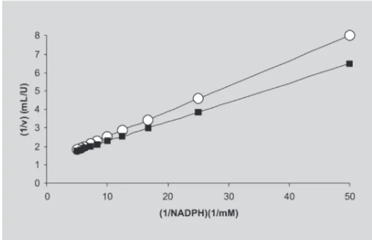
The kinetic constants for XR present in the CH and APII were calculated from the straight lines showed in Figu-res 3 and 4 (plots based on the conventional Lineweaver-Burk’s method). Thus, for XR present in the CH the kinetic constants were (KM)xylose = 11.0mM, (KM)NADPH = 0.119mM,
TABLE IV - Residual activity of xylose reductase (U/mL), present in the CH and APII, submitted up to 50 min at different temperatures.
t T (oC)
(min) 35 40 45 50
0 *0.381 / 0.380** 0.290 / 0.310 - / 0.230 0.299/
-5 - / - - / - - / 0.126 /
-10 0.390 / 0.390 0.275 / 0.270 - / 0.0700 0.268 /
-15 - / - - / - - / 0.0300 /
-20 0.387 / 0.360 0.272 / 0.180 - / 0.0100 0.251 /
-30 0.398 / 0.340 0.254 / 0.150 - / - 0.219 /
-40 0.380 / 0.360 0.244 / 0.109 - / - 0.203 /
-50 0.386 / 0.370 0.235 / 0.090 - / - 0.184 /
-*The first number refers to the residual activity of XR present in the CH; *-*The second number refers to the residual activity of XR present in the APII.
(Vmax)xylose = 0.76 U/mL and (Vmax)NADPH = 0.87 U/mL, whereas for XR in the APII were (KM)xylose = 17.0 mM, (KM)NADPH = 0.085 mM, (Vmax)xylose = 0.81 U/mL and (Vmax)NADPH = 0.81 U/mL. The Vmax values against both xylose and NADPH for XR in the CH and APII differed about 6%, whereas the difference on KM values were more pronounced. It must be borne out that (KM)xylose for XR in the APII was about 35% higher than in CH, meanwhile (KM)NADPH was about 30% lower in the APII than in the CH. This could explain, at least in part, why XR molecules having their conformational structure perturbed (probably by residual organic solvents used in the extraction procedure), as the KM variations indirectly signaled, had quite similar Vmax values. In other words, some kind of catalytic compensation would occur as far as the XR affinity simultaneously diminishes for xylose and increases for NADPH. Such a behavior is not uncommon with enzymes having a Bi-Bi catalytic mechanism like xylose reductase.
CONCLUSION
The main conclusion was the suitability of reversed-micelles procedure in the direct separation of xylose reductase from a crude cell homogenate, although the pH and temperature intervals for enzyme stability were narrowed after extraction. If we remember that this extraction is only the first step of the overall down stream process for purifying XR, so the remaining residues from that procedure will certainly be removed and the full enzyme performance recuperated.
RESUMO
Caracterização da xilose redutase extraída por micelas reversas-CTAB a partir de homogenato de
Candida guilliermondii
A xilose redutase (XR) (E.C.1.1.1.21), produzida por
Candida guilliermondii cultivada em hidrolisado de baga-ço de cana de açúcar, foi separada diretamente do homogenato livre de células através da técnica de micelas reversas feitas com cetil trimetil brometo de amônio (CTAB). Obteve-se um rendimento de recuperação da enzima de 100% e um fator de enriquecimento de 5,6 ve-zes. As condições de extração foram: pH=7,0, condutividade elétrica = 14 mS/cm, T= 5 oC, 5% (w/w) de
hexanol, 22% (w/w) de butanol e 0.15M CTAB. A XR após a extração manteve-se estável no intervalo de pH entre 6.0 e 6.5, sendo a constante de inativação térmica cerca de 6,5 vezes maior do que aquela antes da extração. Os valores de Vmax da XR frente à xilose e NADPH antes e após a extração por micelas reversas diferiram cerca de 6%, enquanto que as diferenças nos valores de KM foram mais pronunciadas. O (KM)xilose para a XR após a extração foi cerca de 35% maior do que antes da extração, enquanto que (KM)NADPH foi 30% menor após do que antes da extra-ção. As variações nos valores de KM indicam, indiretamen-te, que a afinidade da XR simultaneamente diminui para a xilose e aumenta para o NADPH. Este resultado pode-ria explicar a razão pela qual os valores de Vmax antes e após a extração terem sido praticamente iguais.
UNITERMOS:Xilose redutase. Micela reversa. Candida guilliermondii.
ACKNOWLEDGMENTS
This work was supported by Fundação de Amparo à Pesquisa do Estado de São Paulo, Brazil (FAPESP) and CNPq (Conselho Nacional de Desenvolvimento Científico e Tecnológico, Brazil).
REFERENCES
ALVES, L.A.; FELIPE, M.G.A.; ALMEIDA-SILVA, J.B.; SILVA, S.S.; PRATA, A.M.R. Pretreatment of sugar cane bagasse hemicellulose hydrolysate for xylitol production by Candida guilliermondii. Appl.Biochem.Biotechnol., v.70, p.89-98, 1998.
BARBOSA, M.F.S.; MEDEIROS, M.B.; MANCILHA, I.M.; SCHENEIDERS, H.; LEE, H. Screening of yeast for production of xylitol from D-xylose and some factors which affect xylitol yield in Candida guilliermondii.
J.Ind.Microbiol., v.3, p.241-251, 1988
DIETZELMULLER, G.; KUBICEK, C.P.; WOEHRER, W.; ROEHR, M. Xylose metabolism in Pachysolen tannophilus: purification and properties of xylose reductase. Can.J.Microbiol., v.30, p.1330-1336, 1984.
KILIKIAN, B.V.; BASTAZIN, M.R.; MINAMI, N.M.; GONÇALVES, E.M.R.; PESSOA-Jr, A. A liquid-liquid extraction by reversed micelles in biotechnological process. Braz.J.Chem.Eng., v.17, p.29-38, 2000
KOPP, J.; SCHEDE, T. The Swiss-model repository of annotated three-dimensional protein structure homology models. Nucleic Acids Res., v.32, p.230, 2004.
OWUSU, R.K.; MAKHZOUM, J. Heat inactivation of lipase from psychnotropic P.fluorescens P.38: activation parameters and enzyme stability at low or ultra-high temperatures. Food Chem., v.29, n.10, p.2482-2487, 1992.
PESSOA-Jr, A.; VITOLO, M. Separation of inulinase from Kluyveromyces marxianus using reversed mucellar extraction. Biotechnol. Techn., v.11, n.6, p.421-422, 1997.
PESSOA-Jr, A.; VITOLO, M. Recovery of inulinase using BDBAC reversed micelles. Proc. Biochem., v. 33, p.291-297, 1998.
RODRIGUES, E.M.G.; PESSOA-Jr, A.; MILAGRES, A.M.F. Screening of variables in xylanase recovery using BDBAC reversed micelles. Appl. Biochem. Biotechnol., v.77, p.779-788, 1999.
SILVA, S.S.; VITOLO, M.; PESSOA-Jr, A.; FELIPE, M.G.A. Xylose reductase and xylitol dehydrogenase activities of D-xylose-xylitol-fermenting Cândida guilliermondii. J. Basic Microbiol., v.36, n.3, p.187-191, 1996.
VERDUYN, C.; VAN KLEEF, R.; FRANK, J.; SCHREUDER, H.; VAN DIJKEN, J.P.; SCHEFFERS, W.A. Properties of the NADPH-dependent xylose reductase from the xylose fermenting yeast Pichia stipitis.
Biochemistry, v.226, p.669-677, 1985.
VITOLO, M. Reatores com enzimas imobilizadas. In: SCHMIDELL, W.; LIMA, U.A.; AQUARONE, E.; BORZANI, W. Biotecnologia industrial: engenharia bioquímica. V.2, São Paulo: Ed. Edgard Blücher LTDA., 2001, cap.17, p.373-396.